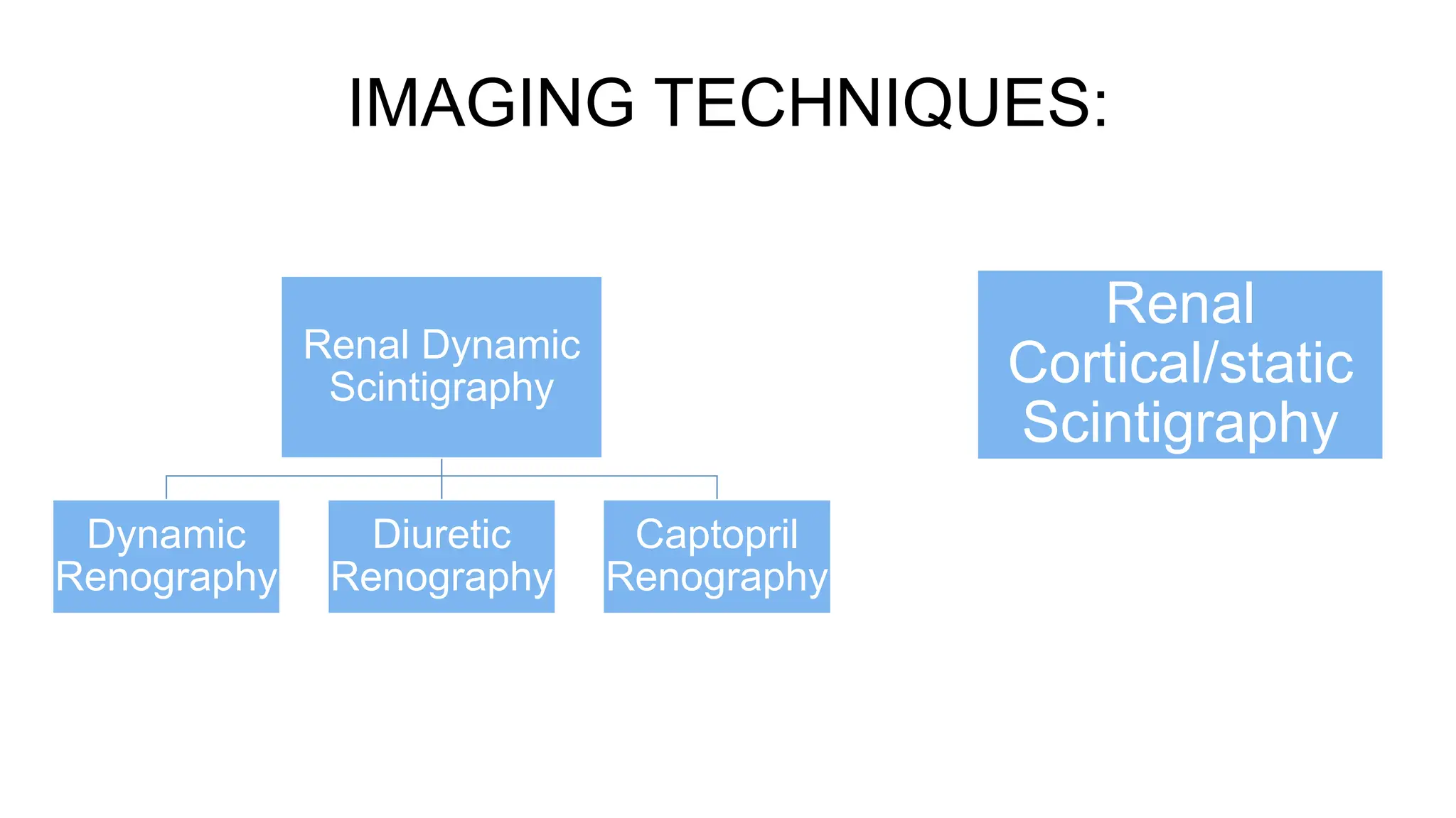
3. INTRODUCTION
• Medical imaging of the kidney using radionuclide material and viewed with a
Gamma camera.
• Also called Nuclear Renography or Radioisotope Renography.
• Done by injecting a radionuclide material into the intravenous system and it’s
progress can be traced using a gamma camera.
• Radionuclides contain radioactive atoms & when they decay , they emit
gamma rays that are detected by the gamma camera.
4. GAMMA CAMERA
Consists of :-
• Collimators – Made of lead, helps maintain image quality.
• Scintillator(Image crystal) – Converts gamma ray photons to visible light.
• Photomultiplier tube(PMT) – Converts the visible light to electrical signals.
• Pre-amplifier – Attached to the back of the PMT to amplify the electrical
signal, and send to the computer for encoding and image formation.
5.
6. POSITIONING
• Posterior - Patient will lie supine or can be seated on a stool where the
detector is brought as close as possible to the patient’s back.
• Right Posterior Oblique(RPO) – To view the right kidney better, with the
patient lying sideward on his/her right side.
• Left Posterior Oblique(LPO) – To view the left kidney better, with the patient
lying sideward on his/her left side.
7. RADIONUCLIDES
• Isotope - Two or more forms of the same element that contain equal numbers
of protons but different numbers of neutrons in their nuclei.
• Isotopes attempting to reach stability by emitting radiation are called
radionuclides/radioisotopes.
• Divided into 2 groups:
» Rapidly eliminated by the kidneys
» Concentrated in the renal parenchyma for a sufficiently long time
8. • Rapidly eliminated by the kidneys:-
» 99mTc-MAG(mercapto-acetyl-triglycine)
» 99mTc-DTPA(diethylene triamine penta-acetic acid)
» 99mTc-GHA(glucoheptonate)
» 99mTc-LLEC(L,L ethylene cystine)
» 123I-OIH(orthoiodohippurate)
• Concentrated in renal parenchyma for a long time:-
» 99mTc-DMSA(dimercaptosuccinic acid)
» 99mTc-GHA
9. 99Tc DTPA
• ECF distribution
• Tracer of choice for dynamic renal scintigraphy.
• 2-6% plasma protien binding
• Extraction fraction 20 %
• 90% excreted in urine in first 4hrs
• Useful in measuring GFR
10. 99Tc - DMSA
• 75% Plasma protein binding.
• Slow renal excretion – retained in renal cortex.
• 40-65% of injected dose concentrated in renal cortex at 6 hours.
• Gives best morphological images.
• Assessment of scarring.
• Differential renal function.
11. 99Tc – MAG3
• It is the most commonly used renal radiopharmaceutical.
• Excreted by active tubular secretion and remaining by glomerular
filtration.
• Preferred in paediatric patients and patients with poor renal
function.
13. DYNAMIC RENOGRAPHY
Indications :-
• Evaluation of obstruction.
• Assessment of renal function following drainage procedures
of the urinary tract.
• Demonstration of vesicoureteric reflux.
• Assessment of renal transplantation.
• Renal trauma.
• Diagnosis of Renal artery stenosis.
Contraindications :-
• Pregnancy
14. • Patient preparation :-
» Should be well hydrated before study
» Patient is asked to empty their bladder before examination
» In suspected cases of obstruction, Foley’s should be
inserted.
» IV line is established to avoid extravasations of the tracer.
• Radiopharmaceuticals used :-
» 99mTc-MAG(Most common).
» 99mTc-DTPA
» 99mTc-LLEC
» 99mTc-GHA
Dose - 0.100 mCi/kg
» 123I-OIH
Dose -0.010
15. Imaging:-
• Examined in supine position with gamma camera placed underneath
the examination table.
• In children, appropriate immobilisation should be attained during
imaging.
• Once appropriately positioned, a rapid IV bolus of the tracer is injected
and simultaneously the acquisition is started.
• 1 frame/2s is recorded for 1 minute, followed by 1 frame/15s for a
duration of 20-30 mins.
16. Parenchymal phase:
Visualized 60-120 seconds after the initial vascular distribution of the tracer and
shows:-
• Relative and absolute size of the functional renal parenchyma units
• Total renal function(kidney/background ratio)
• Relative or split renal function
• Overall renal morphology and redistribution of functioning parenchyma
• Position of the renal units.
17. Cortical transit time :-
• Interval between IV injection of the radiotracer and its first appearance
within the renal collecting system
• Normally, it’s about 3-6 mins.
• Normal cortical transit time indicates that the renal parenchymal
function is not compromised.
• Poorer the renal function, longer is the cortical transit time.
• Conditions which prolong the cortical transit time are:-
» Ureteral obstruction
» Acute and chronic pyelonephritis
» Nephrotoxicity
» Trauma
» Renal artery stenosis
» Renal vein thrombosis
» Acute Tubular necrosis
» Allograft rejection
18. Drainage phase:-
• Passage of radiotracer from the pelvicalyceal system through the ureter into
the bladder
• Most of the radiotracer leaves the renal parenchyma after 20 mins
19.
20. DIURETIC RENOGRAPHY
• Uses 99mTc-MAG combined with IV Furosemide administered 20-30 mins
after injection of the radiotracer.
• Used to distinguish between simple dilation of the collecting system and true
obstruction.
• Rapid washout of the radiotracer from the kidney indicates simple dilation.
• Persistence of the radiotracer indicates a degree of obstruction.
21. CAPTOPRIL RENOGRAPHY
• To evaluate for renal artery stenosis and renovascular hypertension.
• Patient needs to be well hydrated and not eat anything for atleast 4 hours before the exam.
• ACE inhibitors or Angiotension receptor blockers(ARBs) should be stopped prior to exam.
• Baseline study is obtained either one hour after oral captopril(1 mg/kg upto 50 mg) or 15 mins after
IV enalapril(0.03 mg/kg).
• Typical findings of a positive study include:-
» Split renal uptake
» Increase in cortical transit time
» Prolongation of time to peak and retention of tracer in the renal
parenchyma.
22. CORTICAL/STATIC RENAL
SCINTIGRAPHY
• Indications include evaluation of renal scarring or
calculation of differential renal function.
• 99mTc-DMSA is used.
• Imaging occurs 2-4 hours after radiotracer administration.
• Imaging techniques:-
– Planar Renal Scinitigraphy
– Magnified Renal Scintigraphy
– SPECT(Single Photon Emission Computed Tomography)