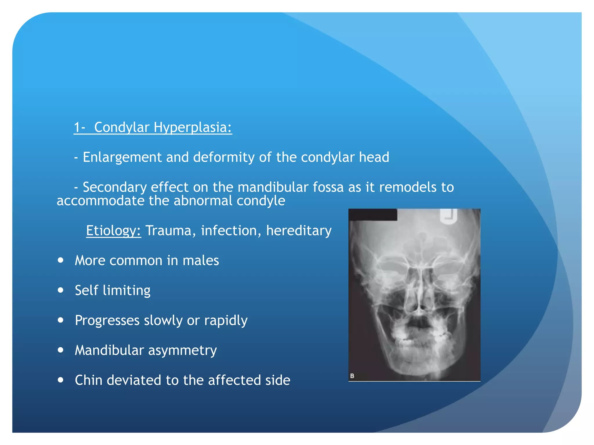
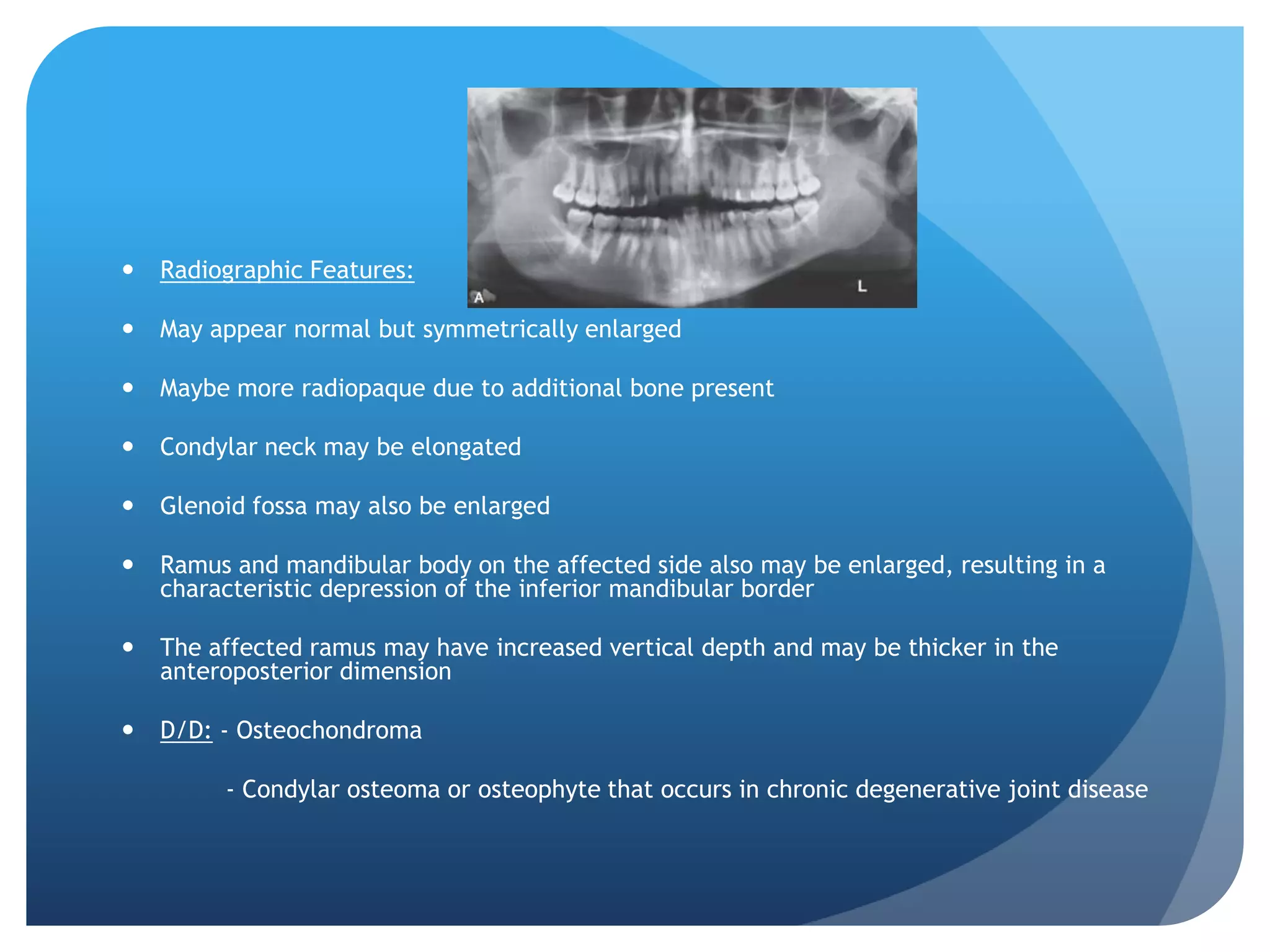
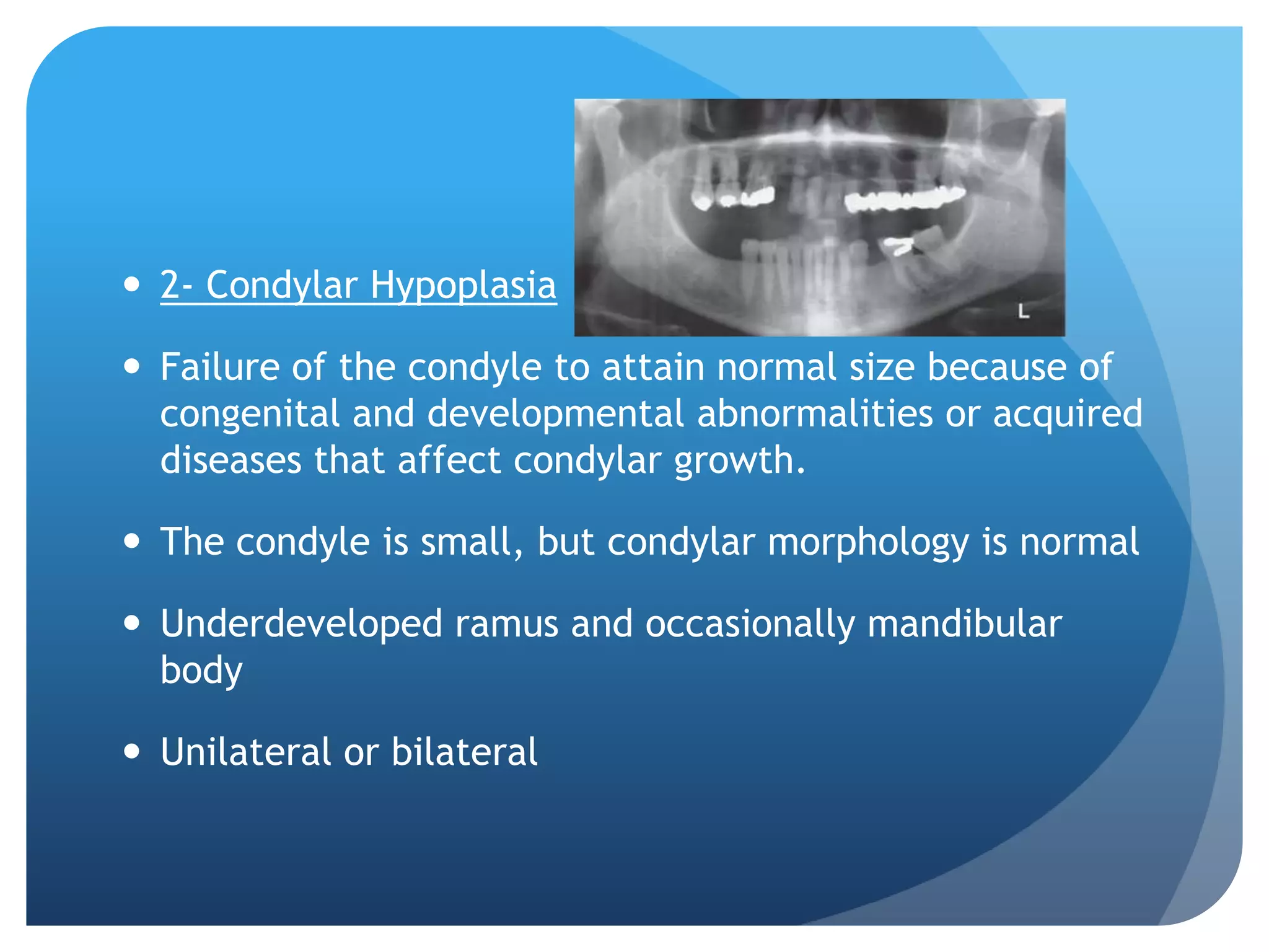
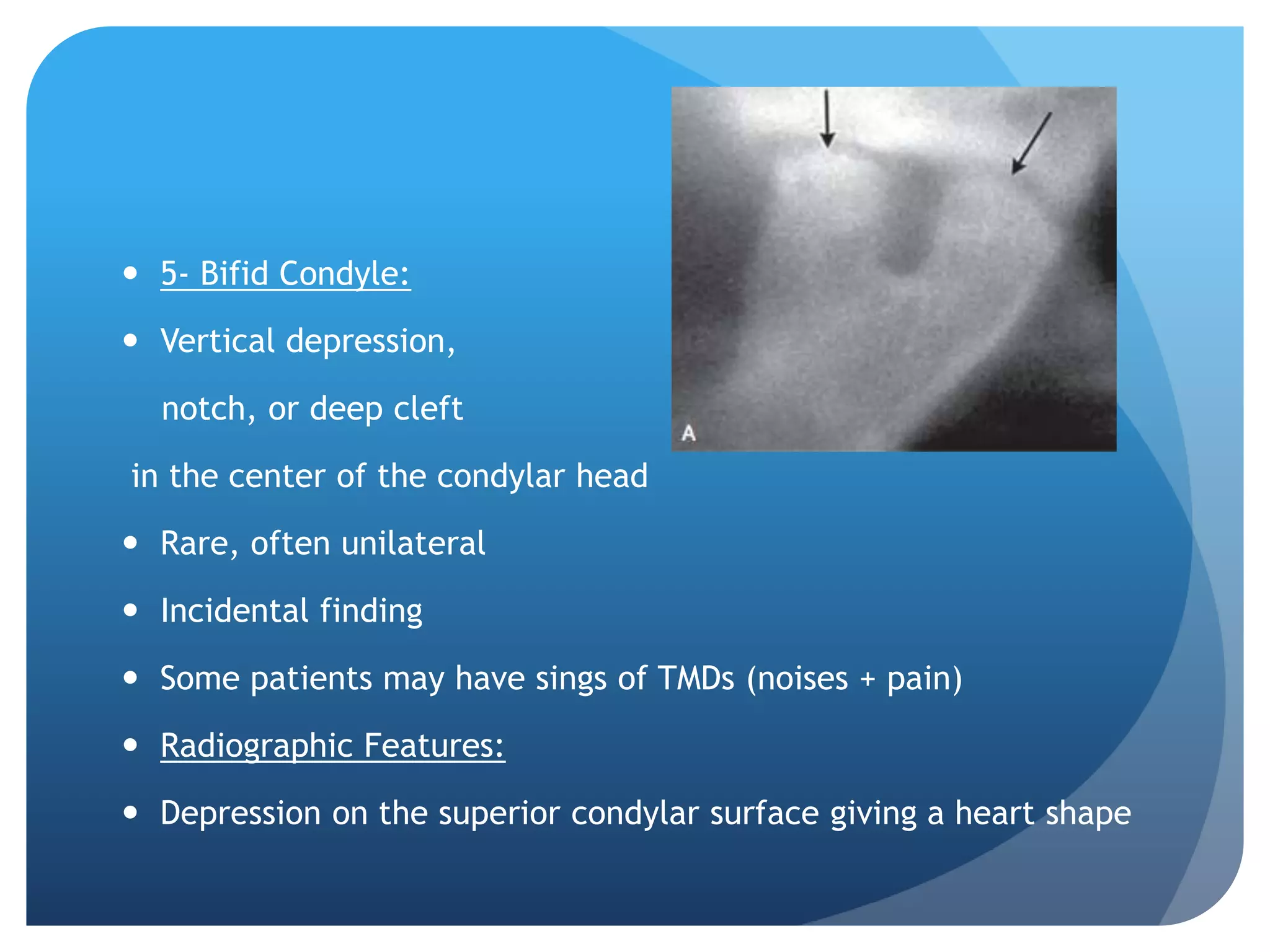
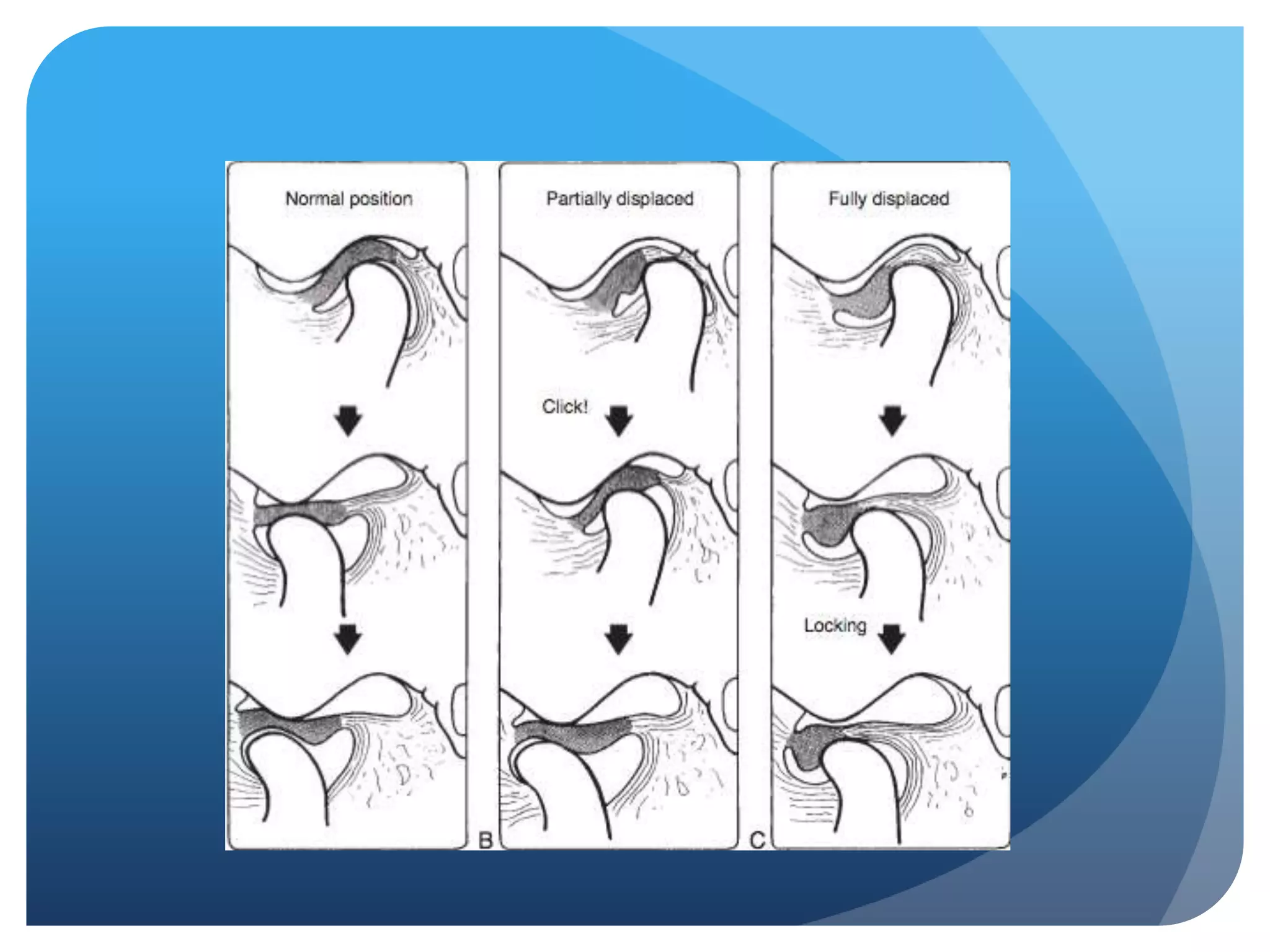
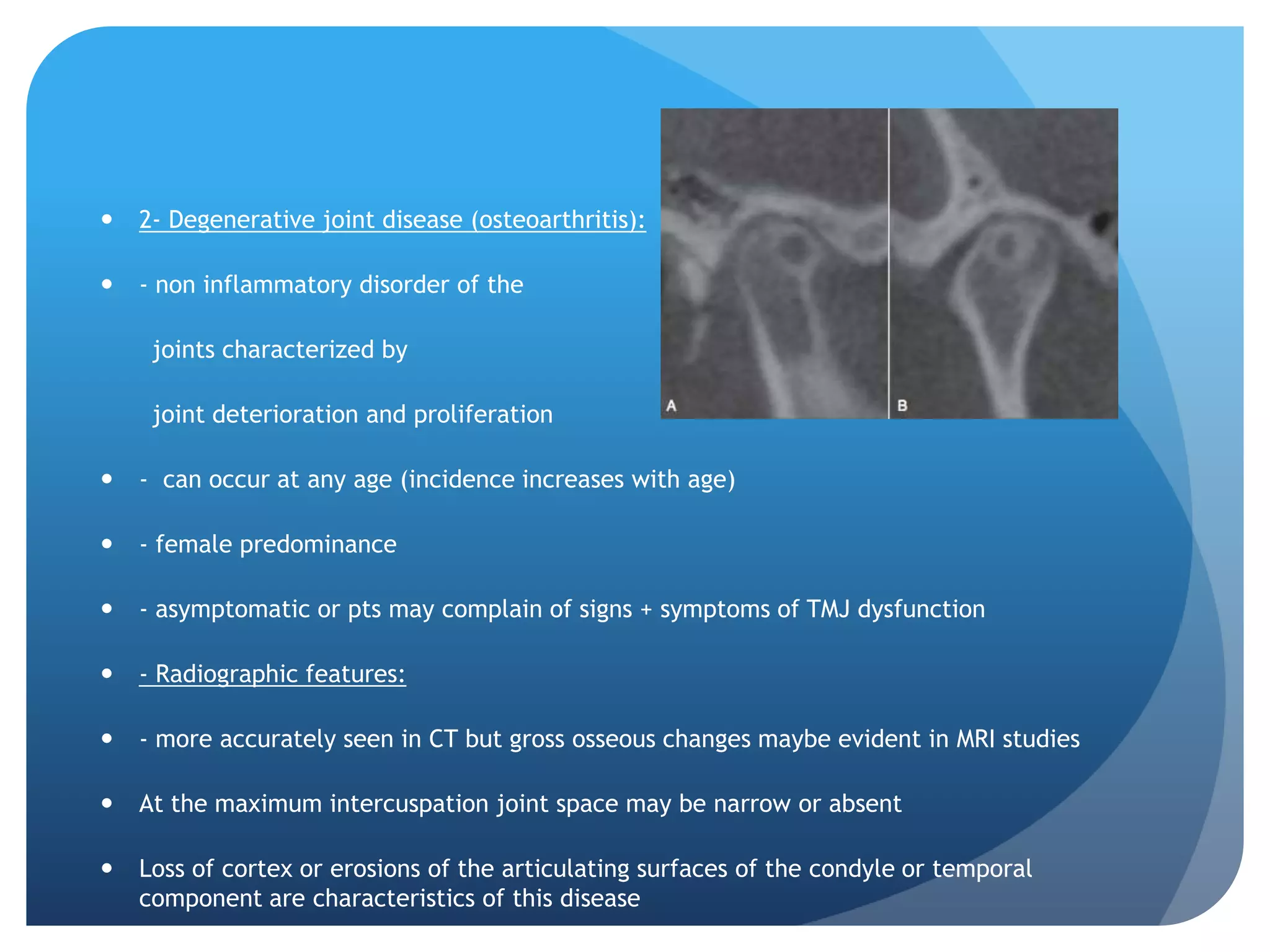
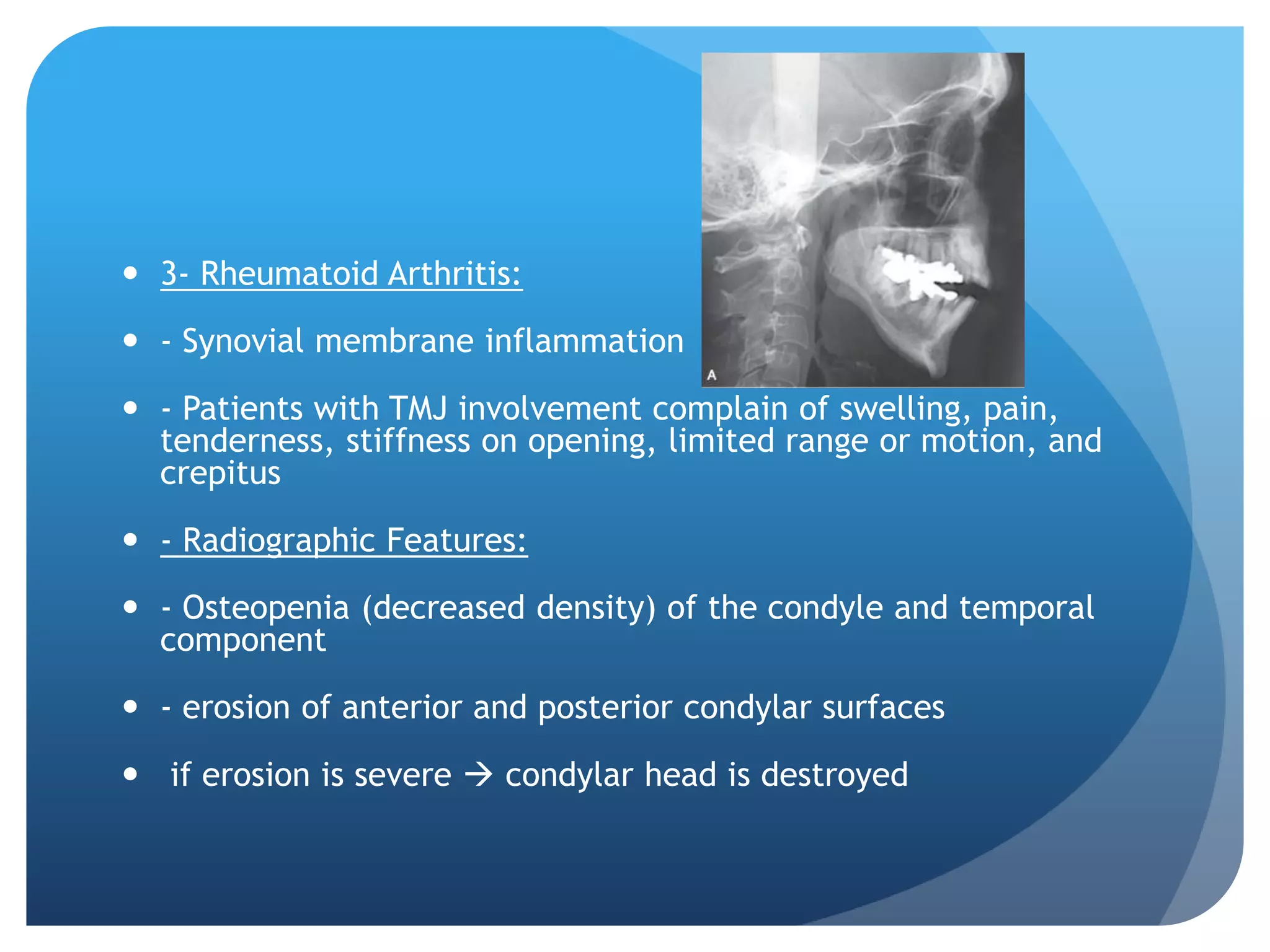
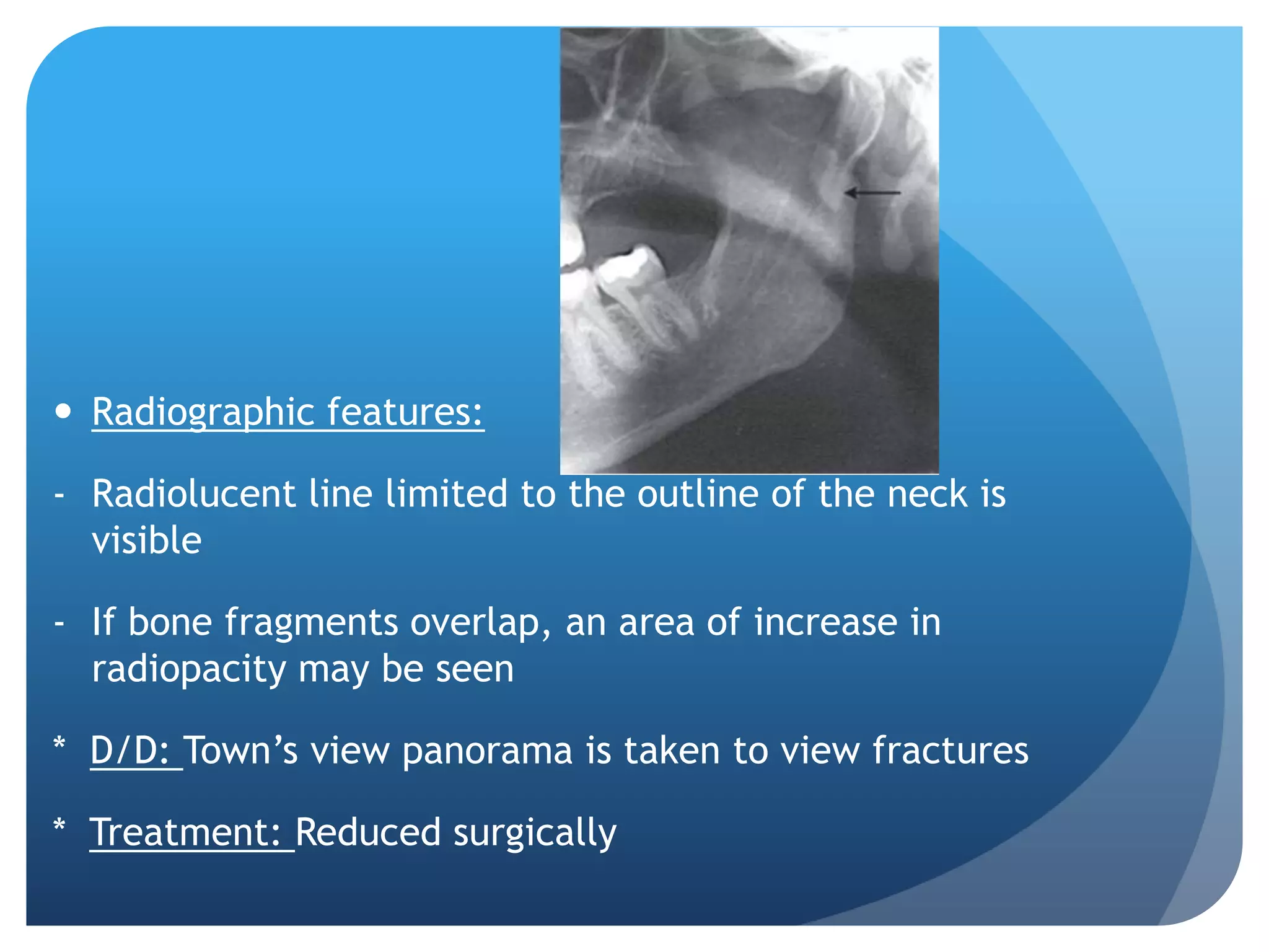
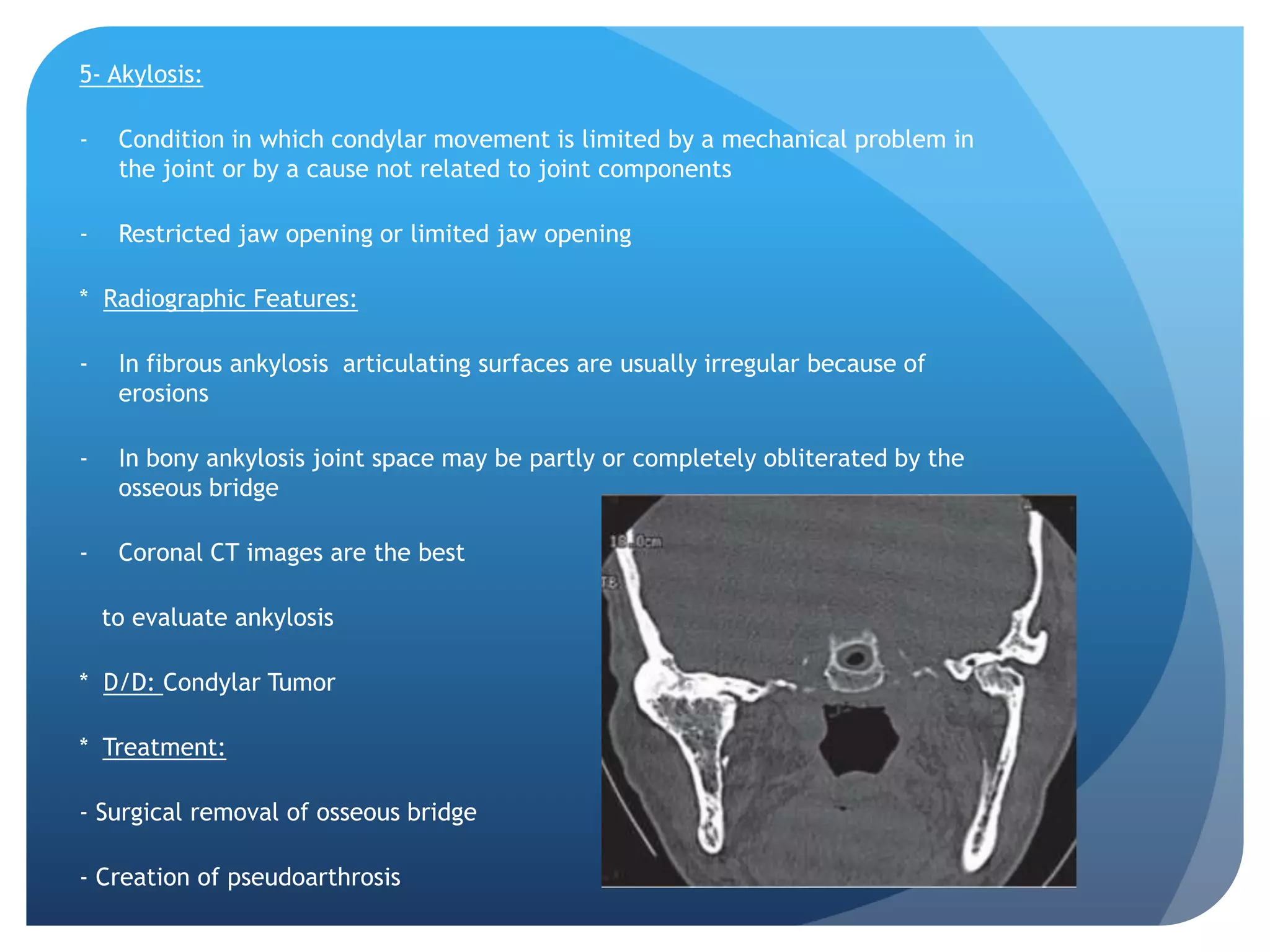
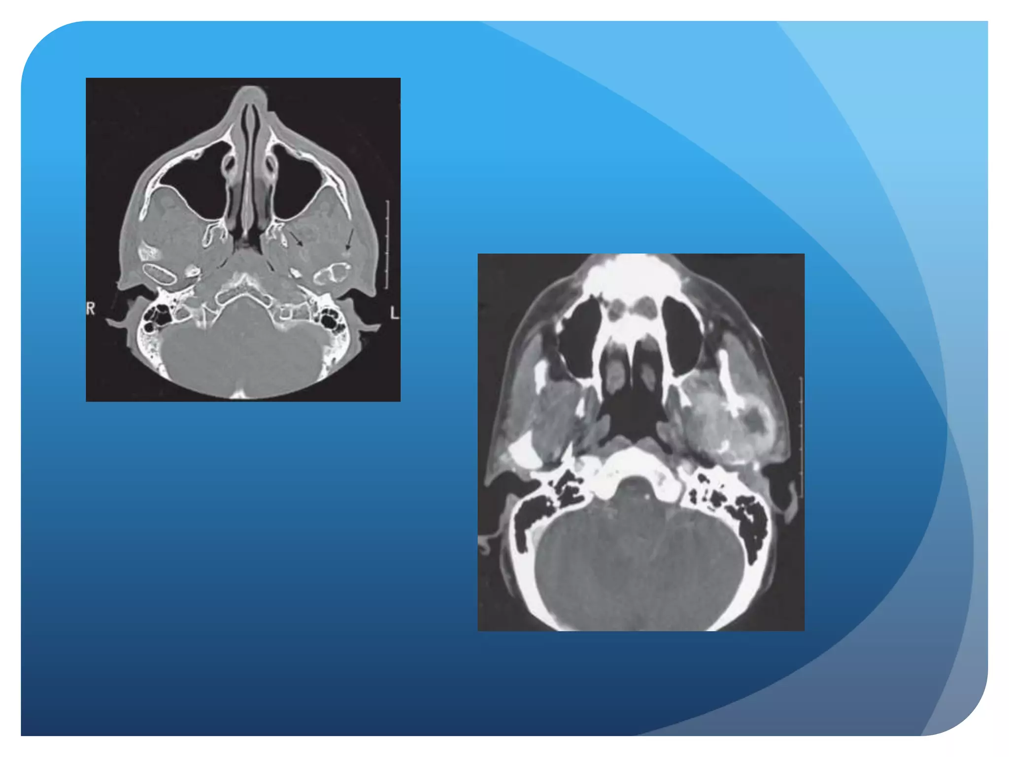
Diagnostic Imaging of the
Temporomandibular Joint
          Fares H. Hanafieh & Fahad F. Salehi
 What is the Temporomandibular joint?


 Unique in that it constitutes of two separate joints
  anatomically and they function together as a single unit
 Consists of: Condyles
              Articular Disc
              Mandibular Fossa


 Has a fibrous capsule that surrounds and encloses the
  joint
 CONDYLE:

-   Shape of condyle varies considerably

-    Superior aspect maybe flattened,

     rounded or markedly convex

-   Mediolateral contour is usually slightly

    convex

-   Variations in shape may cause difficulty

    with radiographic interpretation

-   Extreme aspects of the condyle are the medial

    pole and lateral pole
 MANDIBULAR FOSSA: Composed of the glenoid fossa and atricular eminence.


 INTERARTICULAR DISK: - Between condylar head and

                                 mandibular fossa

                                - Biconcave shape
Disorders of the temporomandibular joint are
abnormalities that interfere with the normal form or
                function of the joint
Disorders of the
      Temporomandibular Joint
1- Developmental Abnormalities

2-   Soft Tissue Abnormalities
 Developmental Abnormalities:

1- Condylar Hyperplasia

2- Condylar Hypoplasia

3- Juvenile Arthrosis

4- Coronoid Hyperplasia

5- Bifid Condyle
1- Condylar Hyperplasia:

  - Enlargement and deformity of the condylar head

  - Secondary effect on the mandibular fossa as it remodels to
accommodate the abnormal condyle

    Etiology: Trauma, infection, hereditary

 More common in males

 Self limiting

 Progresses slowly or rapidly

 Mandibular asymmetry

 Chin deviated to the affected side
   Radiographic Features:

   May appear normal but symmetrically enlarged

   Maybe more radiopaque due to additional bone present

   Condylar neck may be elongated

   Glenoid fossa may also be enlarged

   Ramus and mandibular body on the affected side also may be enlarged, resulting in a
    characteristic depression of the inferior mandibular border

   The affected ramus may have increased vertical depth and may be thicker in the
    anteroposterior dimension

   D/D: - Osteochondroma

         - Condylar osteoma or osteophyte that occurs in chronic degenerative joint disease
 Treatment:

 Orthodontics combined with orthognathic surgery
 2- Condylar Hypoplasia

 Failure of the condyle to attain normal size because of
  congenital and developmental abnormalities or acquired
  diseases that affect condylar growth.

 The condyle is small, but condylar morphology is normal

 Underdeveloped ramus and occasionally mandibular
  body

 Unilateral or bilateral
 Radiographic Features:

 The condylar neck and coronoid process usually are very slender
  and are shortened or elongated in some cases

 The ramus and mandibular body on the affected side may also be
  small, resulting in a mandibular asymmetry and occasional
  dental crowding, depending on the severity of mandibular
  underdevelopment

 D/D: Juvenile rheumatoid arthritis and arthritic conditions

 Treatment: orthognathic surgery

                  bone grafts

                  orthodontic therapy maybe required
 3- Juvenile Arthrosis:

 Manifests as hypoplasia and characteristic morphologic
  abnormalities

 May be a form of condylar hypoplasia

 It affects children and adolescents during the of
  mandibular growth

 More common in females

 Incidental finding in a panoramic projection
 Radiographic appearance:

 Condylar head develops a characteristic “toadstool” appearance

 Condylar neck is shortened or even absent in some cases

 D/D: developmental hypoplasia

          rheumatoid arthritis

* Treatment: orthrognathic surgery

              orthodontic therapy
 4- Coronoid Hyperplasia:

 - acquired or developmental

 - elongation of the coronoid process

 - developmental -> bilateral

 acquired -> uni or bilateral

 - inability to open mouth

 - painless
 - Radiographic features:

 Best seen in panoramic,

  Waters, and lateral tomographic views and on CT scans

 TMJs usually appear normal

 -D/D: Unilateral cases should be differentiated from a tumor of the
  coronoid process (osteochondroma or osteoma)

 Unlike coronoid hyperplasia, tumors have an irregular shape

 -Treatment: surgical removal or the coronoid process and postoperative
  physiotherapy
 5- Bifid Condyle:

 Vertical depression,

  notch, or deep cleft

in the center of the condylar head

 Rare, often unilateral

 Incidental finding

 Some patients may have sings of TMDs (noises + pain)

 Radiographic Features:

 Depression on the superior condylar surface giving a heart shape
 D/D: Vertical fracture through the condylar head

 Treatment: Not indicated unless pain or functional
  impairment is present
Soft Tissue Abnormalities

 Internal Derangements - abnormality in the articular disc and
  may interfere with normal function

 - Cause is unknown

 - Internal derangements can be diagnosed by MRI

 Clinical Features:

 - found in both symptomatic and healthy pts

 - symptomatic pts may have a decreased range of mandibular
  motion

 -displacements may be unilateral or bilateral
 Radiographic Features:

 - MRI is the technique of choice

 Disc Displacement:

 - Anterior displacement is most common

 - The articular disc is located anterior to the condylar head

 Disk reduction and nonreduction:

 - reduction is when an anteriorly displaced disk may reduce to a normal
  relationship with the condylar head during any part of the mouth
  opening movement

 - nonreduction is when the disk remains anteriorly displaced and will
  undergo permanent deformation.
 Perforation and Deformities:

 - perforations between the superior and inferior joint spaces
  most commonly occur in the retrodiskal tissue, just behind the
  posterior band of the disk

 - Not reliably detected with MRI

 Fibrous Adhesions and Effusion:

 - Fibrous adhesions are masses of fibrous or scarred tissue that
  form in the joint space, particularly after TMJ surgery

 - Joint Effusion means fluid in the joint and is considered to be
  and early change that may precede degenerative joint disease

 - Both can be detected by MRI
Remodeling and Arthritic conditions

 1- Remodeling:

 - Adaptive response of cartilage and osseous tissue to forces
  applied to the joint that maybe excessive, resulting in
  alteration of the shape of the condyle and articular eminence

 - no destruction or degeneration of articular soft tissue occurs

 - occurs throughout adult life

 - considered abnormal only if it is accompanied by clinical
  signs and symptoms of pain or dysfunction
 - Radiographic Features:

 - flattening

 - cortical thickening of articulating surfaces

 - subchondral sclerosis

 -D/D: flattening and subchondral sclerosis maybe difficult to
  differentiate from early degenerative joint disease

 - Treatment:

 - Only indicated when signs and symptoms are present. (ex. Splint
  therapy)
   2- Degenerative joint disease (osteoarthritis):

   - non inflammatory disorder of the

     joints characterized by

     joint deterioration and proliferation

   - can occur at any age (incidence increases with age)

   - female predominance

   - asymptomatic or pts may complain of signs + symptoms of TMJ dysfunction

   - Radiographic features:

   - more accurately seen in CT but gross osseous changes maybe evident in MRI studies

   At the maximum intercuspation joint space may be narrow or absent

   Loss of cortex or erosions of the articulating surfaces of the condyle or temporal
    component are characteristics of this disease
 D/D:
 - Erosive appearance  inflammatory arthritides (rheumatoid
  arthritis)
 - Proliferative appearance with extensive osteophyte
  formation  benign tumor  osteoma or osteochondroma
 Treatment:
 - Relieving joint stress (e.g. Splint therapy)
 - relieving secondary inflammation with anti-inflammatory
  drugs
 - Increasing joint mobility and function  physiotherapy
 3- Rheumatoid Arthritis:

 - Synovial membrane inflammation

 - Patients with TMJ involvement complain of swelling, pain,
  tenderness, stiffness on opening, limited range or motion, and
  crepitus

 - Radiographic Features:

 - Osteopenia (decreased density) of the condyle and temporal
  component

 - erosion of anterior and posterior condylar surfaces

 if erosion is severe  condylar head is destroyed
 D/D: severe DJD and psoriatic arthritis and osteopenia

 Treatment:

 - pain relief (analgesics)

 - anti inflammatory drugs

 - physiotherapy

 - surgery (joint replacement)
 4- Juvenile Arthritis:

 - Inflammatory disease that is characterized by chronic, intermittent
  synovial inflammation

 - results in: synovial hypertrophy, joint effusion, and swollen, painful
  joints

 -pain and tenderness of affected joint or joints

 - can be asymptomatic

 - unilateral is common

 - facial appearance known as “bird face”

 - possible mandibular asymmetry if one side is more severely affected
 Radiographic features:

 - Osteopenia (decreased density) maybe only an initial
  radiographic finding

 - Impaired mandibular growth

 - Severe cases: only pencil shaped small condyle
  remains

 - Abnormal disk shape is often observed in patients with
  TMJ involvement
Psoriatic Arthritis and

                               Akylosing Spondylitis

Septic Arthritis: Infection and inflammation of a joint that can result in joint
destruction

-   Affects any age

-   No sex predilection

-   Occurs unilaterally

-   Redness and swelling over joint

-   Trismus

-   Severe pain on opening

-   Inability to occlude the teeth

-   Large, tender cervical lymph nodes

-   Fever and malaise
 Radiographic Features:

- No radiographic signs may be present in early stages of
  the disease

- Osteopenic (radiolucent) changes of the joint
  components and mandibular ramus may be evident (7-10
  days after onset of clinical symptoms)

- Osseous ankylosis may occur after infection subsides
 D/D: radiographic changes caused by septic arthritis
  may mimic those of severe DJD or RA

 Treatment:

- Antimicrobial therapy

- Drainage of effusion and joint rest

- Physiotherapy
Articular Loose Bodies

-   Radiopacities of varying origin located in the joint synoviom, within the
    capsule in the joint spaces, or outside in soft tissue


1- Synovial Chondromatosis:

-   Uncommon disorder characterized by metaplastic formation of multiple
    cartilaginous and osteocartilaginous nodules within connective tissue of the
    synovial membrane of joint

-   Asymptomatic

-   May complain of preauricular swelling, pain, and decreased range of motion

-   Some patients have crepitus or other joint noises
 Radiographic Features:

- Osseous components may appear normal or may exhibit
  osseous changes similar to those in DJD

- Sclerosis of glenoid fossa and condyle may be seen (chronic
  bone reaction to an active lesion)

- MRI may be useful in defining the tissue planes between the
  synovial chondromatosis and surrounding soft tissue

* D/D: DJD with joint mice or chondrosarcoma or osteosarcoma

* Treatment: Arthroscopic or open joint surgery  remove loose
bodies and resection of abnormal synovial tissue
2- Chondrocalcinosis:
- Characterized by acute or chronic synovitis and
  precipitation of calcium pyrophosphate dihydrate
  crystals in the joint space
- Most commonly affected joints are knee, wrist,
  shoulder, and elbow
- TMJ involvement uncommon
- Unilaterally and more common in males
- Asymptomatic or complaints of pain and joint swellings
 Radiographic Features:

-   May simulate synovial chondromatosis

-   Bone erosions and severe increase in condylar bone density

-   Erosions of the glenoid fossa may be present (detected with CT)

-   Soft tissue swelling and edema of the surrounding muscles may be seen with MRI

* D/D: DJD with joint mince or chondrosarcoma or osteosarcoma

* Treatment:

-   Surgical removal of crystalline deposits

-   Steroids, aspirin, and non steroidal anti inflammatory agents may provide relief
Trauma

1- Effusion:

- Influx of fluid into the joint as a result of trauma
  (hemorrhage or inflammation)

- Swelling over affected joint

- Pain in TMJ, preauricular region, and limited range of
  motion
 Radiographic Features:
- Commonly seen in conjunction with internal
  derangements
- Joint space is widened
* D/D: septic arthritis
* Treatment:
- Anti-inflammatory drugs
- Surgical drainage
2- Dislocation:

- Abnormal positioning of the condyle out of the mandibular fossa
  but within the joint capsule

- Unable to close mandible to maximal intercuspation

* Radiographic Features:

- In bilateral cases, both condyles are located anterior and
  superior to summits of articular eminentia

* Treatment:

- Manual manipulation to reduce the dislocation

- Surgery in the case of fracture dislocation
3- Fracture:

- Usually occur at condylar neck and often are
  accompanied by dislocation of the condylar head

- Unilateral fractures more common

- May be accompanied by parasymphyseal or mandibular
  body fracture on contralateral side

- Swelling over TMJ

- Limited range of motion
 Radiographic features:

- Radiolucent line limited to the outline of the neck is
  visible

- If bone fragments overlap, an area of increase in
  radiopacity may be seen

* D/D: Town’s view panorama is taken to view fractures

* Treatment: Reduced surgically
4- Neonatal Fracture:

- Use of forceps during delivery of neonates may result in
  fracture and displacement of the rudimentary condyle

- Severe mandibular hypoplasia

* D/D: Developmental hypoplasia

* Treatment: Combination of orthodontic and
orthognathic surgery
5- Akylosis:

-    Condition in which condylar movement is limited by a mechanical problem in
     the joint or by a cause not related to joint components

-    Restricted jaw opening or limited jaw opening

* Radiographic Features:

-    In fibrous ankylosis articulating surfaces are usually irregular because of
     erosions

-    In bony ankylosis joint space may be partly or completely obliterated by the
     osseous bridge

-    Coronal CT images are the best

    to evaluate ankylosis

* D/D: Condylar Tumor

* Treatment:

- Surgical removal of osseous bridge

- Creation of pseudoarthrosis
Tumors

- Intrinsic or extrinsic

- Intrinsic develop in condyle, temporal bone or coronoid
  process

- Extrinsic tumor may affect the morphology, structure
  and function of the joint without invading the joint
  itself
1- Benign Tumors:

- Osteoma, osteochondroma, Langerhans histocytosis and osteoblastomas

- Chondroblastomas, fibromyxomas, benign giant cell lesions and
  anneurysmal bone cysts also occur

- Benign tumors and cysts of the mandible may involve the entire ramus
  and condyle

- Grow slowly

- TMJ swelling

- Pain and decrease in range of motion

- Tumors of coronoid process are painless but may complain of progressive
  limitation of motion
* Radiographic Features:
- Condylar tumors  condylar enlargement with irregular
  outline
- Osteoma and osteochondroma appear as abnormal,
  pedunculated mass attached to the condyle
* D/D: Condylar neoplasms may simulate condylar
hyperplasia because of condylar enlargement although it
might be irregular in appearance
* Treatment: Surgical excision of tumor and occasionally
excision of condylar head or coronoid process
2- Malignant Tumors:

A- Primary (rare): - Intrinsic

                       - Extrinsic

Intrinsic: Chondrosarcoma

           Osteogenic sarcoma

           Senovial sarcoma

           Fibrosarcoma

Extrinsic: Direct extension of adjacent parotid salivary gland
malignancies
B- Metastatic (more common)

-   May be asymptomatic or patients may have symptoms of TMJ dysfunction
    (pain, limited mandibular opening, mandibular deviation and swelling)

* Radiographic Features:

-   Variant degree of bone destruction with ill defined, irregular margins

-   CT modality of choice

-   MRI useful for displaying extent of involvement into surrounding tissues

* D/D: Osseous destruction of bone seen in severe DJD

* Treatment:

-   Wide surgical removal of tumor

-   May include radiotherapy and chemotherapy
Thank you

Radiographic imaging of TMJ

  • 1.
    Diagnostic Imaging ofthe Temporomandibular Joint Fares H. Hanafieh & Fahad F. Salehi
  • 2.
     What isthe Temporomandibular joint?  Unique in that it constitutes of two separate joints anatomically and they function together as a single unit  Consists of: Condyles Articular Disc Mandibular Fossa  Has a fibrous capsule that surrounds and encloses the joint
  • 3.
     CONDYLE: - Shape of condyle varies considerably - Superior aspect maybe flattened, rounded or markedly convex - Mediolateral contour is usually slightly convex - Variations in shape may cause difficulty with radiographic interpretation - Extreme aspects of the condyle are the medial pole and lateral pole
  • 4.
     MANDIBULAR FOSSA:Composed of the glenoid fossa and atricular eminence.  INTERARTICULAR DISK: - Between condylar head and mandibular fossa - Biconcave shape
  • 5.
    Disorders of thetemporomandibular joint are abnormalities that interfere with the normal form or function of the joint
  • 6.
    Disorders of the Temporomandibular Joint 1- Developmental Abnormalities 2- Soft Tissue Abnormalities
  • 7.
     Developmental Abnormalities: 1-Condylar Hyperplasia 2- Condylar Hypoplasia 3- Juvenile Arthrosis 4- Coronoid Hyperplasia 5- Bifid Condyle
  • 8.
    1- Condylar Hyperplasia: - Enlargement and deformity of the condylar head - Secondary effect on the mandibular fossa as it remodels to accommodate the abnormal condyle Etiology: Trauma, infection, hereditary  More common in males  Self limiting  Progresses slowly or rapidly  Mandibular asymmetry  Chin deviated to the affected side
  • 9.
    Radiographic Features:  May appear normal but symmetrically enlarged  Maybe more radiopaque due to additional bone present  Condylar neck may be elongated  Glenoid fossa may also be enlarged  Ramus and mandibular body on the affected side also may be enlarged, resulting in a characteristic depression of the inferior mandibular border  The affected ramus may have increased vertical depth and may be thicker in the anteroposterior dimension  D/D: - Osteochondroma - Condylar osteoma or osteophyte that occurs in chronic degenerative joint disease
  • 10.
     Treatment:  Orthodonticscombined with orthognathic surgery
  • 11.
     2- CondylarHypoplasia  Failure of the condyle to attain normal size because of congenital and developmental abnormalities or acquired diseases that affect condylar growth.  The condyle is small, but condylar morphology is normal  Underdeveloped ramus and occasionally mandibular body  Unilateral or bilateral
  • 12.
     Radiographic Features: The condylar neck and coronoid process usually are very slender and are shortened or elongated in some cases  The ramus and mandibular body on the affected side may also be small, resulting in a mandibular asymmetry and occasional dental crowding, depending on the severity of mandibular underdevelopment  D/D: Juvenile rheumatoid arthritis and arthritic conditions  Treatment: orthognathic surgery bone grafts orthodontic therapy maybe required
  • 13.
     3- JuvenileArthrosis:  Manifests as hypoplasia and characteristic morphologic abnormalities  May be a form of condylar hypoplasia  It affects children and adolescents during the of mandibular growth  More common in females  Incidental finding in a panoramic projection
  • 14.
     Radiographic appearance: Condylar head develops a characteristic “toadstool” appearance  Condylar neck is shortened or even absent in some cases  D/D: developmental hypoplasia rheumatoid arthritis * Treatment: orthrognathic surgery orthodontic therapy
  • 15.
     4- CoronoidHyperplasia:  - acquired or developmental  - elongation of the coronoid process  - developmental -> bilateral  acquired -> uni or bilateral  - inability to open mouth  - painless
  • 16.
     - Radiographicfeatures:  Best seen in panoramic, Waters, and lateral tomographic views and on CT scans  TMJs usually appear normal  -D/D: Unilateral cases should be differentiated from a tumor of the coronoid process (osteochondroma or osteoma)  Unlike coronoid hyperplasia, tumors have an irregular shape  -Treatment: surgical removal or the coronoid process and postoperative physiotherapy
  • 17.
     5- BifidCondyle:  Vertical depression, notch, or deep cleft in the center of the condylar head  Rare, often unilateral  Incidental finding  Some patients may have sings of TMDs (noises + pain)  Radiographic Features:  Depression on the superior condylar surface giving a heart shape
  • 18.
     D/D: Verticalfracture through the condylar head  Treatment: Not indicated unless pain or functional impairment is present
  • 19.
    Soft Tissue Abnormalities Internal Derangements - abnormality in the articular disc and may interfere with normal function  - Cause is unknown  - Internal derangements can be diagnosed by MRI  Clinical Features:  - found in both symptomatic and healthy pts  - symptomatic pts may have a decreased range of mandibular motion  -displacements may be unilateral or bilateral
  • 21.
     Radiographic Features: - MRI is the technique of choice  Disc Displacement:  - Anterior displacement is most common  - The articular disc is located anterior to the condylar head  Disk reduction and nonreduction:  - reduction is when an anteriorly displaced disk may reduce to a normal relationship with the condylar head during any part of the mouth opening movement  - nonreduction is when the disk remains anteriorly displaced and will undergo permanent deformation.
  • 22.
     Perforation andDeformities:  - perforations between the superior and inferior joint spaces most commonly occur in the retrodiskal tissue, just behind the posterior band of the disk  - Not reliably detected with MRI  Fibrous Adhesions and Effusion:  - Fibrous adhesions are masses of fibrous or scarred tissue that form in the joint space, particularly after TMJ surgery  - Joint Effusion means fluid in the joint and is considered to be and early change that may precede degenerative joint disease  - Both can be detected by MRI
  • 23.
    Remodeling and Arthriticconditions  1- Remodeling:  - Adaptive response of cartilage and osseous tissue to forces applied to the joint that maybe excessive, resulting in alteration of the shape of the condyle and articular eminence  - no destruction or degeneration of articular soft tissue occurs  - occurs throughout adult life  - considered abnormal only if it is accompanied by clinical signs and symptoms of pain or dysfunction
  • 24.
     - RadiographicFeatures:  - flattening  - cortical thickening of articulating surfaces  - subchondral sclerosis  -D/D: flattening and subchondral sclerosis maybe difficult to differentiate from early degenerative joint disease  - Treatment:  - Only indicated when signs and symptoms are present. (ex. Splint therapy)
  • 25.
    2- Degenerative joint disease (osteoarthritis):  - non inflammatory disorder of the joints characterized by joint deterioration and proliferation  - can occur at any age (incidence increases with age)  - female predominance  - asymptomatic or pts may complain of signs + symptoms of TMJ dysfunction  - Radiographic features:  - more accurately seen in CT but gross osseous changes maybe evident in MRI studies  At the maximum intercuspation joint space may be narrow or absent  Loss of cortex or erosions of the articulating surfaces of the condyle or temporal component are characteristics of this disease
  • 26.
     D/D:  -Erosive appearance  inflammatory arthritides (rheumatoid arthritis)  - Proliferative appearance with extensive osteophyte formation  benign tumor  osteoma or osteochondroma  Treatment:  - Relieving joint stress (e.g. Splint therapy)  - relieving secondary inflammation with anti-inflammatory drugs  - Increasing joint mobility and function  physiotherapy
  • 27.
     3- RheumatoidArthritis:  - Synovial membrane inflammation  - Patients with TMJ involvement complain of swelling, pain, tenderness, stiffness on opening, limited range or motion, and crepitus  - Radiographic Features:  - Osteopenia (decreased density) of the condyle and temporal component  - erosion of anterior and posterior condylar surfaces  if erosion is severe  condylar head is destroyed
  • 28.
     D/D: severeDJD and psoriatic arthritis and osteopenia  Treatment:  - pain relief (analgesics)  - anti inflammatory drugs  - physiotherapy  - surgery (joint replacement)
  • 29.
     4- JuvenileArthritis:  - Inflammatory disease that is characterized by chronic, intermittent synovial inflammation  - results in: synovial hypertrophy, joint effusion, and swollen, painful joints  -pain and tenderness of affected joint or joints  - can be asymptomatic  - unilateral is common  - facial appearance known as “bird face”  - possible mandibular asymmetry if one side is more severely affected
  • 30.
     Radiographic features: - Osteopenia (decreased density) maybe only an initial radiographic finding  - Impaired mandibular growth  - Severe cases: only pencil shaped small condyle remains  - Abnormal disk shape is often observed in patients with TMJ involvement
  • 31.
    Psoriatic Arthritis and Akylosing Spondylitis Septic Arthritis: Infection and inflammation of a joint that can result in joint destruction - Affects any age - No sex predilection - Occurs unilaterally - Redness and swelling over joint - Trismus - Severe pain on opening - Inability to occlude the teeth - Large, tender cervical lymph nodes - Fever and malaise
  • 32.
     Radiographic Features: -No radiographic signs may be present in early stages of the disease - Osteopenic (radiolucent) changes of the joint components and mandibular ramus may be evident (7-10 days after onset of clinical symptoms) - Osseous ankylosis may occur after infection subsides
  • 33.
     D/D: radiographicchanges caused by septic arthritis may mimic those of severe DJD or RA  Treatment: - Antimicrobial therapy - Drainage of effusion and joint rest - Physiotherapy
  • 34.
    Articular Loose Bodies - Radiopacities of varying origin located in the joint synoviom, within the capsule in the joint spaces, or outside in soft tissue 1- Synovial Chondromatosis: - Uncommon disorder characterized by metaplastic formation of multiple cartilaginous and osteocartilaginous nodules within connective tissue of the synovial membrane of joint - Asymptomatic - May complain of preauricular swelling, pain, and decreased range of motion - Some patients have crepitus or other joint noises
  • 35.
     Radiographic Features: -Osseous components may appear normal or may exhibit osseous changes similar to those in DJD - Sclerosis of glenoid fossa and condyle may be seen (chronic bone reaction to an active lesion) - MRI may be useful in defining the tissue planes between the synovial chondromatosis and surrounding soft tissue * D/D: DJD with joint mice or chondrosarcoma or osteosarcoma * Treatment: Arthroscopic or open joint surgery  remove loose bodies and resection of abnormal synovial tissue
  • 36.
    2- Chondrocalcinosis: - Characterizedby acute or chronic synovitis and precipitation of calcium pyrophosphate dihydrate crystals in the joint space - Most commonly affected joints are knee, wrist, shoulder, and elbow - TMJ involvement uncommon - Unilaterally and more common in males - Asymptomatic or complaints of pain and joint swellings
  • 37.
     Radiographic Features: - May simulate synovial chondromatosis - Bone erosions and severe increase in condylar bone density - Erosions of the glenoid fossa may be present (detected with CT) - Soft tissue swelling and edema of the surrounding muscles may be seen with MRI * D/D: DJD with joint mince or chondrosarcoma or osteosarcoma * Treatment: - Surgical removal of crystalline deposits - Steroids, aspirin, and non steroidal anti inflammatory agents may provide relief
  • 38.
    Trauma 1- Effusion: - Influxof fluid into the joint as a result of trauma (hemorrhage or inflammation) - Swelling over affected joint - Pain in TMJ, preauricular region, and limited range of motion
  • 39.
     Radiographic Features: -Commonly seen in conjunction with internal derangements - Joint space is widened * D/D: septic arthritis * Treatment: - Anti-inflammatory drugs - Surgical drainage
  • 40.
    2- Dislocation: - Abnormalpositioning of the condyle out of the mandibular fossa but within the joint capsule - Unable to close mandible to maximal intercuspation * Radiographic Features: - In bilateral cases, both condyles are located anterior and superior to summits of articular eminentia * Treatment: - Manual manipulation to reduce the dislocation - Surgery in the case of fracture dislocation
  • 41.
    3- Fracture: - Usuallyoccur at condylar neck and often are accompanied by dislocation of the condylar head - Unilateral fractures more common - May be accompanied by parasymphyseal or mandibular body fracture on contralateral side - Swelling over TMJ - Limited range of motion
  • 42.
     Radiographic features: -Radiolucent line limited to the outline of the neck is visible - If bone fragments overlap, an area of increase in radiopacity may be seen * D/D: Town’s view panorama is taken to view fractures * Treatment: Reduced surgically
  • 43.
    4- Neonatal Fracture: -Use of forceps during delivery of neonates may result in fracture and displacement of the rudimentary condyle - Severe mandibular hypoplasia * D/D: Developmental hypoplasia * Treatment: Combination of orthodontic and orthognathic surgery
  • 44.
    5- Akylosis: - Condition in which condylar movement is limited by a mechanical problem in the joint or by a cause not related to joint components - Restricted jaw opening or limited jaw opening * Radiographic Features: - In fibrous ankylosis articulating surfaces are usually irregular because of erosions - In bony ankylosis joint space may be partly or completely obliterated by the osseous bridge - Coronal CT images are the best to evaluate ankylosis * D/D: Condylar Tumor * Treatment: - Surgical removal of osseous bridge - Creation of pseudoarthrosis
  • 45.
    Tumors - Intrinsic orextrinsic - Intrinsic develop in condyle, temporal bone or coronoid process - Extrinsic tumor may affect the morphology, structure and function of the joint without invading the joint itself
  • 46.
    1- Benign Tumors: -Osteoma, osteochondroma, Langerhans histocytosis and osteoblastomas - Chondroblastomas, fibromyxomas, benign giant cell lesions and anneurysmal bone cysts also occur - Benign tumors and cysts of the mandible may involve the entire ramus and condyle - Grow slowly - TMJ swelling - Pain and decrease in range of motion - Tumors of coronoid process are painless but may complain of progressive limitation of motion
  • 47.
    * Radiographic Features: -Condylar tumors  condylar enlargement with irregular outline - Osteoma and osteochondroma appear as abnormal, pedunculated mass attached to the condyle * D/D: Condylar neoplasms may simulate condylar hyperplasia because of condylar enlargement although it might be irregular in appearance * Treatment: Surgical excision of tumor and occasionally excision of condylar head or coronoid process
  • 49.
    2- Malignant Tumors: A-Primary (rare): - Intrinsic - Extrinsic Intrinsic: Chondrosarcoma Osteogenic sarcoma Senovial sarcoma Fibrosarcoma Extrinsic: Direct extension of adjacent parotid salivary gland malignancies
  • 51.
    B- Metastatic (morecommon) - May be asymptomatic or patients may have symptoms of TMJ dysfunction (pain, limited mandibular opening, mandibular deviation and swelling) * Radiographic Features: - Variant degree of bone destruction with ill defined, irregular margins - CT modality of choice - MRI useful for displaying extent of involvement into surrounding tissues * D/D: Osseous destruction of bone seen in severe DJD * Treatment: - Wide surgical removal of tumor - May include radiotherapy and chemotherapy
  • 52.

Editor's Notes

  • #14 But is thought to differ in that the affected condyle at one time was normal, becoming abnormal during growth.
  • #18 Heart shape: anteroposterior silhouette
  • #20 Disc is most often displaced in an anterior direction, but maybe be displaced anetromedially, medially, or anterolaterally. (lateral and posterior RARE)
  • #21 A- position and movement of the disk during jaw opening B- mildly displaced anteriorly with reductionC- Severely displaced anteriorly without reduction
  • #22 Reduction: appear normal in MRINonreduction: false interpretation because of the fibrotic changes on the bilaminar zone
  • #23 Fibrous adhesions: Low signal intensityAdhesion: High signal intensity
  • #25 A- The right temporal component shows subchondral sclerosis and flattening (arrow)B- The right condyle shows mild flattening of the lateral aspect and subchondral sclerosis of the medial aspect (arrow) C- Cadaver specimen. Note the flattening of the temporal component (black arrows) and large perforation posterior to a residual deformed disk (white arrow)
  • #26 Deterioration: characterized by loss of articular cartilage and bone erosion Proliferation: proliferative component is characterized by new bone formation at the articular surface and in the subchondral region* Signs and sympts: pain on palpation + movement, joint noises (crepitus), limited range of motion and muscle spasm
  • #28 Bilateral destruction of condyles I \\/anterior open biteChin appears receded
  • #30 2- TMJ involvement occurs in approximately 40% of pts. Unilateral or bilateral 6- Contralateral involvement may occur as the disease progresses 7- Because pts have micrognathic + posteroinferior chin rotation
  • #31 During quiescent periods the cortex of joint surface may appear, and the surfaces will be flattened
  • #33 Coronal reformat CT image of a case of septic arthritis involving the right joint. Note the erosions, sclerosis and periosteal reaction that extends along the back of the condyle and lateral neck of the condyle
  • #36 Cropped panoramic image of a right joint involved with osteochondramatosis
  • #38 CT axial image bone algorithm. Note the calcifications anterior to the right condyle and large erosions involving the medial pole of the condyle.
  • #45 Right condyle and ramus are markedly enlarged
  • #47 Most common benign tumor osteochondroma
  • #49 Axial bone algorithm. CT image of an osteochondroma extending from the anterior surface of the left condylar head (arrow)
  • #51 1-chondrosarcoma (CT axial section bone algorithm) radiolucent destructive lesion present in the left condylar head and faint radiopacities (soft tissue calcifications) are visible anterior to the condylar head (arrows)2- Axial soft tissue algorithm CT image of a metastatic lesion from a carcinoma of the thyroid gland that has destroyed all of the left mandibular condyle.
  • #52 Unfortunately some patients are treated occasionally for temporomandibular joint dysfunction without recognition that the underlying condition is a malignancy