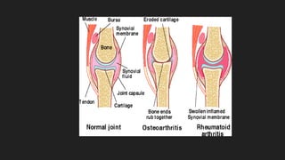
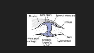
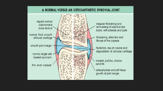
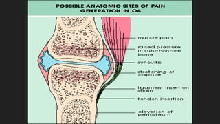
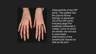
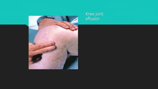
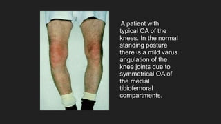
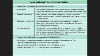
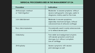
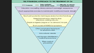
Osteoarthritis
DEFINITION
 Osteoarthritis is a non-inflammatory,
degenerative condition of joints
Characterized by degeneration of articular
cartilage and formation of new bone i.e.
osteophytes.
 Common in weight-bearing joints such as hip and knee.
 Also seen in cervical , lumbosacral spine and in hands (DIP, PIP, base of thumb).
 Both male and females are affected.
 But more common in older women i.e. above 50 yrs,particularly in postmenopausal
age.
JOINT PROTECTIVE MECHANISMS
 SYNOVIAL FLUID
 JOINT CAPSULE AND LIGAMENTS
 MUSCLE
 SENSORY AFFERENTS
 UNDERLYING BONE
SYNOVIAL FLUID
 Reduces friction between articulating cartilage surfaces
 Hyaluronic acid + lubricin
 Concentration of the above – reduced in injury/ inflammation of synovium
LIGAMENTS, SKIN, TENDONS
 Contain mechanoceptor sensory afferent nerves
 Provide feedback via spinal cord to muscles + tendons
CARTILAGE
 Impact absorbing capacity along with synovium
 Earliest OA changes occur here
 Healthy cartilage – avascular due to angiogenesis inhibitors in cartilage
 Diseased cartilage – invasion of blood vessels into cartilage from bone and proliferation
in synovium
 VEGF synthesis in bone + cartilage is implicated
 OA cartilage – depletion of aggrecan, loss of type 2 collagen, unfurling of collagen
matrix
PATHOLOGY
 OA is a degenerative condition primarily affecting the articular cartilage.
 1.articular cartilage
 2.Bone
 3.Synovial membrane
 4.capsule
 5.Ligament
 6.muscle
Articular Cartilage
o Cartilage - 1st structure to be affected.
o Erosion occurs, often central & frequently in wt. bearing areas.
o Fibrillation - causes softening, splitting and fragmentation of cartilage,in both wt.
bearing & non-wt. bearing areas.
o Collagen fibres split , disorganisation of proteoglycan-collagen relationship
o Further softening and flaking.
o Flakes of cartilage break off, may be impacted b/w jt.surfaces  locking and
inflammation.
Right: Early OA with
area of cartilage loss in
the center.
Left: More advanced
changes with extensive
cartilage loss and
exposed underlying
bone
Arthroscopic
appearances in OA of
the knee joint: fibrillated
surface of the cartilage
on the medial femoral
condyle
Bone(Eburnation)
 Bone surface become hard & polished as there is loss of protection from the cartilage.
 Cystic cavities form in the subchondral bone because eburnated bone is brittle and
microfractures occur.
 Venous congestion in the subchondral bone.
Gross superior view of a
femoral head from a
patient with radiographic
stage I OA. This shows an
area of complete cartilage
loss, with polishing or
eburnation of the
underlying bone.
 Osteophytes form at the margin of the articular surface,which may get projected into
the jt. Or into capsule & ligament,bone of the wt.-bearing jt.
 There is alteration in the shape of the femoral head which becomes flat and
mushroom shaped.
 Tibial condyles become flatened.
Osteophyte at margin of
articular surface
Synovial Membrane
o Synovial membrane undergo hypertrophy and become oedematous (which can
lead to ‘cold’ effusions).
o Reduction of synovial fluid secretion results in loss of nutrition and lubricating action of
articular cartilage.
o Capsule
o It undergoes fibrous degeneration and there are low-grade chronic inflammatory
changes
Ligament
Undergoes fibrous degeneration
There is low grade chronic inflammatory changes and acc.to the aspect joint become contracted
or elongated.
Muscles
Undergoes atrophy, as pt. is not able to use the jt. Because of pain which further limits movts. and
function.
CLINICAL FEATURES
 JOINT PAIN – ACTIVITY RELATED
 IN EARLY DISEASE – Episodic, triggered by overuse of diseased joint
 Progressively, continuous pain, even disturbs sleep at night
 Stiffness of the joint.
 Morning stiffness – brief ( <30min)
 In knee- buckling, catching, locking
 Pain with activities requiring knee flexion ( climbing stairs, rising from a chair )
Osteoarthritis of the DIP
joints. This patient has
the typical clinical
findings of advanced
OA of the DIP joints,
including large firm
swellings (Heberden’s
nodes), some of which
are tender and red due
to associated
inflammation of the
periarticular tissues as
well as the joint.
Knee joint
effusion
A patient with
typical OA of the
knees. In the normal
standing posture
there is a mild varus
angulation of the
knee joints due to
symmetrical OA of
the medial
tibiofemoral
compartments.
Radiographic Classification
Stage 1
Stage 2
Stage 3
Stage 4
Bony spur only
Narrowing of jt.
Space,less than half of
the normal jt. space
Narrowing of jt.
Space,more than half
of the normal jt. space
Obliteration of jt. space
Stage 5 Subluxation or
sec.lateral arthrosis
Special Investigations
• Blood tests: Normal
• Radiological features:
– Cartilage loss
– Subchondral sclerosis
– Cysts
– Osteophytes
Treatment Principles
• Education
• Physiotherapy
– Exercise program
– Pain relief modalities
• Aids and appliances
• Medical Treatment
• Surgical Treatment
Aids and appliances
• Braces / splints
• Special shoes/insoles
• Mobility aids
• Aids: dressing, reaching, tap openers,
kitchen aids
• Taping of patella in patello femoral OA
Use of a cane, stick or other walking aid. This patient,
who has hip OA, has found that she can reduce the
pain in her damaged left hip by leaning on the stick in
the right hand as she walks. The reduction in loading
can be huge, and the effect on symptoms and
confidence with walking very beneficial.
Joint replacement surgery
• Indications: pain affecting work, sleep,
walking and leisure activities
• Complications
– sepsis
– loosening
– lifespan of materials (mechanical failure)
Osteoarthritis
Osteoarthritis

Osteoarthritis

  • 1.
  • 2.
    DEFINITION  Osteoarthritis isa non-inflammatory, degenerative condition of joints Characterized by degeneration of articular cartilage and formation of new bone i.e. osteophytes.
  • 3.
     Common inweight-bearing joints such as hip and knee.  Also seen in cervical , lumbosacral spine and in hands (DIP, PIP, base of thumb).  Both male and females are affected.  But more common in older women i.e. above 50 yrs,particularly in postmenopausal age.
  • 4.
    JOINT PROTECTIVE MECHANISMS SYNOVIAL FLUID  JOINT CAPSULE AND LIGAMENTS  MUSCLE  SENSORY AFFERENTS  UNDERLYING BONE
  • 5.
    SYNOVIAL FLUID  Reducesfriction between articulating cartilage surfaces  Hyaluronic acid + lubricin  Concentration of the above – reduced in injury/ inflammation of synovium
  • 6.
    LIGAMENTS, SKIN, TENDONS Contain mechanoceptor sensory afferent nerves  Provide feedback via spinal cord to muscles + tendons
  • 7.
    CARTILAGE  Impact absorbingcapacity along with synovium  Earliest OA changes occur here  Healthy cartilage – avascular due to angiogenesis inhibitors in cartilage  Diseased cartilage – invasion of blood vessels into cartilage from bone and proliferation in synovium  VEGF synthesis in bone + cartilage is implicated  OA cartilage – depletion of aggrecan, loss of type 2 collagen, unfurling of collagen matrix
  • 9.
    PATHOLOGY  OA isa degenerative condition primarily affecting the articular cartilage.  1.articular cartilage  2.Bone  3.Synovial membrane  4.capsule  5.Ligament  6.muscle
  • 10.
    Articular Cartilage o Cartilage- 1st structure to be affected. o Erosion occurs, often central & frequently in wt. bearing areas. o Fibrillation - causes softening, splitting and fragmentation of cartilage,in both wt. bearing & non-wt. bearing areas. o Collagen fibres split , disorganisation of proteoglycan-collagen relationship o Further softening and flaking. o Flakes of cartilage break off, may be impacted b/w jt.surfaces  locking and inflammation.
  • 11.
    Right: Early OAwith area of cartilage loss in the center. Left: More advanced changes with extensive cartilage loss and exposed underlying bone
  • 12.
    Arthroscopic appearances in OAof the knee joint: fibrillated surface of the cartilage on the medial femoral condyle
  • 13.
    Bone(Eburnation)  Bone surfacebecome hard & polished as there is loss of protection from the cartilage.  Cystic cavities form in the subchondral bone because eburnated bone is brittle and microfractures occur.  Venous congestion in the subchondral bone.
  • 14.
    Gross superior viewof a femoral head from a patient with radiographic stage I OA. This shows an area of complete cartilage loss, with polishing or eburnation of the underlying bone.
  • 15.
     Osteophytes format the margin of the articular surface,which may get projected into the jt. Or into capsule & ligament,bone of the wt.-bearing jt.  There is alteration in the shape of the femoral head which becomes flat and mushroom shaped.  Tibial condyles become flatened.
  • 16.
    Osteophyte at marginof articular surface
  • 17.
    Synovial Membrane o Synovialmembrane undergo hypertrophy and become oedematous (which can lead to ‘cold’ effusions). o Reduction of synovial fluid secretion results in loss of nutrition and lubricating action of articular cartilage. o Capsule o It undergoes fibrous degeneration and there are low-grade chronic inflammatory changes
  • 19.
    Ligament Undergoes fibrous degeneration Thereis low grade chronic inflammatory changes and acc.to the aspect joint become contracted or elongated. Muscles Undergoes atrophy, as pt. is not able to use the jt. Because of pain which further limits movts. and function.
  • 22.
    CLINICAL FEATURES  JOINTPAIN – ACTIVITY RELATED  IN EARLY DISEASE – Episodic, triggered by overuse of diseased joint  Progressively, continuous pain, even disturbs sleep at night  Stiffness of the joint.  Morning stiffness – brief ( <30min)  In knee- buckling, catching, locking  Pain with activities requiring knee flexion ( climbing stairs, rising from a chair )
  • 24.
    Osteoarthritis of theDIP joints. This patient has the typical clinical findings of advanced OA of the DIP joints, including large firm swellings (Heberden’s nodes), some of which are tender and red due to associated inflammation of the periarticular tissues as well as the joint.
  • 25.
  • 26.
    A patient with typicalOA of the knees. In the normal standing posture there is a mild varus angulation of the knee joints due to symmetrical OA of the medial tibiofemoral compartments.
  • 27.
    Radiographic Classification Stage 1 Stage2 Stage 3 Stage 4 Bony spur only Narrowing of jt. Space,less than half of the normal jt. space Narrowing of jt. Space,more than half of the normal jt. space Obliteration of jt. space Stage 5 Subluxation or sec.lateral arthrosis
  • 29.
    Special Investigations • Bloodtests: Normal • Radiological features: – Cartilage loss – Subchondral sclerosis – Cysts – Osteophytes
  • 31.
    Treatment Principles • Education •Physiotherapy – Exercise program – Pain relief modalities • Aids and appliances • Medical Treatment • Surgical Treatment
  • 33.
    Aids and appliances •Braces / splints • Special shoes/insoles • Mobility aids • Aids: dressing, reaching, tap openers, kitchen aids • Taping of patella in patello femoral OA
  • 34.
    Use of acane, stick or other walking aid. This patient, who has hip OA, has found that she can reduce the pain in her damaged left hip by leaning on the stick in the right hand as she walks. The reduction in loading can be huge, and the effect on symptoms and confidence with walking very beneficial.
  • 37.
    Joint replacement surgery •Indications: pain affecting work, sleep, walking and leisure activities • Complications – sepsis – loosening – lifespan of materials (mechanical failure)