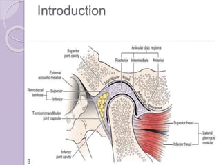
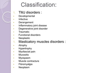
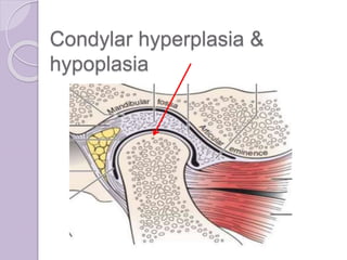
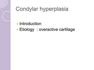
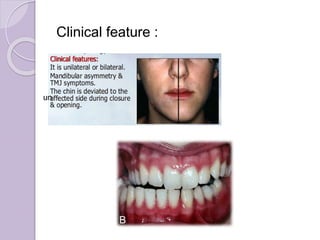
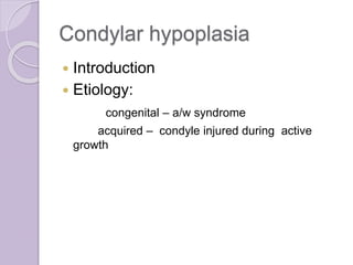
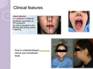
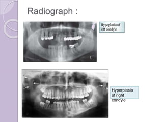
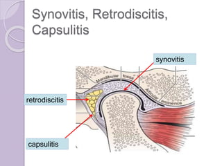
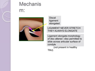
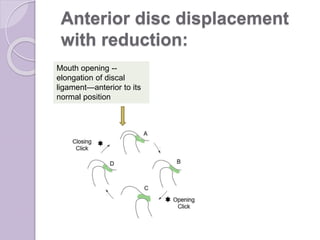
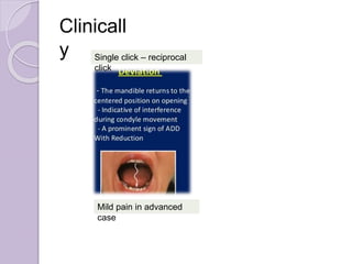
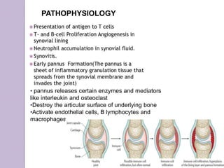
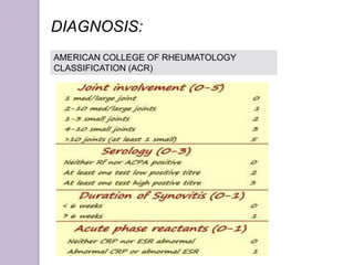
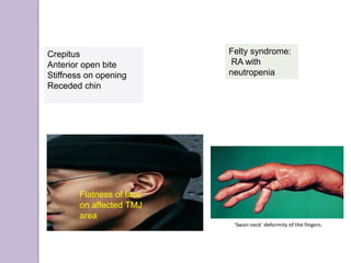
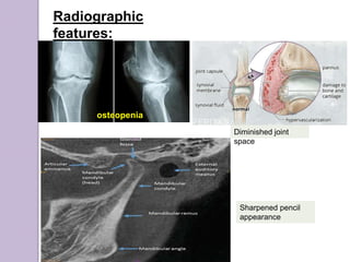
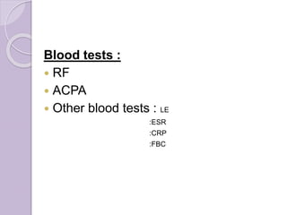
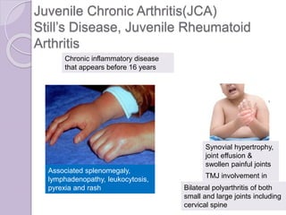
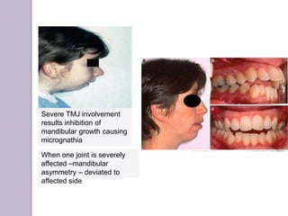
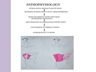
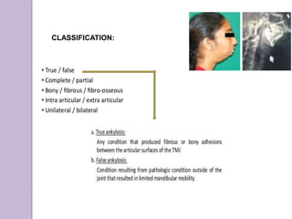
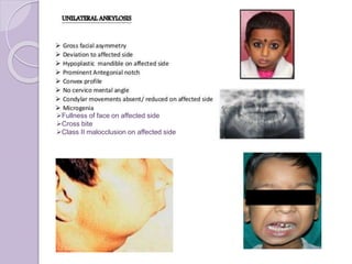
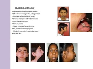
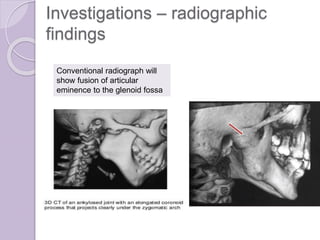
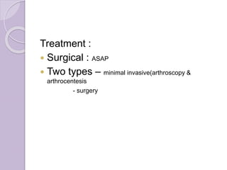
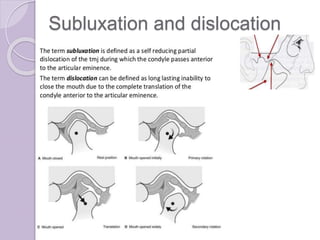
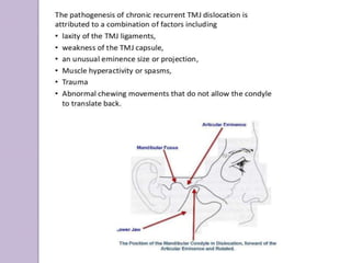
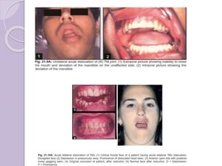
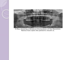
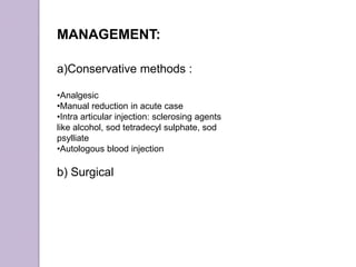
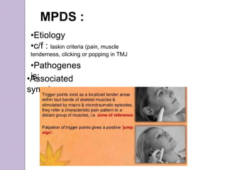
TEMPOROMANDIBULAR
JOINT DISORDERS
Submitted by :
Nischala Chaulagain
BDS 4th year (5th
batch)
Guided by:
Dr. Peeyush Shivhare
Department of Oral Medicine
and Radiology
Contents :
๏‚— Introduction
๏‚— Classification of TMJ disorders
๏‚— Condylar hypoplasia / hyperplaisa
๏‚— Synovitis, retrodiscitis, capsulitis
๏‚— Disc derangement
๏‚— Arthritis affecting TMJ(osteoarthritis, rheumatoid
arthritis, juvenile chronic arthritis)
๏‚— Ankylosis
๏‚— Subluxation and dislocation
๏‚— Neoplastic diseases of TMJ
๏‚— Myofascial pain dysfunction syndrome (MPDS)
Introduction
Classification:
1. TMJ disorders :
๏‚— Developmental
๏‚— Infective
๏‚— Derangement
๏‚— Inflammatory joint disease
๏‚— Degenerative joint disorder
๏‚— Traumatic
๏‚— Functional disorders
๏‚— Neoplastic
1. Masticatory muscles disorders :
๏‚— Atrophy
๏‚— Hypertrophy
๏‚— Myofascial pain
๏‚— Mucositis
๏‚— Myospasm
๏‚— Muscle contracture
๏‚— Fibromyalgia
๏‚— Neoplasm
Condylar hyperplasia &
hypoplasia
Condylar hyperplasia
๏‚— Introduction
๏‚— Etiology : overactive cartilage
Clinical feature :
un
Condylar hypoplasia
๏‚— Introduction
๏‚— Etiology:
congenital โ€“ a/w syndrome
acquired โ€“ condyle injured during active
growth
Clinical features
Due to underdeveloped
ramus and mandibular
body
Radiograph :
Hyperplasia
of right
condyle
๏‚— Treatment : orthodontics combined with
orthognathic surgey
Synovitis, Retrodiscitis,
Capsulitis
synovitis
capsulitis
retrodiscitis
๏‚— Aetiology : result as a consequence of
acute trauma or natural strains
Synovitis & Retrodiscitis : They are
interrelated.
Clinical symptoms:
๏‚— joint pain (clench)
๏‚— Acute malocclusion (due to anterior positioning
of condyle due to oedema accumulation )
๏‚— Pain
๏‚— Limitation of movement
Capsulitis:
Capsular ligament acts to resist forces that tend to
separate or dislocate the articular surface. So, painful
movement of joint.
Clinical symptom:
๏‚— Pain on translatory movement (stretch inflammed
capsuleโ€”pain)
๏‚— Palpable tenderness
๏‚— Fluctuant swelling
๏‚— Ipsilateral posterior open bite due to inferior
displacement of condyle.
๏‚— Restricted movement.
๏‚— No increase in pain on clenching
Management :
Disc Derangement
It is an abnormal relationship among the disc, the
mandibular condyle and the articular eminence,
resulting from the elongation or tearing of the discal
ligament.
TYPES:
1. Anterior disc
displacement:
stage I
stage II
stage III
2. Posterior disc
displacement:
Etiology :
๏‚— Macrotrauma
๏‚— Microtrauma
Mechanis
m:
Discal
ligament
elongated
LIGAMENT NEVER STRETCH
THEY ALWAYS ELONGATE
Ligament elongate-morphology
of disc altered โ€“disc permitted to
slide across articular surface of
condyle
(not present in healthy
TMJ)
Anterior disc displacement
with reduction:
Mouth opening --
elongation of discal
ligamentโ€”anterior to its
normal position
Single click โ€“ reciprocal
click
Mild pain in advanced
case
Clinicall
y
RADIOGRAPHICALL
Y (MRI)/
ARTHROGRAPHY :
On mouth opening :
anterior position of disc
over condyle
TREATMENT:
No pain โ€“no tt
required
Prevents the
condyle from
closing posterior
to the disc
Anterior disc displacement without
reduction (CLOSED LOCK )
Excessive elongation of discal
ligament โ€“ articular disc anterior to
condyle even on closed mouth.
When trying to open mouthโ€”
movement restricted by articular disc
Localized pain on
TMJ
Restricted mouth opening
Clinically:
painfu
l
TREATMENT:
Deposition of intraarticular
corticosteroidsSodium hyaluronate to increase joint
lubrication
Posterior disc displacement
Abnormal backward position of
articular disc to condyle.
During mouth opening, condyle
is slipping over anterior rim of
disc.
Disc folded in the posterior part
of joint , preventing full mouth
closure
No
restriction
on mouth
opening
Arthritis affecting TMJ
๏‚— Osteoarthritis
๏‚— rheumatoid arthritis
๏‚— juvenile chronic arthritis
๏‚— Septic arthritis
๏‚— Gouty arthritis
Degenerative joint disease
(DJD)/ Osteoarthritis
Multiple erosive
fragments (joint
mice)present within
joint space
Types:
๏‚— Primary : a/w ^ age
: older age
๏‚— Secondary : younger patients
:joint change occur in response to
factors like traumatic, endocrinal ,
hematological
Enlargement of fossa and
reduced condylar head size โ€“
condyle moves forward &
superiorly into abnormal
anterior position.
Clinically
:
RADIOGRAPHICALLY:
Degenerative
component
Ely cysts
Erosions of condylar head , posterior
slope of articular eminence.
Large glenoid fossa
Small, round , radiolucent
areas with irregular margins
surrounded by areas of
increased density, deep to the
articulating surfaces
Sharp pencil shaped
condyle
proliferative component/ osteophyte
Rheumatoid arthritis
TMJ involvement 40 to 80%
Flatness of face.
Crepitus
Anterior open bite and
receded chin due to
bilateral destruction of
condyle and anterosuperior
positioning of condyle
PATHOPHYSIOLOGY
โ€ข pannus releases certain enzymes and mediators
like interleukin and osteoclast
โ€ขDestroy the articular surface of underlying bone
โ€ขActivate endothelial cells, B lymphocytes and
macrophages
DIAGNOSIS:
AMERICAN COLLEGE OF RHEUMATOLOGY
CLASSIFICATION (ACR)
Flatness of face
on affected TMJ
area
Felty syndrome:
RA with
neutropenia
Crepitus
Anterior open bite
Stiffness on opening
Receded chin
Radiographic
features:
osteopenia
Diminished joint
space
Sharpened pencil
appearance
Blood tests :
๏‚— RF
๏‚— ACPA
๏‚— Other blood tests : LE
:ESR
:CRP
:FBC
Juvenile Chronic Arthritis(JCA)
Stillโ€™s Disease, Juvenile Rheumatoid
Arthritis
Synovial hypertrophy,
joint effusion &
swollen painful joints
Associated splenomegaly,
lymphadenopathy, leukocytosis,
pyrexia and rash
TMJ involvement in
40%
Chronic inflammatory disease
that appears before 16 years
Bilateral polyarthritis of both
small and large joints including
cervical spine
Severe TMJ involvement
results inhibition of
mandibular growth causing
micrognathia
When one joint is severely
affected โ€“mandibular
asymmetry โ€“ deviated to
affected side
RADIOGRAPHICALLY:
Erosion of condyle
Deepened antegonial
notch
Bending of ramus and
condylar head
Osteopenia
ANKYLOSIS
SYNDROMES :
T.C
:
P.R
CLASSIFICATION:
๏ƒ˜Fullness of face on affected side
๏ƒ˜Cross bite
๏ƒ˜Class II malocclusion on affected side
Investigations โ€“ radiographic
findings
Conventional radiograph will
show fusion of articular
eminence to the glenoid fossa
Treatment :
๏‚— Surgical : ASAP
๏‚— Two types โ€“ minimal invasive(arthroscopy &
arthrocentesis
- surgery
Subluxation and dislocation
Precipitating factors :
๏‚— Trauma
๏‚— Forceful wide opening โ€ฆ
Clinical features :
a) Subluxation
Normal mouth opening but with a click
Painless
Deviation
b) Dislocation
โ€ขInability to close mouth โ€œOPEN LOCKโ€
โ€ขDifficulty in speech
โ€ขDrooling saliva
โ€ขLip incompetency
โ€ขIn acute, pain in preauricular region
โ€ขUsually bilateral
โ€ขWhen palpated over preauricular region,-
emptiness in joint space
โ€ขPatient looks anxious
MANAGEMENT:
a)Conservative methods :
โ€ขAnalgesic
โ€ขManual reduction in acute case
โ€ขIntra articular injection: sclerosing agents
like alcohol, sod tetradecyl sulphate, sod
psylliate
โ€ขAutologous blood injection
b) Surgical
Downward,backward,upward
MPDS :
โ€ขEtiology
โ€ขc/f : laskin criteria (pain, muscle
tenderness, clicking or popping in TMJ
โ€ขPathogenes
is:โ€ขAssociated
symptoms:
TMJ (temporomandibular joint) disorders

TMJ (temporomandibular joint) disorders