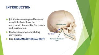
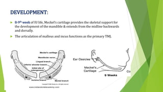
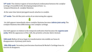
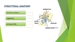
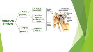
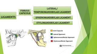
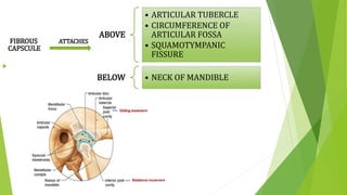
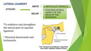
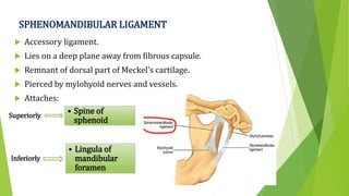
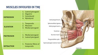
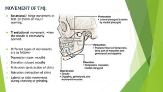
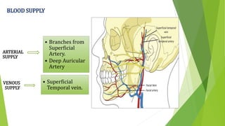
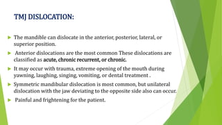
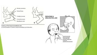
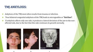
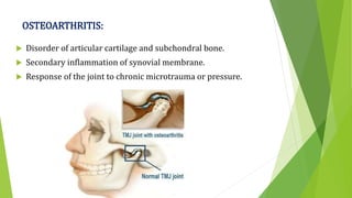
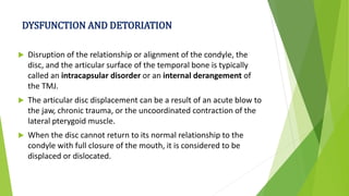
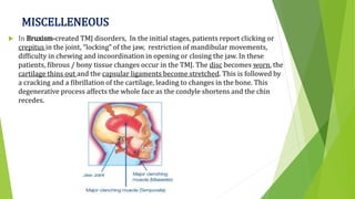
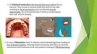
2. INDEX:
INTODUCTION
PECULARITY OF TMJ
DEVELOPMENT
STRUCTURAL ANATOMY
RELATIONS OF TMJ
MOVEMENTS OF TMJ
AGE CHANGES AND DISORDERS
EXAMINATION AND PALPATION OF TMJ
CONCLUSION
REFERENCES
3. INTRODUCTION:
Joint between temporal bone and
mandible that allows the
movement of mandible for speech
and mastication.
Produces rotation and sliding
movements.
It is GINGLYMOARTROIDAL JOINT.
4. TMJ IS UNIQUE!
Bilateral diarthrosis – right & left function together
Articular surface covered by fibrocartilage instead of hyaline
cartilage
Only joint in human body to have a rigid endpoint of closure
(that of the teeth making occlusal contact).
Only mobile joint of skull.
5. DEVELOPMENT:
8-9th week of IU life, Meckel’s cartilage provides the skeletal support for
the development of the mandible & extends from the midline backwards
and dorsally.
The articulation of malleus and incus functions as the primary TMJ.
6. 10th week -Two distinct regions of mesenchymal condensation between the condylar
cartilage of mandible & the developing temporal bone i.e..
temporal blastema & condylar blastema
At the same time lateral pterygoid muscle attaches to condyle.
12th weeks- Two slit like joint cavities & an intervening disc appear.
1st cleft appears immediately above condylar blastema becomes inferior joint cavity. The
condylar blastema then differentiates into condylar cartilage.
2nd cleft appears in relation to the temporal ossification that becomes the superior joint
cavity. With the appearance of this cleft, the primitive articular disk is formed.
16th week-Malleus & Incus begin its transformation into middle ear bones &
disappearance of primary joint starts.
18th-20th week- Secondary joint becomes functional & Meckel’s Cartilage loses its
function & disappears.
11. LATERAL LIGAMENT
ATTACHES
* It reinforce and strengthen
the lateral part of capsular
ligament.
* Directed downwards and
backwards.
ABOVE • ARTICULAR TUBERCLE
BELOW
• POSTEROLATERAL
ASPECT OF THE
NECK OF THE
MANDIBLE.
12. SPHENOMANDIBULAR LIGAMENT
Accessory ligament.
Lies on a deep plane away from fibrous capsule.
Remnant of dorsal part of Meckel’s cartilage.
Pierced by mylohyoid nerves and vessels.
Attaches:
Superiorly
• Spine of
sphenoid
Inferiorly
• Lingula of
mandibular
foramen
13. STYLOMANDIBULAR LIGAMENT
Represents a thickened part of deep cervical fascia.
Separates parotid and submandibular gland.
Pierced by external carotid artery.
Accessory ligament.
Above
• Lateral surfaces of
styloid process.
Below • Angle and posterior
border of ramus
14. ARTICULAR DISC
Oval fibrous plates divides joint into:
Upper compartment – Gliding movement
Lower compartment – Rotatory + Gliding movement
Composed of Dense connective fibrous tissues devoid of blood
vessels and nerve fibres.
Consist : Type 1 & 2 collagen fibres, few elastic fibres.
Stabilises the joint.
Aids lubrication.
15. RELATIONS OF TMJ
LATERAL
• Skin & fascia
• Parotid gland
• Temporal branches of the facial
nerve
MEDIAL
• The tympanic plate separates the
joint from the internal carotid artery
• Spine of the sphenoid with upper
end of the sphenomandibular
ligament attached to it.
• Auriculotemporal and chorda
tympani nerves
• Middle meningeal artery
ANTERIOR
• Lateral pterygoid
• Masseteric nerve and artery
16. POSTERIOR
• The parotid gland
separates the joint from the
external auditory meatus
• Superficial temporal
vessels
• Auriculotemporal nerve
SUPERIOR
• Middle cranial fossa
• Middle meningeal vessels
INFERIOR • Maxillary artery and vein
18. MOVEMENT OF TMJ:
Rotational/ hinge movement in
first 20-25mm of mouth
opening.
Translational movement: when
the mouth is excessively
opened.
Different types of movements
are as follows-
1. Depression (open mouth)
2. Elevation (closed mouth)
3. Protrusion (protraction of chin)
4. Retrusion (retraction of chin)
5. Lateral or side movements
during chewing or grinding.
23. AGE CHANGES IN TMJ
Condyle
1. Becomes more flattened.
2. Fibrous capsule becomes thicker.
3. Osteoporosis of underlying bone.
4. Thinning or absence of cartilaginous zone
Disk
1. Becomes thinner
2. Shows hyalinization and chondroid changes
Synovial fold
1. Becomes fibrotic with thick basement membrane
Blood vessels and nerves
1. Walls of blood vessels thickened
2. Nerves decrease in number
24. AGE CHANGES LEAD TO:
Decrease in synovial fluid formation.
Impairment of motion due to decrease in disc and
capsule extensibility.
Decrease the resilience during mastication due to
chondroid changes into collagenous elements.
Dysfunction in older people.
25. EXAMINATION OF TMJ:
CLINICALLY
• Inspection
• Auscultation
• Palpation of TMJ & Musculature with mandibular
movements.
• Functional Analysis of Mandibular Movements.
RADIOGRAPHY
• Transcranial &Transpharyngeal Projections
• Panaromic Projections
• Transorbital Projections
• Submentovertex Projections
• Computed Tomography
• Arthrography
• Magnetic Resonance Imaging
26. INSPECTION OF TMJ:
Area surrounding TMJ is inspected for any signs of inflammation.
Any Gross Asymmetry
Any Swelling/Growth
Depression
Discharge
Color change of skin over TMJ
Surface of the overlying skin in Pre-Auricular area.
Mouth Opening: Normal/Restricted
Deviation/Deflection
27.
28. PALPATION OF TMJ:
Pain or Pressure of condylar areas.
Right and left condyles checked for synchrony of action.
LATERAL PALPATION- Slight pressure on condylar process
with an index finger.
POSTERIOR PALPATION- Position of little finger in external
auditory meatus and palpate the posterior surface of condyle
during opening and closing.
29. AUSCULATIONS:
Sounds made by TMJ can be examined with a stethoscope.
CREPITATIONS: A grafting or scalping noise that occurs on jaw
movements. Sound is like when sand paper is rubbed against a surface.
It is very uncommon and asymptomatic. It may caused by roughened,
irregular anterior surface.
May be early sign of degenerative joint disease.
CLICKING: occurs due to uncoordinated movement of condylar head and
TMJ disc.
CLICK • Single sound of short duration
POP • Loud Click
30. Etiologic considerations for TMD.
5 major factors associated with TMD
Occlusal condition
Trauma
Emotional stress
Deep pain input
Para-functional activities
31. TMJ DISLOCATION:
The mandible can dislocate in the anterior, posterior, lateral, or
superior position.
Anterior dislocations are the most common These dislocations are
classified as acute, chronic recurrent, or chronic.
It may occur with trauma, extreme opening of the mouth during
yawning, laughing, singing, vomiting, or dental treatment .
Symmetric mandibular dislocation is most common, but unilateral
dislocation with the jaw deviating to the opposite side also can occur.
Painful and frightening for the patient.
32.
33. TMJ ANKYLOSIS:
Ankylosis of the TMJ most often results from trauma or infection.
True bilateral congenital ankylosis of the TMJ leads to micrognathia or “bird face”.
If ankylosis affects only one side, it produces a lateral deviation of the jaw to the non-
affected side, due to the fact that this side continues its growth normally.
34. OSTEOARTHRITIS:
Disorder of articular cartilage and subchondral bone.
Secondary inflammation of synovial membrane.
Response of the joint to chronic microtrauma or pressure.
35. DYSFUNCTION AND DETORIATION
Disruption of the relationship or alignment of the condyle, the
disc, and the articular surface of the temporal bone is typically
called an intracapsular disorder or an internal derangement of
the TMJ.
The articular disc displacement can be a result of an acute blow to
the jaw, chronic trauma, or the uncoordinated contraction of the
lateral pterygoid muscle.
When the disc cannot return to its normal relationship to the
condyle with full closure of the mouth, it is considered to be
displaced or dislocated.
36. MISCELLENEOUS
In Bruxism-created TMJ disorders, In the initial stages, patients report clicking or
crepitus in the joint, “locking” of the jaw, restriction of mandibular movements,
difficulty in chewing and incoordination in opening or closing the jaw. In these
patients, fibrous / bony tissue changes occur in the TMJ. The disc becomes worn, the
cartilage thins out and the capsular ligaments become stretched. This is followed by
a cracking and a fibrillation of the cartilage, leading to changes in the bone. This
degenerative process affects the whole face as the condyle shortens and the chin
recedes.
37. In Unilateral mastication, the favored side wears down due to
overuse. This results in uneven teeth that twist to one side,
resulting in facial asymmetry, also can develop muscle
hypertrophy. On contralateral side, it leads to weakening of
joint and muscle atrophy.
In case of Edentulism, there is absence of periodontal ligament, leading to
loss of proprioception, reducing tactile sensitivity, therefore no resilient
connection exists between teeth and jawbone leading to TMJ Disharmony.
38. CONCLUSION:
Nature has blessed us with a marvelously dynamic
masticatory system , allowing us to function and therefore
exist.
Articulatory system is an important part of the masticatory
system of our body.
So as a dental care provider to treat the patients of TMDs
before knowing the pathology, this is essential to know the
normal anatomy and physiology of TMJ.
39. REFERENCES:
TEXTBOOK OF ORAL MEDICINE.
A GOVINDRAO, G LODAM, AG SAVITA - 2019 - JAYPEE BROTHERS MEDICAL P
Textbook of Oral & Maxillofacial Surgery-
SM Balaji, Padma Preetha Balaji
Human anatomy
BD Chaurasia - 2004 - CBS Publisher
Gray's anatomy : the anatomical basis of clinical practice
Susan Standring Elsevier Health Sciences, 2015
Text Book of Carranza’s Clinical Periodontology
MG Newman, HH Takei
Editor's Notes
Assist lat. Pte in trans n rot. mvt
Limits the excessive protrusive mvt of mandible.
Disc is biconcave, located in sup. Position. The central thin part is consistently lying between the condyle and tubercle. Opening: rot in upper. More of trans in lower