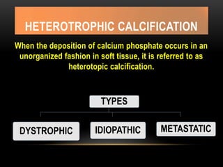
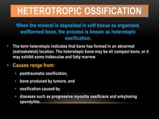
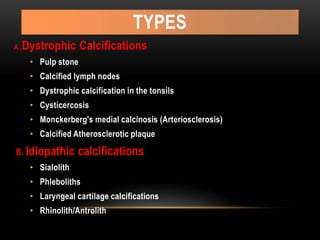
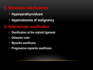
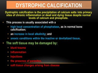
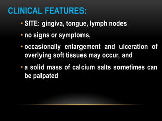
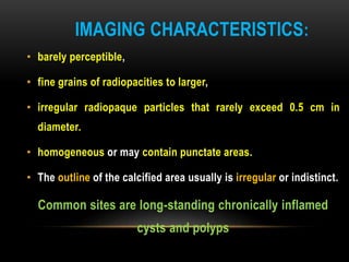
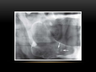
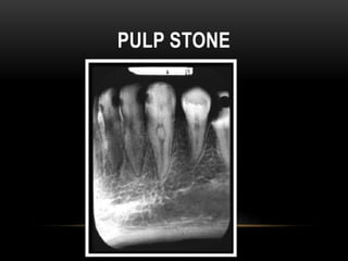
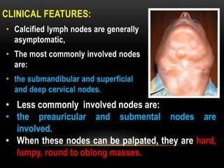
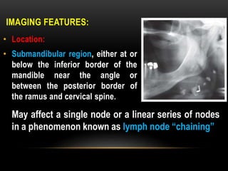
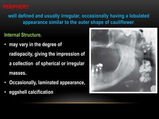
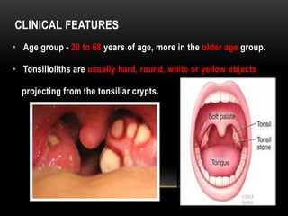
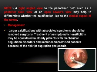
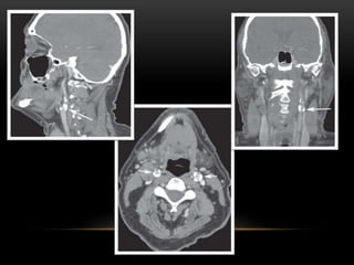
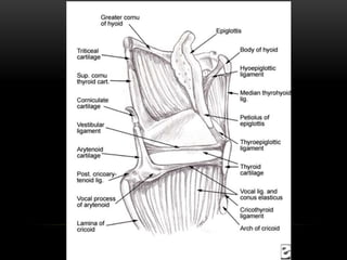
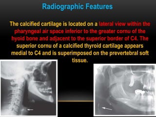
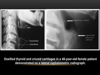
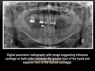
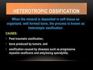
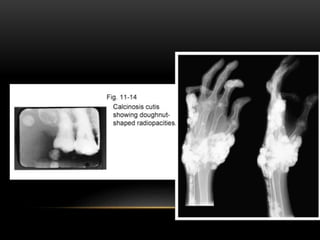
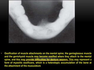
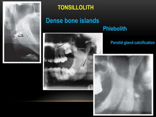
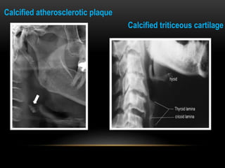
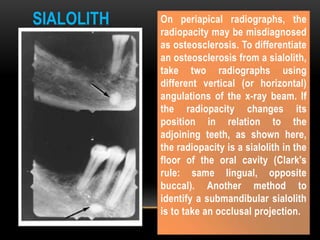
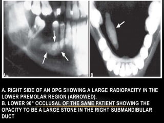
Call Girl Bangalore Nandini 7001305949 Independent Escort Service Bangalore
Soft tissue calcification orofacial region
1. PREPARED BY: Dr. Monali Prajapati (PG Part III)
UNDER THE GUIDANCE OF: Dr. Jigna Shah (Professor and Head)
Oral Medicine and Radiology department
GDCH, Ahmedabad
DATE: 3-5-16
2. REFERENCES
• White SC, Pharoah MJ. Oral Radiology: Principles
and Interpretation. 6th ed. Saint Louis: Mosby;
2007
• Frenny Karjodkar, Textbook of dental and
maxillofacial radiology, 2nd edition
• Langland OE, Langlais RP, Nortje. Diagnostic imaging of
jaws, soft tissue opacities, pg 617
3. HETEROTROPHIC CALCIFICATION
When the deposition of calcium phosphate occurs in an
unorganized fashion in soft tissue, it is referred to as
heterotopic calcification.
TYPES
DYSTROPHIC IDIOPATHIC METASTATIC
4. HETEROTROPIC OSSIFICATION
When the mineral is deposited in soft tissue as organized,
wellformed bone, the process is known as heterotopic
ossification.
• The term heterotopic indicates that bone has formed in an abnormal
(extraskeletal) location. The heterotopic bone may be all compact bone, or it
may exhibit some trabeculae and fatty marrow.
• Causes range from:
• posttraumatic ossification,
• bone produced by tumors, and
• ossification caused by
• diseases such as progressive myositis ossificans and ankylosing
spondylitis.
5. TYPES
A .Dystrophic Calcifications
• Pulp stone
• Calcified lymph nodes
• Dystrophic calcification in the tonsils
• Cysticercosis
• Monckerberg's medial calcinosis (Arteriosclerosis)
• Calcified Atherosclerotic plaque
B. Idiopathic calcifications
• Sialolith
• Phleboliths
• Laryngeal cartilage calcifications
• Rhinolith/Antrolith
6. C. Metastatic calcifications
• Hyperparathyroidusm
• Hypercalcemia of malignancy
D. Heterotropic ossification
• Ossification of the styloid ligament
• Osteoma cutis
• Myositis ossificans
• Progressive myositis ossificans
7. DYSTROPHIC CALCIFICATION
Dystrophic calcification is the precipitation of calcium salts into primary
sites of chronic inflammation or dead and dying tissue despite normal
levels of calcium and phosphate.
• This process is usually associated with a
• high local concentration of phosphatase, as in normal bone
calcification;
• an increase in local alkalinity; and
• anoxic conditions within the inactive or devitalized tissue.
• The soft tissue may be damaged by:
• blunt trauma
• Inflammation
• Injections
• the presence of parasites,
• soft tissue changes arising from disease
8. CLINICAL FEATURES:
• SITE: gingiva, tongue, lymph nodes
• no signs or symptoms,
• occasionally enlargement and ulceration of
overlying soft tissues may occur, and
• a solid mass of calcium salts sometimes can
be palpated
9. IMAGING CHARACTERISTICS:
• barely perceptible,
• fine grains of radiopacities to larger,
• irregular radiopaque particles that rarely exceed 0.5 cm in
diameter.
• homogeneous or may contain punctate areas.
• The outline of the calcified area usually is irregular or indistinct.
Common sites are long-standing chronically inflamed
cysts and polyps
12. LYMPH NODE CALCIFICATION
Dystrophic calcification occurs in lymph
nodes that have been chronically inflamed
because of various diseases, frequently
granulomatous disorders.
• The presence of calcifications in lymph nodes implies disease,
either active or the result of previously treated pathosis
• The lymphoid tissue becomes replaced by hydroxyapatite-like
calcium salts, nearly effacing all nodal architecture.
13. • Most common disease:
• scrofula or cervical tuberculous adenitis).
• bacille Calmette-Guérin vaccination,
• sarcoidosis,
• cat-scratch disease,
• rheumatoid arthritis
• systemic sclerosis,
• lymphoma previously treated with radiation therapy,
• fungal infections,
• malignancy (treated Hodgkin’s lymphoma and metastases
from distant calcifying neoplasms, most notably metastatic
thyroid carcinoma).
14. CLINICAL FEATURES:
• Calcified lymph nodes are generally
asymptomatic,
• The most commonly involved nodes
are:
• the submandibular and superficial
and deep cervical nodes.
• Less commonly involved nodes are:
• the preauricular and submental nodes are
involved.
• When these nodes can be palpated, they are hard,
lumpy, round to oblong masses.
15. IMAGING FEATURES:
• Location:
• Submandibular region, either at or
below the inferior border of the
mandible near the angle or
between the posterior border of
the ramus and cervical spine.
May affect a single node or a linear series of nodes
in a phenomenon known as lymph node “chaining”
16. PERIPHERY:
well defined and usually irregular, occasionally having a lobulated
appearance similar to the outer shape of cauliflower.
Internal Structure.
• may vary in the degree of
radiopacity, giving the impression of
a collection of spherical or irregular
masses.
• Occasionally, laminated appearance,
• eggshell calcification
17. Management:
• Calcified lymph nodes usually do not
require treatment;
• however, the underlying cause should be
established in case treatment is required,
such as in the case of active disease.
18. (TONSILLAR CALCULI, TONSIL CONCRETIONS,
TONSILLOLITHS)
• Tonsillar calculi are formed when repeated
bouts of inflammation enlarge the tonsillar
crypts.
• Incomplete resolution of dead bacteria and pus
serve as the nidus for dystrophic calcification.
19. CLINICAL FEATURES
• Age group - 20 to 68 years of age, more in the older age group.
• Tonsilloliths are usually hard, round, white or yellow objects
projecting from the tonsillar crypts.
20. • The small calculi may not produce any signs or symptoms.
• In case of larger calcifications:
• pain,
• swelling,
• fetor oris
• dysphagia and
• a foreign body feeling on swallowing has been reported.
• In rare cases there may be giant tonsilloliths, which stretch the
lymphoid tissue, resulting in ulcerations and extrusion
21. RADIOGRAPHIC FEATURES
• On the panoramic film,
tonsilloliths appear as
single or multiple
radiopacities that overlap
the mid portion of the
mandibular ramus in the
region where the image of
the dorsal surface of the
tongue crosses the ramus
in the palatoglossal air
spaces.
22. • It appears as clusters of
multiple small ill-
defined radiopacities.
This may vary from 0.5
cm to 14.5 cm in
diameter.
• The radiopacity is of
the same density as
that of cortical bone,
and a little more
radiopaque than
cancellous bone.
23. On axial computed tomographic (CT) images, they appear in the soft
tissue medial to the mandibular ramus and next to the lateral wall of
the oropharyngeal air space.
24. NOTE:- A right angled view to the panoramic field such as a
posterior skull view or an open Towne's view may help to
differentiate whether the calcification lies to the medial aspect of
the ramus.
• Management
• Larger calcifications with associated symptoms should be
removed surgically. Treatment of asymptomatic tonsilloliths
may be considered in elderly patients with mechanical
deglutition disorders and immunocompromised patients
because of the risk for aspiration pneumonia.
25. CYSTICERCOSIS
• eggs or gravid proglottids from Taenia Solium (pork
tapeworm)
• ingested by human,
• their covering is digested in the stomach and the larval
form (cysticercus cellulosae) of the parasite is hatched
• larvae penetrate the mucosa,
26. • enter the blood vessels and lymphatics and are distributed in
the tissues all over the body, but preferentially locate to the
brain, muscle, skin and heart.
• They are also found in the oral and perioral tissues, especially
the muscles of mastication.
• After the larva die, they are treated as foreign bodies causing
granuloma formation, scarring and calcification, this takes
approximately 3 months.
• These areas in the tissues are called cysticerci.
27. CLINICAL FEATURES
• Multiple small nodules may be felt in the region of the masseter
and suprahyoid muscles and in the buccal mucosa and lip.
• Examination of the head and neck region may disclose palpable,
well circumscribed soft fluctuant swellings, which resemble a
mucocele.
• Mild cases are completely asymptomatic.
• Moderate cases have symptoms that range from mild to severe
gastrointestinal upset with epigastric pain and severe nausea and
vomiting.
• Invasion of the brain may result in seizures, headaches, visual
disturbances, acute obstructive hydrocephalus, irritability and loss
of consciousness.
29. RADIOGRAPHIC FEATURES
• When alive the larva is not visible radiographically.
• They are usually found in the muscles of mastication and facial
expression, the suprahyoid muscle, and the postcervical
musculature, tongue, buccal mucosa and lip
• They appear as multiple, well-defined, elliptical, homogeneous,
radiopacities, which resemble grains of rice.
30. MANAGEMENT
• basic sanitation (proper preparation of pork and avoiding fecal
contamination of water supplies and vegetables) is needed to
extinguish this source of infection,
• Medical management by using an antihelmintic (albendazole or
praziquantel), in the initial stage.
• Adjunctive corticosteroids help stem the inflammatory reaction,
and anticonvulsants may prevent epileptic seizures.
• After the larvae have settled and calcified in the oral tissues,
they are harmless.
32. MONCKERBERG'S MEDIAL CALCINOSIS
(ARTERIOSCLEROSIS)
• This is characterized by the fragmentation,
degeneration and eventual loss of elastic fibers
followed by the deposition of calcium within the
medial coat of the vessel.
• These calcium deposits do not narrow the vessel
lumen or interfere with flow.
33. CLINICAL FEATURES
• Age: 35 to 50 years
• Femoral, popliteal and radial arteries are affected most
• Initially most patients are asymptomatic.
• Eventually they may develop cutaneous gangrene, peripheral
vascular disease and myositis due to vascular insufficiency.
• Patients with Sturge-Weber syndrome also develop intracranial
arterial calcifications.
34. RADIOGRAPHIC FEATURES
• Calcinosis involving the facial or the carotid artery may be seen
on the panoramic radiographs.
• Calcification occurs in form of closely spaced fine concenteric
rings.
• The calcific deposits in the walls of the artery outline
an image of the artery.
From the side, it may appear as
a parallel pair of thin,
radiopaque lines, that may
have a straight or tortuous
path (pipe stem or tram track
appearance).
36. MANAGEMENT
• Evaluation of the patient for occlusive arterial
disease and peripheral vascular disease may be
appropriate.
• In addition, hyperparathyroidism may be
considered because medial calcinosis frequently
develops as a metastatic calcification in patients
with this condition.
37. CALCIFIED ATHEROSCLEROTIC PLAQUE
• Arterial plaque is a form of intimal arteriosclerosis referred to as
atherosclerotic plaque.
• This consists of focal deposits of fat, primarily cholesterol in the
arterial intima.
• It is associated with an inflammatory response resulting from
fibroblastic proliferation and calcium salt encrustations.
• The abdoMinal aorta, iliac, femoral and popliteal arteries are
involved most often.
• Head and neck region, carotid artery is involved in the area
opposite to upper border of thyroid cartilage where they bifurcate
to external and internal carotid arteries.
38. RADIOGRAPHIC FEATURES
This first develops at the arterial bifurcation as a result of increased
endothelial damage at these sites.
When calcification occurs,
these lesions may be visible on
the panoramic radiograph in
the soft tissues of the neck
adjacent to the greater cornu of
the hyoid bone and the cervical
vertebrae C3, C4 or the
intervertebral space between
them.
39. • The soft tissue calcifications are usually seen as
heterogeneous radiopacities with radiolucent voids,
which are multiple and irregular in shape, sharply
defined from the surrounding soft tissues and have a
vertical linear distribution
40.
41. MANAGEMENT
• The patient should be referred to the physician
for cerebrovascular and cardiovascular workup.
42. IDIOPATHIC CALCIFICATION
Idiopathic calcification (or calcinosis) results from
deposition of calcium in normal tissue despite
normal serum calcium and phosphate levels.
EXAMPLES: Chondrocalcinosis, Phleboliths
43. • Sialolithiasis is the formation of calcified obstruction
within the salivary duct resulting in chronic retrograde
infection because of a decreased salivary flow.
• Sialoliths may also form in any of the major or minor
salivary glands (glandular sialolith) or their ducts (ductal
sialolith), usually only one gland is involved.
44. • Mechanical conditions contributing to the slow flow rate and
physiochemical characteristics of the gland secretions both
contribute to the formation of a nidus and subsequent
precipitation of calcium and phosphate salts.
• Accordingly, the submandibular gland and ductal system lie in a
dependent position. The Wharton duct is long and has an
irregular tortuous course, an uphill flow in the proximal portion
and the orifice is much smaller than the lumen. The salivary
secretion of the submandibular gland is more viscous and has
higher mineral content.
45. • sialoliths consist of:
• hydroxyapatite,
• amorphous carbonated calcium
phosphate,
• carbonated apatite, and
• whitlockite in combination with fibrous
proteins such as mucins.
46. • These are common in the middle age with a slight
predilection for men.
• The submandibular gland and the Wharton duct are by
far the most frequently involved (83% of the cases),
followed by the parotid (10%) and sublingual (7%)
glands.
• About half of the submandibular stones lie in the distal
portion of the Wharton's duct, 20% in the proximal
portion, and 30% in the gland.
Clinical Features
47. • The patient may be asymptomatic, or they may
history of pain and swelling in the floor of the
mouth and in the involved gland. Intra glandular
stones cause less severe symptoms than the
extra glanular or the intraductal types.
• The discomfort may intensify at meal times, when
the salivary flow is stimulated.
48. • If the blockage is partial, then the pain and swelling gradually
subsides.
• 9% of patients have recurrent sialolithiasis and 10% have
nephrolithiasis.
• Pus may exude from the duct orifice, the surrounding soft
tissue may be inflamed, and tender, and the overlying mucosa
may ulcerate.
• Stones in the more peripheral portion of the duct may be
palpated, if it is of sufficient size.
49.
50.
51. • homogeneously radiopaque,
• Round, oval or cylindericle
• Stones that form in the hilus of the submandibular gland tend to
be larger and more irregularly shaped.
Radiographic Features
52. RADIOGRAPHIC INVESTIGATIONS
• Periapical views: placed in the buccal vestibule, with reduced
exposure and time and the central ray directed through the cheek,
helps to demonstrate stones in the parotid gland duct.
• Occlusal views
• Lateral oblique
• panoramic
• Puffed cheek AP skull: of the patient with "blow-out" cheek , or an open-
mouth lateral skull projection, helps to demonstrate stones in the parotid duct.
When producing radiographs to detect sialoliths, the exposure time should be
reduced to about half of normal, this helps to detect stones that are highly
calcified.
• CT scan: also helps to detect minimally calcified sialoliths which are not
visible on plain films.
• Ultrasonography: is of limited use in the diagnosis of inflammatory and
obstructive diseases, but if the stone is large (2 mm), it will be detected as
a characteristic acoustic shadow showing echodense spots
53. • If non calcified stone are suspected, then Sialography; is helpful in
locating obstructions that are undetectable with plain radiography.
The contrast agent usually flows around the sialolith, filling the
duct proximal to the obstruction.
• Radiolucent sialoliths appear as ductal filling defects.
• Sialography should not be performed if the radiopaque stone is
shown by plain radiography to be in the distal portion of the duct,
because the procedure may displace it proximally into the ductal
system, complicating its subsequent removal.
54. Sialogram showing an obstruction in the Wharton's duct
preventing the flow of the radiopaque dye into the submandibular
salivary gland.
55. • Milking by bimanual palpation
• sialogogues
• sialolithotomy using intracorporeal lithotriptors
• piezoelectric extracorporeal shock wave
lithotripsy or
• surgical removal is indicated.
Management:
56. PHEBOLITHS
Phleboliths are calcified thrombi found in veins, venulae, or
the sinusoidal vessels of hemangiomas (especially the
cavernous type).
• Intravascular thrombi, which arise secondary to venous
stagnation, may get organized or mineralized.
• The mineralization begins at the core of the thrombus
and consists of crystals of apatite with calcium
phosphate and calcium carbonate.
57. CLINICAL FEATURES
• In the head and neck region , phleboliths always indicate the
presence of a hemangioma.
• In an adult it may be the sole residua of a childhood
hemangioma, which has long since regressed.
• The involved soft tissue may be swollen, throbbing or discolored
by the presence of veins or a soft tissue hemangioma, which
often fluctuate in size, associated with changes in body position
or during a Valsalva maneuver.
• blanching or change in color on applying pressure.
• Auscultation may reveal bruit in case of cavernous hemangioma
but not in the capillary type.
58. • These are commonly found in hemangiomas.
• It may be homogeneously radiopaque but more commonly
has the appearance of laminations, giving phleboliths a
bull's eye or target appearance.
• A radiolucent center may be seen, which may represent
the remaining patent portion of the vessel.
Radiographic Features
59.
60. LARYNGEAL CARTILAGE CALCIFICATIONS
• Except the epiglottis and vocal
processes of the arytenoid
cartilages (fibroelastic cartilages),
all are hyaline cartilage.
• Enchondral calcification and
ossification of the hyaline
laryngeal cartilages begins on
attainment of skeletal maturity
and progresses thereafter as a
physiologic process.
Both the thyroid and the triticeous (means grain of wheat) cartilages
(found within the lateral thyrohyoid ligaments) consist of hyaline
cartilage, which has a tendency to calcify or ossify with advancing age.
61.
62. Radiographic Features
The calcified cartilage is located on a lateral view within the
pharyngeal air space inferior to the greater cornu of the
hyoid bone and adjacent to the superior border of C4. The
superior cornu of a calcified thyroid cartilage appears
medial to C4 and is superimposed on the prevertebral soft
tissue.
63. Ossified thyroid and cricoid cartilages in a 40-year-old female patient
demonstrated on a lateral cephalometric radiograph.
64. Digital panoramic radiography with image suggesting triticeous
cartilage on both sides (between the greater horn of the hyoid and
superior horn of the thyroid cartilage).
65. RHINOLITH/ANTROLITH
Hard calcified bodies or stones that occur in the nose (rhinoliths) or
the antrum (antroliths) arise from the deposition of mineral salts
such as calcium phosphate, calcium carbonate, and magnesium
around a nidus.
• In case of a rhinolith the nidus is usually an exogenous foreign
body (coin, beads etc) whereas the nidus for an antrolith is
usually endogenous (root tip, bone fragment, masses of
stagnated mucus, etc.).
66. CLINICAL FEATURES
• The patient may be asymptomatic initially.
• With the increase in size of the expanding mass, it may impinge
on the mucosa, producing pain, congestion and ulceration.
• The patient may develop a unilateral purulent rhinorrhea,
sinusitis, headache, epistaxis, nasal obstruction, anosmia, fetor,
fever and facial pain.
67. RADIOGRAPHIC FEATURES
• variety of shapes and sizes
• well-defined smooth or irregular borders
• homogeneous or heterogeneous radiopacities, depending on the
nature of the nidus and sometimes have laminations. Occasionally
the density may exceed the surrounding bone.
• Antroliths occur within the maxillary sinus above the floor of the
antrum and may be seen on the periapical, occlusal and panoramic
radiographs.
• Rhinoliths are seen in the nasal fossae. A posteroanterior skull
view will help to identify the location of a rhinolith.
68. • Antrolith (stone in maxillary sinus) on the floor of the sinus.
• It is asymptomatic.
69.
70. • A. Rhinolith in the maxillary sinus, seen on an OPG. The rhinolith is seen located at the posterior wall of
the right maxillary sinus (arrow), B. Rhinolith in the maxillary sinus of the same patient, seen on Waters
Projection, this projection also reveals the presence of chronic sinusitis
72. HETEROTROPIC OSSIFICATION
When the mineral is deposited in soft tissue as
organized, well formed bone, the process is known as
heterotopic ossification.
CAUSES:
• Post traumatic ossification,
• bone produced by tumors, and
• ossification caused by diseases such as progressive
myositis ossificans and ankylosing spondylitis.
73. OSSIFICATION OF THE STYLOID LIGAMENT
• Normal length of styloid process- 25mm
• Ossification of the styloid ligament usually extends downwards
from the base of the skull and commonly occurs bilaterally.
• In rare cases the ossification begins at the lesser horn of the hyoid
or in the central area of the ligament. The associated conditions
are Eagle's Syndrome, Styloid Syndrome and Styloid Chain
Ossification.
74.
75. CLINICAL FEATURES
• Throat pain
• Fish bone stuck in throat
• Pain on swallowing
• Refered pain to ear of affected side
• Tinnitus
• Pain in anterior cervical triangle
• Pain on turning head
• Headache in orbital area
• Cervical pain
76. Acc to Camarada et al
EAGLES
SYNDROME
Any age
History of trauma or
neck surgery
Radiographic
evidence
STYLOHYOID
SYNDROME
Any age but usually
more than 40yrs
No trauma
Radiographic
evidence
PSEUDOSTYLO
HYOID
SYNDROME
More than 40yrs of
age
No radiographic sign
77. RADIOGRAPHIC FEATURES
In the panoramic image it is seen as a linear, long, tapering, thin, radiopaque
process that is thicker at its base, extending forward from the region of the
mastoid process and crosses the posteroinferior aspect of the ramus towards the
hyoid bone. The hyoid bone is positioned approximately parallel to or
superimposed on the posterior aspect of the inferior cortex of the mandible.
78. OSTEOMA CUTIS
• These are sites of normal bone formation in abnormal locations. It
is a rare soft tissue calcification in the skin.
• It may develop secondary to acne of long duration. In a scar or
chronic inflammatory dermatosis.
• Histologically these are seen as areas of dense viable bone in the
dermis or subcutaneous tissue.
• They are occasionally found in diffuse scleroderma, replacing the
altered collagen in the dermis and subcutaneous septa.
79. CLINICAL FEATURES
• • It may occur on the face (extraoral) in the cheek and lip region,
and tongue (intraoral) where it may be called osteoma mucosae or
osseous choristoma.
• • It does not cause any visible change in the overlying skin, except
in some cases where the color may change to yellowish white.
• • It varies in size from 0.1 mm to 5 cm in diameter, if the lesion is
large. The individual osteoma may be palpated. A needle inserted
into one of the papules usually meets with stone like resistance.
80.
81. RADIOGRAPHIC FEATURES
• An intraoral film placed between the cheek and the alveolar bone
gives accurate localization.
• A posteroanterior skull view with the cheek blown outward using a
soft tissue technique of 60 kVp helps localize osteomas of the
skin.
• If present in the cheek or lip region the shadow may be
superimposed over a tooth root or alveolar process, giving the
appearance of dense bone.
• The osteoma cutis appears as smoothly outlined, radiopaque,
washer-shaped images.
82. • •The single or multiple radiopacities of various sizes.
• • It appears as a homogeneous radiopacity with a radiolucent
center that represents normal fatty marrow, giving the lesion a
dough-nut appearance radiographically.
• • Trabeculae usually develop in the marrow cavity of the larger
osteomas.
• • Lesions of calcified cystic acne resemble a snowflake radiopacity
which corresponds to the clinical location of the scar.
83.
84. MANAGEMENT:
• They may be removed for cosmetic reasons.
The methods used are excision, resurfacing of
the skin with erbium:YAG laser using tretinoin
cream (especially in cases of multiple miliary
osteomacutis).
85. MYOSITIS OSSIFICANS
• In this case, the fibrous tissue and heterotopic bone form
within the interstitial tissue of the muscle and associated
tendons and ligaments.
• There is secondary destruction and atrophy of the muscle
as the fibrous tissue and bone interdigitate and separate
the muscle fibers. It is of two types:
• – Localized myositis ossificans
• – Progressive myositis ossificans
86. LOCALIZED MYOSITIS OSSIFICANS (POST-TRAUMATIC
MYOSITIS, MYOSITIS OSSIFICANS, SOLITARY MYOSITIS)
• This results due to acute or chronic trauma, heavy muscular strain,
muscle injury which may lead to considerable hemorrhage into the
muscle or associated tendons or fascia. The hemorrhage organizes
and undergoes progressive scarring.
• During the healing process, heterotopic bone and in some cases
cartilage is formed. There is no inflammation (the term myositis is
thus misleading).
• The fibrous tissue and bone form within the interstitial tissue of the
muscle, there is no actual ossification of the muscle fibers.
87. CLINICAL FEATURES
• • It may develop at any age, in either gender, but is more common in young
men who engage in vigorous activities.
• The commonly involved oral sites are, the masseter, sterno-cleidomastoid and
lateral pterygoid muscle.
• • The site of the precipitating trauma remains swollen, tender and painful for a
long time. The overlying skin may be red and inflamed.
• • If the lesion involves a muscle of mastication, opening the jaw may be
difficult.
• • After a period of 2-3 weeks the area of ossification may become apparent, as
a firm, intramuscular palpable mass, which enlarges slowly, but eventually
stops growing. The lesion may appear fixed or may be freely movable on
palpation.
88. RADIOGRAPHIC FEATURES
• The most commonly involved muscles of the head and neck are the masseter
and sternocleidomastoid.
• other muscles of mastication may be involved, such as the medial and lateral
pterygoid, buccinator, and temporalis muscles.
• The anterior attachments of the temporalis and the medial pterygoid muscles
are at risk of injury on administration of mandibular block anesthesia.
• A radiolucent band usually can be seen between the area of ossification and
adjacent bone, and the heterotopic bone may lie along the long axis of the
muscle
• Masses generally measure less than 6 cm in greatest dimension.
• The periphery commonly is more radiopaque than the internal structure.
• There is a variation in shape from irregular oval to linear streaks
(pseudotrabeculae) running in
• the same direction as the normal muscle fibers.
89.
90. • Ossification of muscle attachments on the mental spine; the genioglossus muscle
and the geniohyoid muscle may become ossified where they attach to the mental
spine, and this may provide difficulties for denture wearers. This may represent a
form of myositis ossificans, which is a heterotopic accumulation of the bone at
the attachment of the musculature
91. PROGRESSIVE MYOSITIS OSSIFICANS
• This is a rare disease of unknown cause that usually affects
children before 6 years of age, and occasionally as early as
infancy.
• Progressive formation of heterotopic bone occurs within the
interstitial tissue of muscles, tendons, ligaments and fascia, and
the involved muscle atrophies.
• This condition may be inherited or may be a spontaneous mutation
affecting the mesenchyma.
92. CLINICAL FEATURES
• It may affect the striated muscles including the heart and
diaphragm.
• • It starts in the muscles of the neck and upper back and moves
to the extremities.
• • It begins as a soft tissue swelling that is tender and painful and
may show redness and heat, indicating the presence of
inflammation.
• • As the acute symptoms subside, a firm mass remains in the
tissue.
93. • •Sometimes the spread of ossification is limited, in other cases it
may be very extensive; affecting, almost all the muscles of the
body, resulting in stiffness and limitation of motion of the neck,
chest, back and extremities (especially the shoulders), which
gradually increases.
• • Advanced stages of the disease result in the “petrified man” like
appearance.
• • During the third decade the process may spontaneously arrest,
however most of the patients die young during the 3rd or 4th
decade, due to respiratory embarrassment or from inanition
through the involvement of the muscles of mastication.
94. RADIOGRAPHIC FEATURES
• The radiographic appearance is similar to that of localized
myositis ossificans.
• The heterotopic bone more commonly is oriented along the long
axis of the involved muscle,
• The bone that is laid down does not show normal bone structure,
and appears as a rather structure less mass of variable density.
• Osseous malformations of the regions of muscle attachment,
such as the mandibular condyles, may also be seen.
• • Skeleton becomes osteoporotic because of lack of function as
muscles atrophy and joints become ankylosed.
100. On periapical radiographs, the
radiopacity may be misdiagnosed
as osteosclerosis. To differentiate
an osteosclerosis from a sialolith,
take two radiographs using
different vertical (or horizontal)
angulations of the x-ray beam. If
the radiopacity changes its
position in relation to the
adjoining teeth, as shown here,
the radiopacity is a sialolith in the
floor of the oral cavity (Clark's
rule: same lingual, opposite
buccal). Another method to
identify a submandibular sialolith
is to take an occlusal projection.
SIALOLITH
101. A. RIGHT SIDE OF AN OPG SHOWING A LARGE RADIOPACITY IN THE
LOWER PREMOLAR REGION (ARROWED).
B. LOWER 90° OCCLUSAL OF THE SAME PATIENT SHOWING THE
OPACITY TO BE A LARGE STONE IN THE RIGHT SUBMANDIBULAR
DUCT