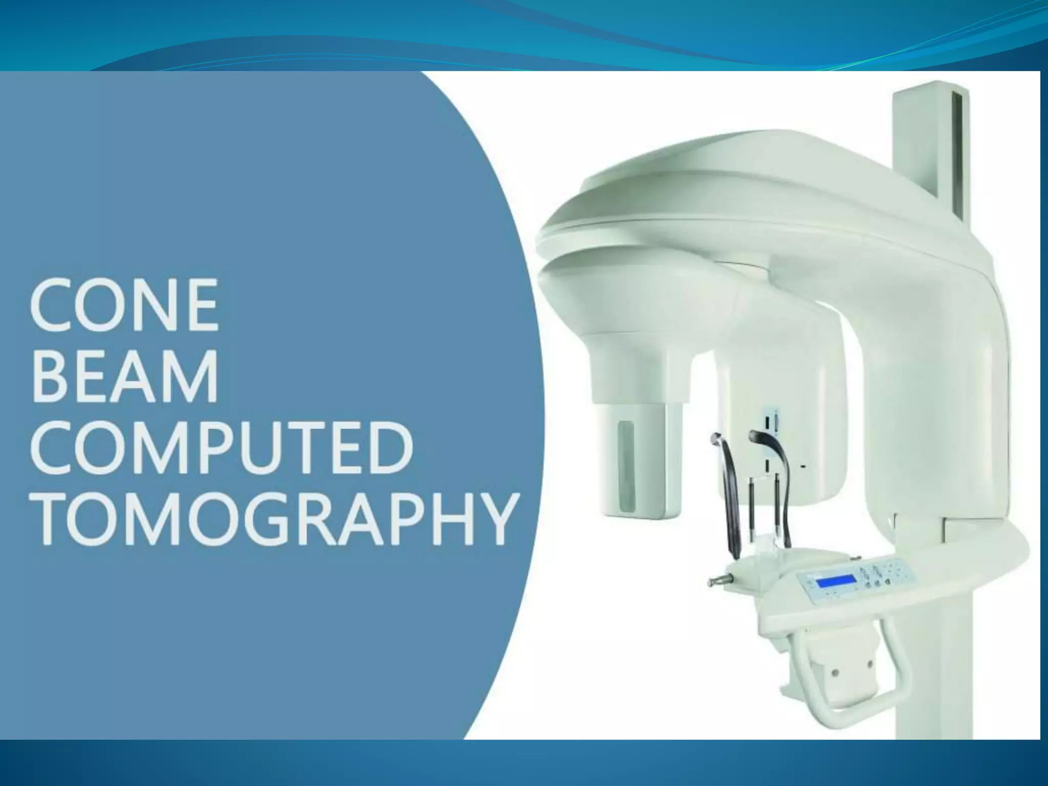
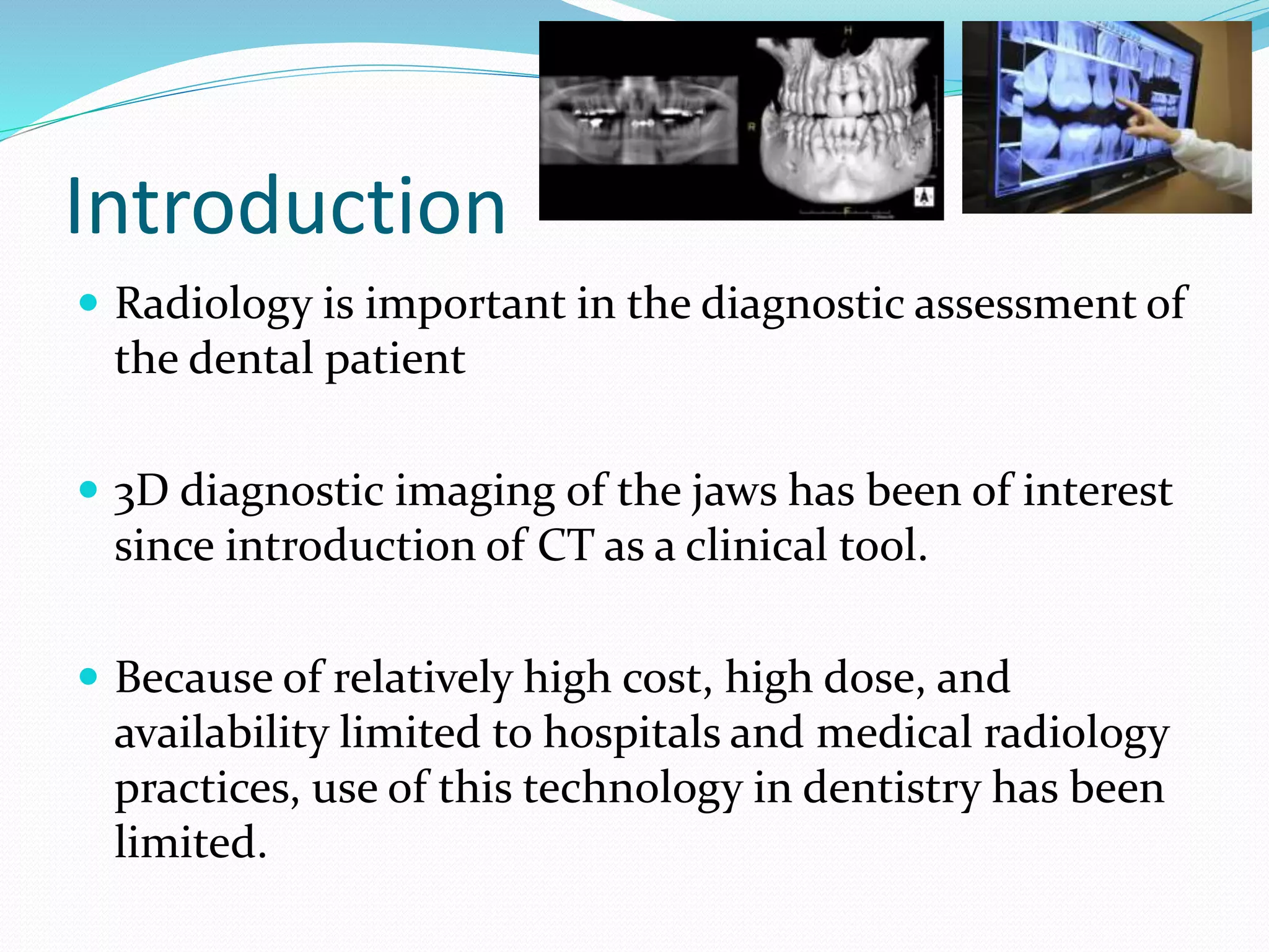
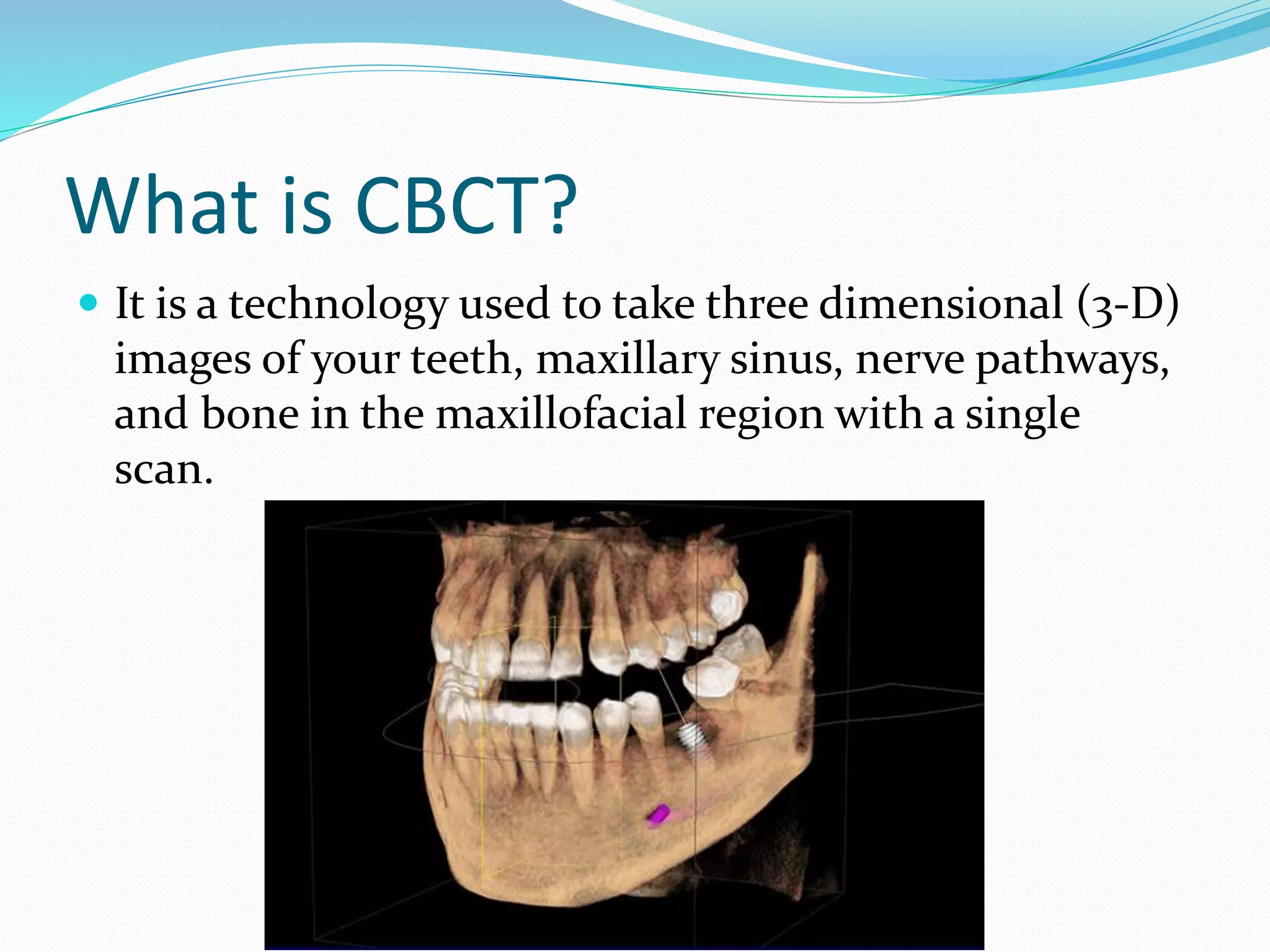
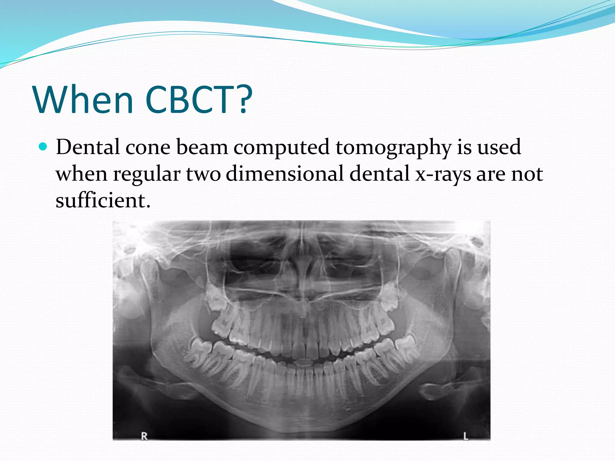
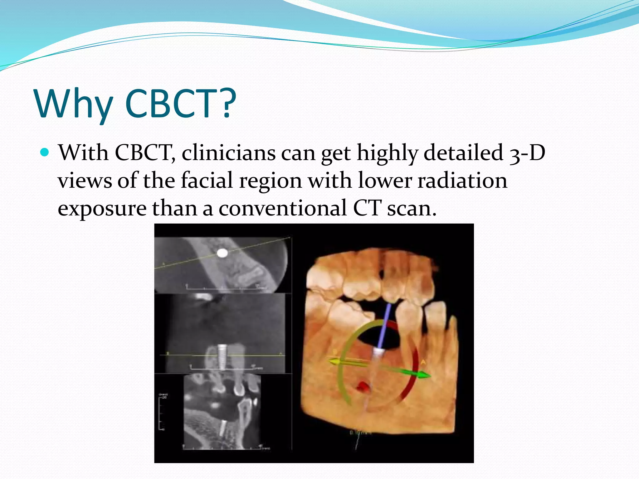
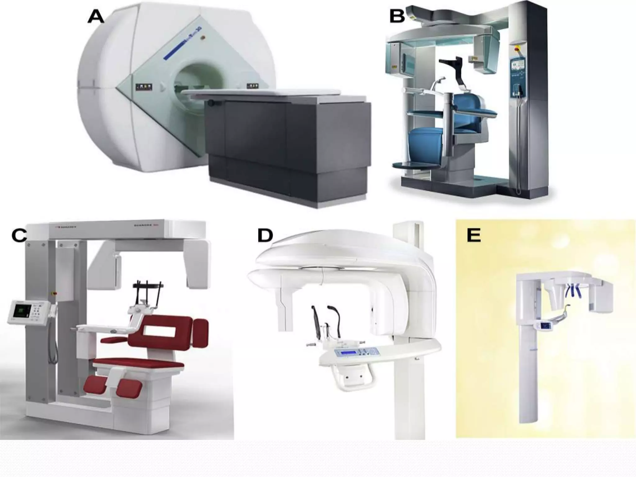
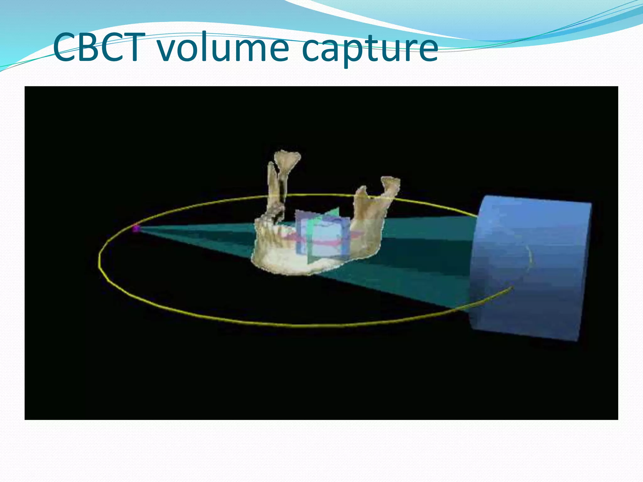
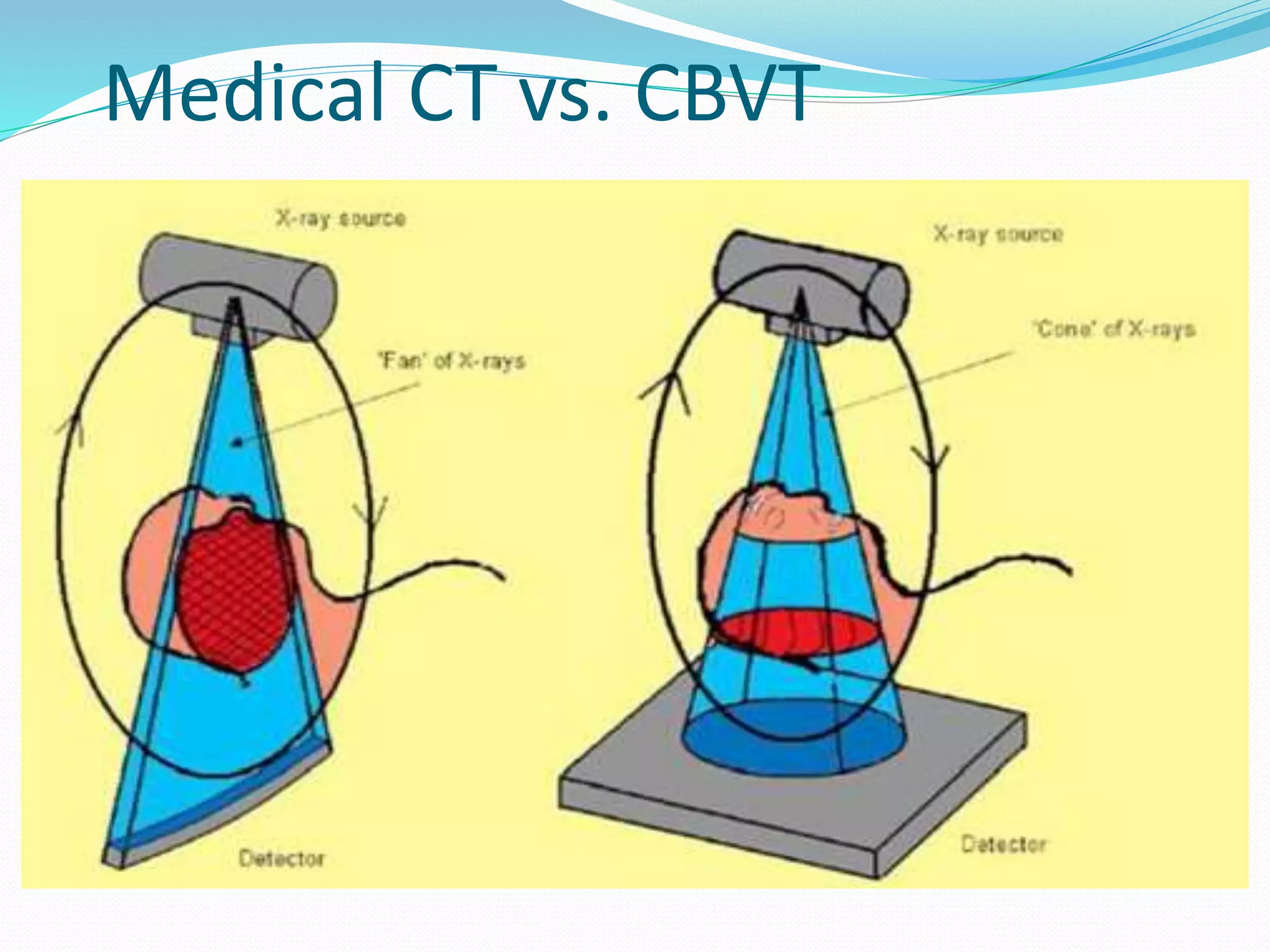
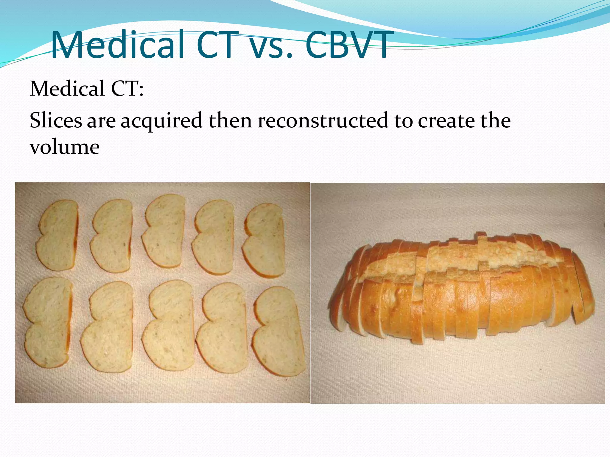
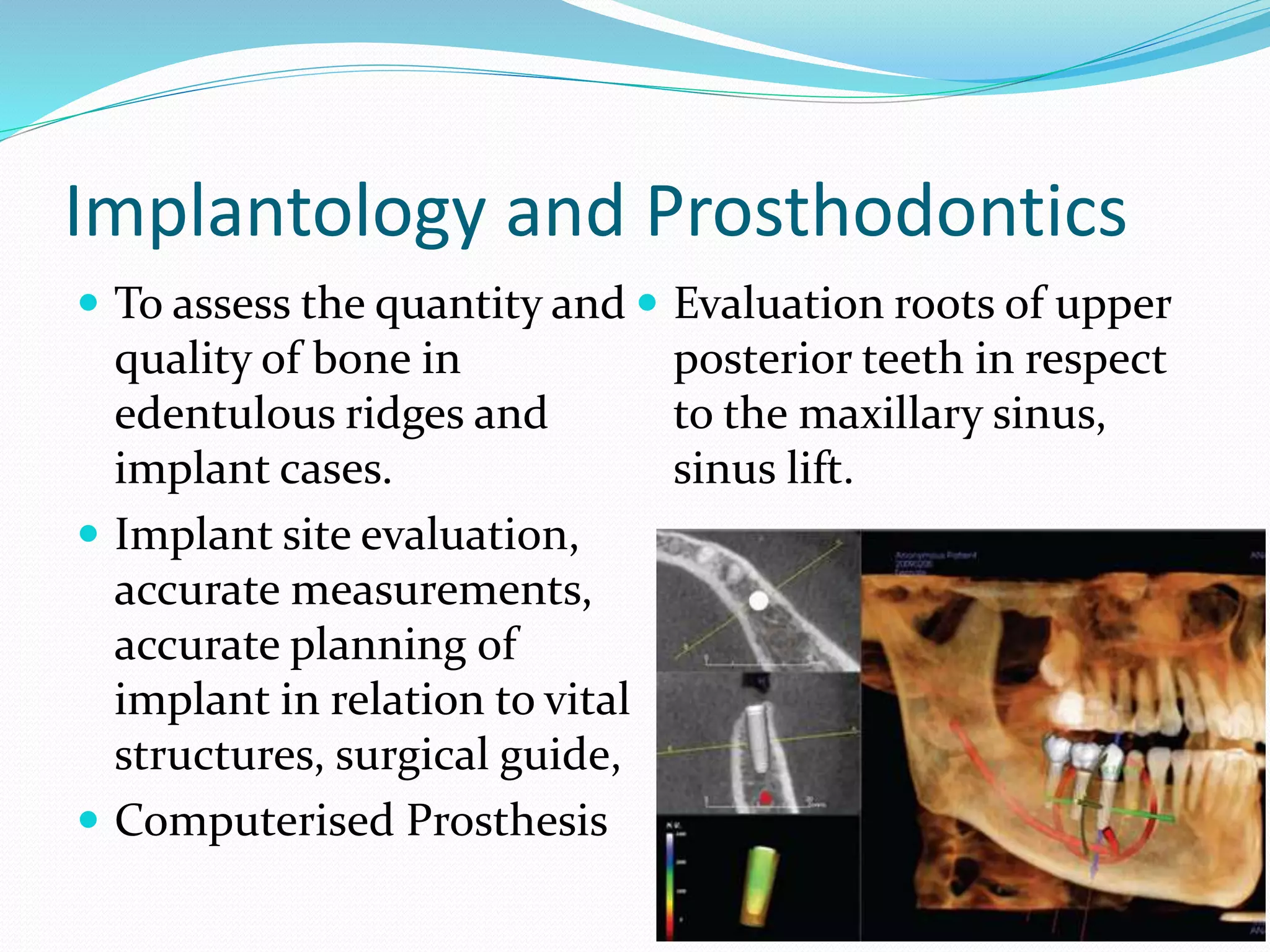
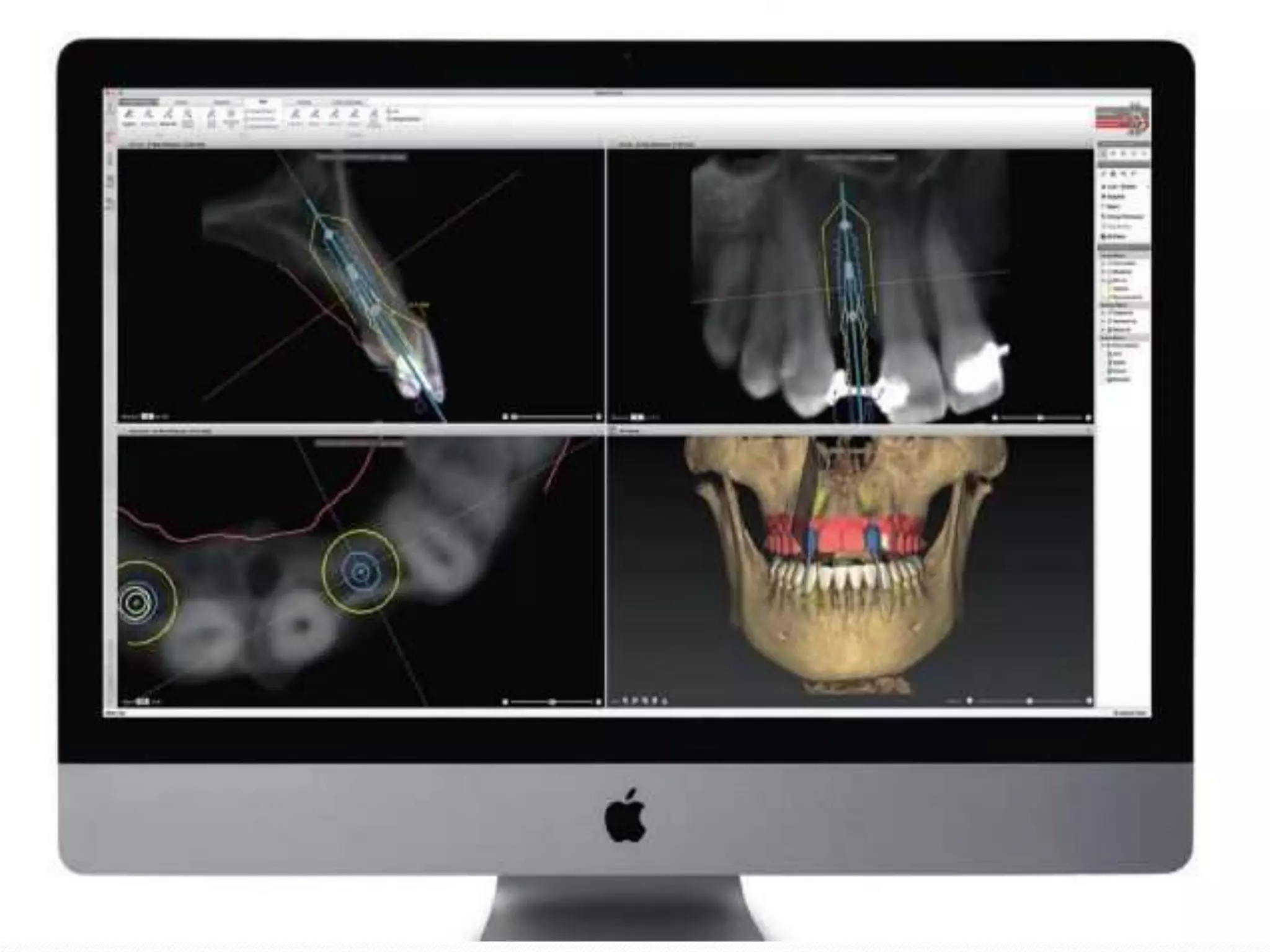
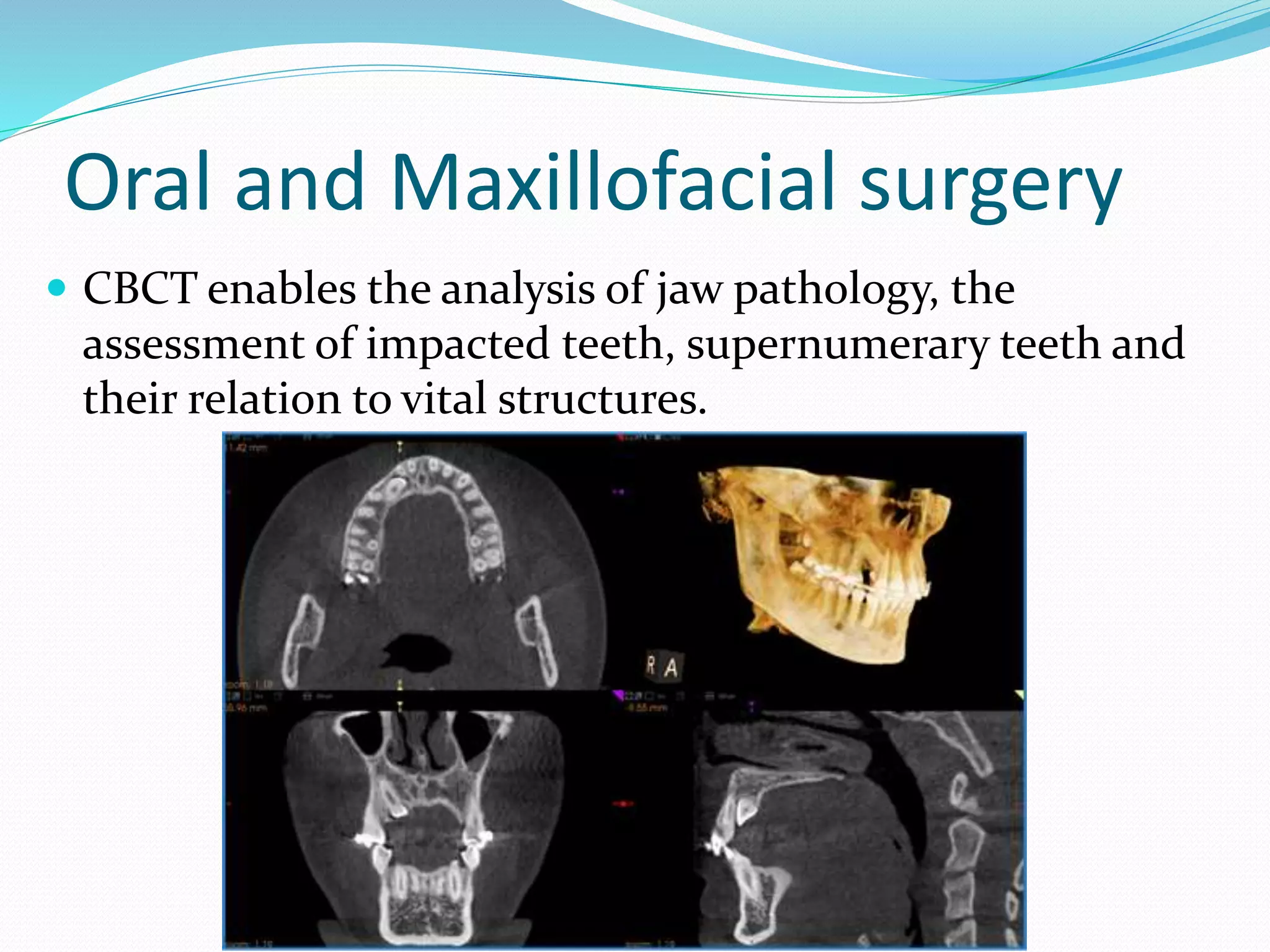
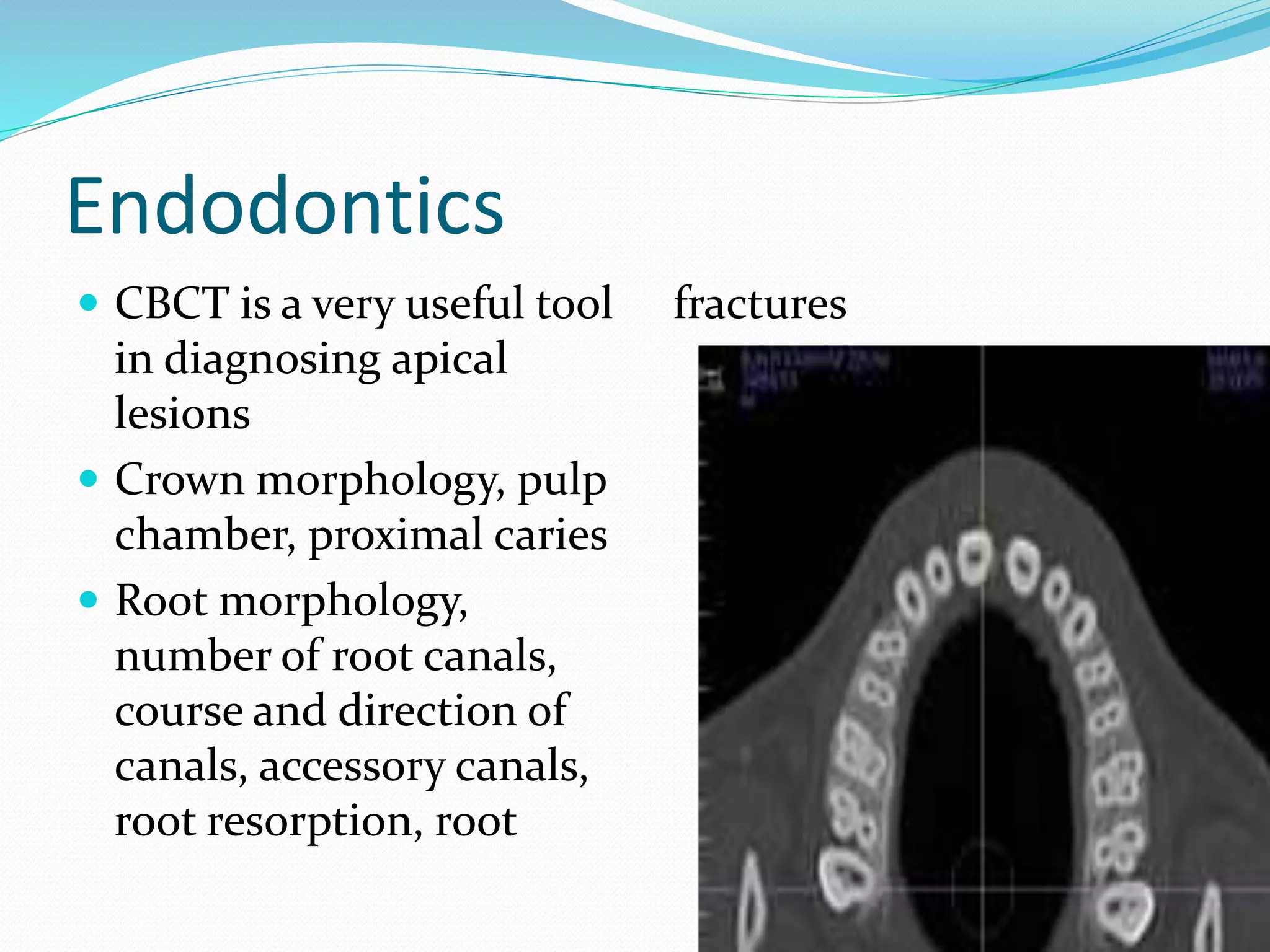
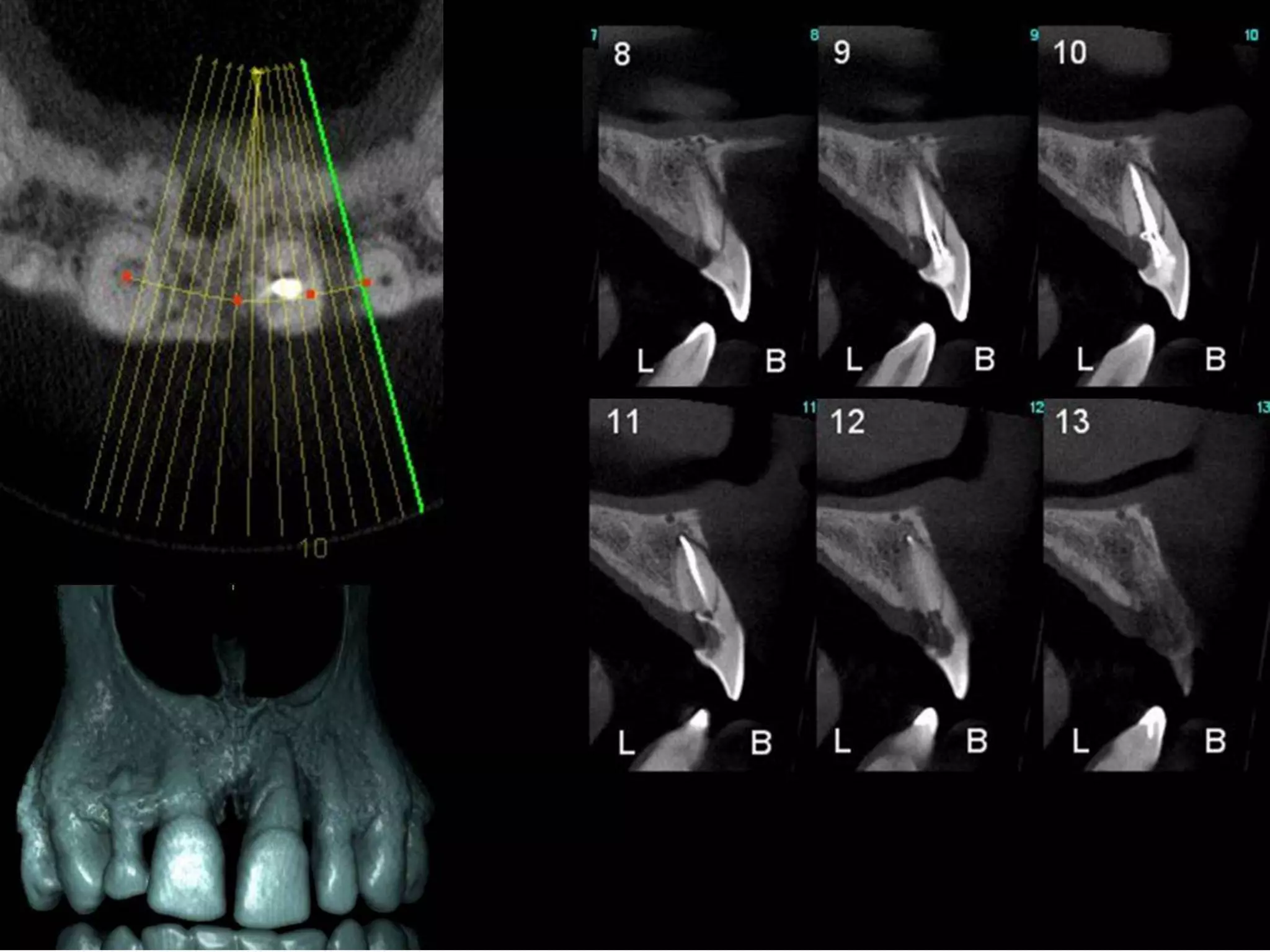
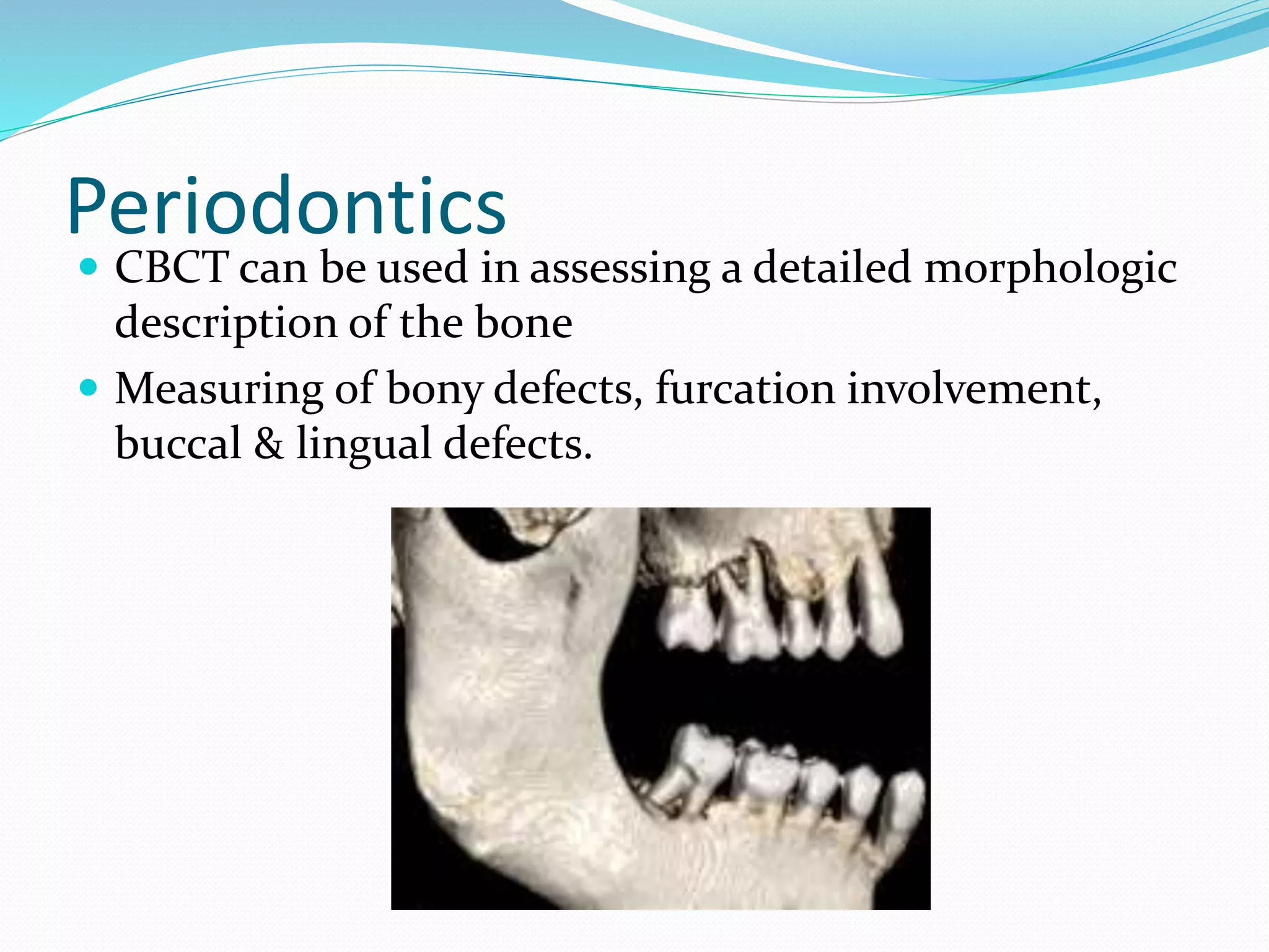
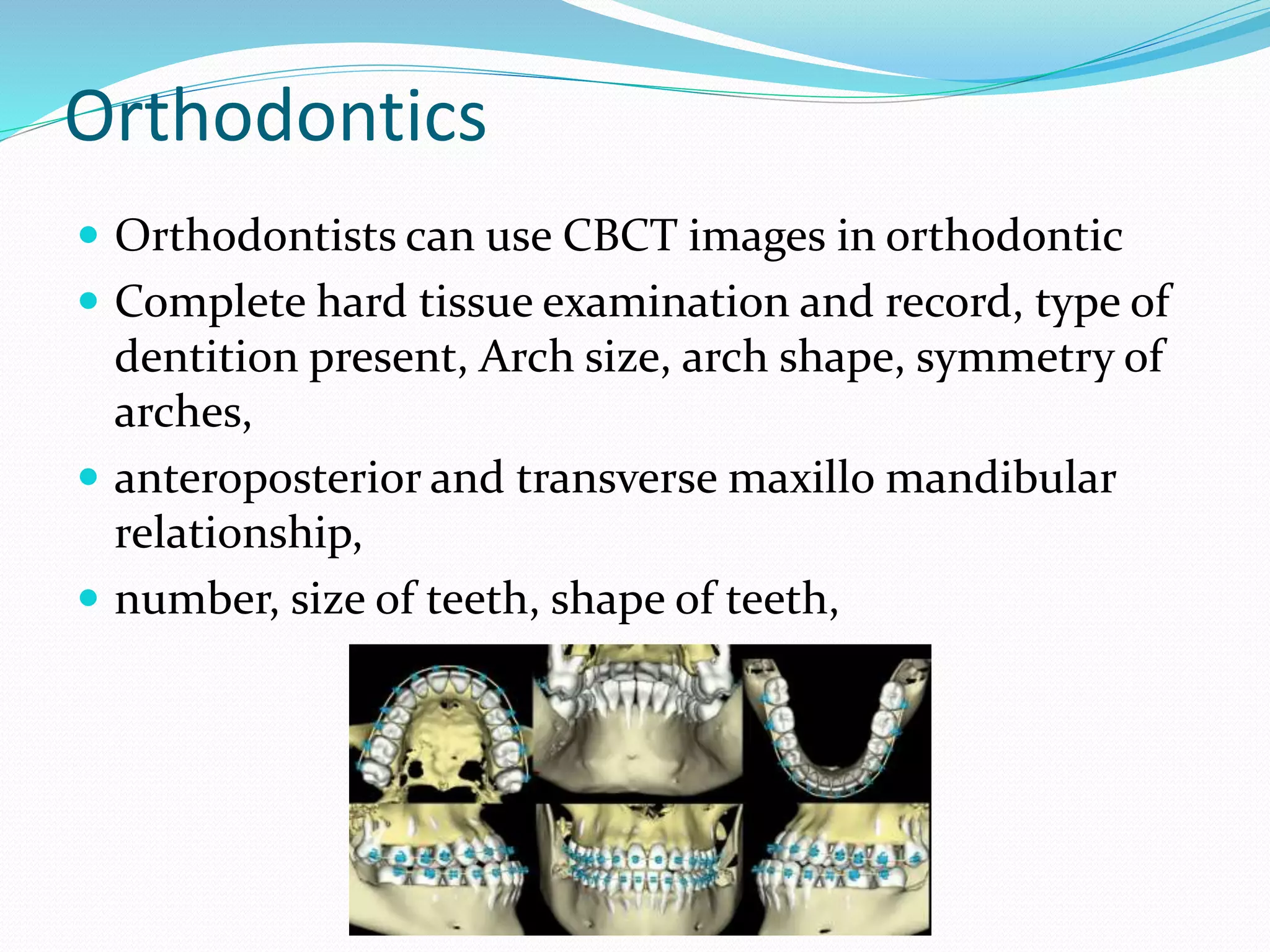
CBCT stands for cone beam computed tomography. It is a 3D imaging technique that uses a cone-shaped X-ray beam to capture volumetric images of the teeth, jaws, and surrounding structures. CBCT provides more detailed views than conventional 2D X-rays and exposes patients to less radiation than traditional medical CT scans. It has various applications in dentistry, including implant planning, endodontics, surgery, and orthodontics by allowing visualization of hard tissues and their relationship to anatomical structures.
brief description about CONTENTS Introduction Principles of panoramic imaging Image layer Panoramic machines Panoramic film Patient positioning Interpreting the panoramic imaging INDICATION Advantages Disadvantages Conclusion References
3. INTRODUCTION • Panoramic imaging also called pantomography is a technique for producing a single tomographic image of facial structures that includes both the maxillary and mandibular dental arches and their supporting structures . • This is a curvilinear variant of conventional tomography.
4. PRINCIPLES OF PANORAMIC IMAGE FORMATION • Patero and Numata - describe the principles of panoramic radiography • based on the principle of reciprocal movement of x-ray source and an image receptor around a central point or plane called the image layer, in which the OBJECT of image is located. • OBJECT in front or behind this image are not clearly captured because of their movement relative to the centre of rotation of the receptor and the x-ray source.
5. The film and x-ray tubehead move around the patient in opposite directions in panoramic radiography
6. ROTATION CENTER The pivotal point or axis around which the cassette carrier and tube head rotate is termed rotation center Three basic rotation center used in panoramic radiography Double centre rotation Triple centre rotation moving centre rotation The location and number of rotational centers INFLUENCE size and shape of focal trough
7. IMAGE LAYER • Also known as focal trough • It is a three dimensional curved zone where the structures lying within this layer are reasonably well defined on final panoramic image. • The structures seen on a panoramic image are primarily those located within image layer. • OBJECTSoutside the image layer are blurred magnified are reduced in size. Even distorted to the extent of not being recognizable. • This shape of image layer varies with the brand of equipment used.
8. FOCAL TROUGH
9. FACTORS AFFECTING SIZE OF IMAGE LAYER: Arc path Velocity of receptor and X-ray tube head Alignment of x-ray beam Collimator width The location of image layer change with extensive machine used so recalibration may be necessary if consistently suboptimal images are produced. As a position of object is moved within the image layer size and shape of image layer change.
10. PANORAMIC UNIT
11. A, Orthophos XG Plus extraoral x-ray machine. B, Orthoralix 8500 extraoral x-ray machine. C, Example of a digital panoramic system
12. PARTS OF PANORAMIC UNITS a. x-ray tube head b. head positioner: chin rest notched bite block forehead rest lateral head support c. exposure controls
13. X-RAY TUBE HEAD: • Similar to intraoral x-ray tube head • Each has a filament to produce electrons and a target to produce x-rays • Collimator is a lead plate with narrow vertical slit • Narrow x-ray beam emerges from collimator minimize patient exposure to radiation
1
this contains the occlusal radiography methods for both maxillary and mandibular different occusal radiographic techniques, principles, classification, indications
IDEAL IMAGE CHARACTERISTICS
FACTORS RELATED TO THE RADIATION BEAM
FACTORS RELATED TO THE OBJECT
FACTORS RELATED TO THE TECHNIQUE
FACTORS RELATED TO RECORDING OF THE ROENTGEN IMAGE OF THE OBJECT
DARK/ LIGHT IMAGE IDEAL IMAGE
IDEAL QUALITY CRIETRIA
This is a presentation describing in brief regarding the physics behind MRI and it's application from dental point of view. It contains few videos as well.
ORN is an inflammatory condition of bone that occurs after the bone has been exposed to therapeutic doses of radiation usually given for a malignancies.
Unveiling the Intricacies Exploring Normal Anatomy on Dental CT scans - CT De...online Marketing
In the realm of dental CT scan imaging, normal anatomy emerges as a testament to the intricate design of the human oral cavity. From the subtle contours of alveolar bone to the complex architecture of temporomandibular joints, each structure plays a vital role in oral health and function. As technology continues to evolve, so too does our understanding of normal anatomy, paving the way for advancements in dental diagnostics and treatment modalities. please visit: https://ct-dent.co.uk/unveiling-the-intricacies-exploring-normal-anatomy-on-dental-ct-scans/
is a diagnostic imaging modality that provide high quality ,CBCT uses systems that are ideal in capturing images of hard tissues especially in the maxillofacial region
A very precise and intimate description on radiographic considerations in dental implants, since the advent of the first radiographic modality in 1905, the dental health care professionals have been striving to achieve clarity & excellence in the development & usage of dental radiographic imaging modalities.
I hope this presentation will make this wonderful topic more understandable and easier to digest in the minds of young and experienced dental health care professionals.
by Dr Ishaan Adhaulia
Dental Cone Beam CT scans (CBCT Scans) provide high resolution, 3D volumetric images, used in Dental Implant Planning, Orthodontics and Maxillofacial surgery. allowing for more accurate analysis of bone structure and dental/tooth orientation. The accuracy of a CBCT scan is comparable to medical CT scans but uses a much lower radiation dose but with the added advantage of greater accuracy of bone structures and dental/tooth orientation. Please visit: https://ct-dent.co.uk/cone-beam-cbct-scans/
Recent prosthetic management of hemimaxillectomy /certified fixed orthodontic...Indian dental academy
The Indian Dental Academy is the Leader in continuing dental education , training dentists in all aspects of dentistry and offering a wide range of dental certified courses in different formats.
Indian dental academy provides dental crown & Bridge,rotary endodontics,fixed orthodontics,
Dental implants courses.for details pls visit www.indiandentalacademy.com ,or call
0091-9248678078
Medical emergencies in the dental practiceRuhi Kashmiri
Medical emergencies do, can and will occur in any dental practice, oral health professionals need to know how to diagnose and manage any such situation when required.
Medical conditions that can directly affect the provision of dental care and/...Ruhi Kashmiri
Medical conditions that can directly affect the provision of dental care and/or consequences of dental treatment. In paediatric dentistry, such children are known as children with special needs and require extra attention for maintainence of optimum oral health.
The prostate is an exocrine gland of the male mammalian reproductive system
It is a walnut-sized gland that forms part of the male reproductive system and is located in front of the rectum and just below the urinary bladder
Function is to store and secrete a clear, slightly alkaline fluid that constitutes 10-30% of the volume of the seminal fluid that along with the spermatozoa, constitutes semen
A healthy human prostate measures (4cm-vertical, by 3cm-horizontal, 2cm ant-post ).
It surrounds the urethra just below the urinary bladder. It has anterior, median, posterior and two lateral lobes
It’s work is regulated by androgens which are responsible for male sex characteristics
Generalised disease of the prostate due to hormonal derangement which leads to non malignant enlargement of the gland (increase in the number of epithelial cells and stromal tissue)to cause compression of the urethra leading to symptoms (LUTS
These lecture slides, by Dr Sidra Arshad, offer a quick overview of physiological basis of a normal electrocardiogram.
Learning objectives:
1. Define an electrocardiogram (ECG) and electrocardiography
2. Describe how dipoles generated by the heart produce the waveforms of the ECG
3. Describe the components of a normal electrocardiogram of a typical bipolar leads (limb II)
4. Differentiate between intervals and segments
5. Enlist some common indications for obtaining an ECG
Study Resources:
1. Chapter 11, Guyton and Hall Textbook of Medical Physiology, 14th edition
2. Chapter 9, Human Physiology - From Cells to Systems, Lauralee Sherwood, 9th edition
3. Chapter 29, Ganong’s Review of Medical Physiology, 26th edition
4. Electrocardiogram, StatPearls - https://www.ncbi.nlm.nih.gov/books/NBK549803/
5. ECG in Medical Practice by ABM Abdullah, 4th edition
6. ECG Basics, http://www.nataliescasebook.com/tag/e-c-g-basics
MANAGEMENT OF ATRIOVENTRICULAR CONDUCTION BLOCK.pdfJim Jacob Roy
Cardiac conduction defects can occur due to various causes.
Atrioventricular conduction blocks ( AV blocks ) are classified into 3 types.
This document describes the acute management of AV block.
micro teaching on communication m.sc nursing.pdfAnurag Sharma
Microteaching is a unique model of practice teaching. It is a viable instrument for the. desired change in the teaching behavior or the behavior potential which, in specified types of real. classroom situations, tends to facilitate the achievement of specified types of objectives.
Ozempic: Preoperative Management of Patients on GLP-1 Receptor Agonists Saeid Safari
Preoperative Management of Patients on GLP-1 Receptor Agonists like Ozempic and Semiglutide
ASA GUIDELINE
NYSORA Guideline
2 Case Reports of Gastric Ultrasound
HOT NEW PRODUCT! BIG SALES FAST SHIPPING NOW FROM CHINA!! EU KU DB BK substit...GL Anaacs
Contact us if you are interested:
Email / Skype : kefaya1771@gmail.com
Threema: PXHY5PDH
New BATCH Ku !!! MUCH IN DEMAND FAST SALE EVERY BATCH HAPPY GOOD EFFECT BIG BATCH !
Contact me on Threema or skype to start big business!!
Hot-sale products:
NEW HOT EUTYLONE WHITE CRYSTAL!!
5cl-adba precursor (semi finished )
5cl-adba raw materials
ADBB precursor (semi finished )
ADBB raw materials
APVP powder
5fadb/4f-adb
Jwh018 / Jwh210
Eutylone crystal
Protonitazene (hydrochloride) CAS: 119276-01-6
Flubrotizolam CAS: 57801-95-3
Metonitazene CAS: 14680-51-4
Payment terms: Western Union,MoneyGram,Bitcoin or USDT.
Deliver Time: Usually 7-15days
Shipping method: FedEx, TNT, DHL,UPS etc.Our deliveries are 100% safe, fast, reliable and discreet.
Samples will be sent for your evaluation!If you are interested in, please contact me, let's talk details.
We specializes in exporting high quality Research chemical, medical intermediate, Pharmaceutical chemicals and so on. Products are exported to USA, Canada, France, Korea, Japan,Russia, Southeast Asia and other countries.
Title: Sense of Taste
Presenter: Dr. Faiza, Assistant Professor of Physiology
Qualifications:
MBBS (Best Graduate, AIMC Lahore)
FCPS Physiology
ICMT, CHPE, DHPE (STMU)
MPH (GC University, Faisalabad)
MBA (Virtual University of Pakistan)
Learning Objectives:
Describe the structure and function of taste buds.
Describe the relationship between the taste threshold and taste index of common substances.
Explain the chemical basis and signal transduction of taste perception for each type of primary taste sensation.
Recognize different abnormalities of taste perception and their causes.
Key Topics:
Significance of Taste Sensation:
Differentiation between pleasant and harmful food
Influence on behavior
Selection of food based on metabolic needs
Receptors of Taste:
Taste buds on the tongue
Influence of sense of smell, texture of food, and pain stimulation (e.g., by pepper)
Primary and Secondary Taste Sensations:
Primary taste sensations: Sweet, Sour, Salty, Bitter, Umami
Chemical basis and signal transduction mechanisms for each taste
Taste Threshold and Index:
Taste threshold values for Sweet (sucrose), Salty (NaCl), Sour (HCl), and Bitter (Quinine)
Taste index relationship: Inversely proportional to taste threshold
Taste Blindness:
Inability to taste certain substances, particularly thiourea compounds
Example: Phenylthiocarbamide
Structure and Function of Taste Buds:
Composition: Epithelial cells, Sustentacular/Supporting cells, Taste cells, Basal cells
Features: Taste pores, Taste hairs/microvilli, and Taste nerve fibers
Location of Taste Buds:
Found in papillae of the tongue (Fungiform, Circumvallate, Foliate)
Also present on the palate, tonsillar pillars, epiglottis, and proximal esophagus
Mechanism of Taste Stimulation:
Interaction of taste substances with receptors on microvilli
Signal transduction pathways for Umami, Sweet, Bitter, Sour, and Salty tastes
Taste Sensitivity and Adaptation:
Decrease in sensitivity with age
Rapid adaptation of taste sensation
Role of Saliva in Taste:
Dissolution of tastants to reach receptors
Washing away the stimulus
Taste Preferences and Aversions:
Mechanisms behind taste preference and aversion
Influence of receptors and neural pathways
Impact of Sensory Nerve Damage:
Degeneration of taste buds if the sensory nerve fiber is cut
Abnormalities of Taste Detection:
Conditions: Ageusia, Hypogeusia, Dysgeusia (parageusia)
Causes: Nerve damage, neurological disorders, infections, poor oral hygiene, adverse drug effects, deficiencies, aging, tobacco use, altered neurotransmitter levels
Neurotransmitters and Taste Threshold:
Effects of serotonin (5-HT) and norepinephrine (NE) on taste sensitivity
Supertasters:
25% of the population with heightened sensitivity to taste, especially bitterness
Increased number of fungiform papillae
Report Back from SGO 2024: What’s the Latest in Cervical Cancer?bkling
Are you curious about what’s new in cervical cancer research or unsure what the findings mean? Join Dr. Emily Ko, a gynecologic oncologist at Penn Medicine, to learn about the latest updates from the Society of Gynecologic Oncology (SGO) 2024 Annual Meeting on Women’s Cancer. Dr. Ko will discuss what the research presented at the conference means for you and answer your questions about the new developments.
Couples presenting to the infertility clinic- Do they really have infertility...Sujoy Dasgupta
Dr Sujoy Dasgupta presented the study on "Couples presenting to the infertility clinic- Do they really have infertility? – The unexplored stories of non-consummation" in the 13th Congress of the Asia Pacific Initiative on Reproduction (ASPIRE 2024) at Manila on 24 May, 2024.
ARTIFICIAL INTELLIGENCE IN HEALTHCARE.pdfAnujkumaranit
Artificial intelligence (AI) refers to the simulation of human intelligence processes by machines, especially computer systems. It encompasses tasks such as learning, reasoning, problem-solving, perception, and language understanding. AI technologies are revolutionizing various fields, from healthcare to finance, by enabling machines to perform tasks that typically require human intelligence.
Tom Selleck Health: A Comprehensive Look at the Iconic Actor’s Wellness Journeygreendigital
Tom Selleck, an enduring figure in Hollywood. has captivated audiences for decades with his rugged charm, iconic moustache. and memorable roles in television and film. From his breakout role as Thomas Magnum in Magnum P.I. to his current portrayal of Frank Reagan in Blue Bloods. Selleck's career has spanned over 50 years. But beyond his professional achievements. fans have often been curious about Tom Selleck Health. especially as he has aged in the public eye.
Follow us on: Pinterest
Introduction
Many have been interested in Tom Selleck health. not only because of his enduring presence on screen but also because of the challenges. and lifestyle choices he has faced and made over the years. This article delves into the various aspects of Tom Selleck health. exploring his fitness regimen, diet, mental health. and the challenges he has encountered as he ages. We'll look at how he maintains his well-being. the health issues he has faced, and his approach to ageing .
Early Life and Career
Childhood and Athletic Beginnings
Tom Selleck was born on January 29, 1945, in Detroit, Michigan, and grew up in Sherman Oaks, California. From an early age, he was involved in sports, particularly basketball. which played a significant role in his physical development. His athletic pursuits continued into college. where he attended the University of Southern California (USC) on a basketball scholarship. This early involvement in sports laid a strong foundation for his physical health and disciplined lifestyle.
Transition to Acting
Selleck's transition from an athlete to an actor came with its physical demands. His first significant role in "Magnum P.I." required him to perform various stunts and maintain a fit appearance. This role, which he played from 1980 to 1988. necessitated a rigorous fitness routine to meet the show's demands. setting the stage for his long-term commitment to health and wellness.
Fitness Regimen
Workout Routine
Tom Selleck health and fitness regimen has evolved. adapting to his changing roles and age. During his "Magnum, P.I." days. Selleck's workouts were intense and focused on building and maintaining muscle mass. His routine included weightlifting, cardiovascular exercises. and specific training for the stunts he performed on the show.
Selleck adjusted his fitness routine as he aged to suit his body's needs. Today, his workouts focus on maintaining flexibility, strength, and cardiovascular health. He incorporates low-impact exercises such as swimming, walking, and light weightlifting. This balanced approach helps him stay fit without putting undue strain on his joints and muscles.
Importance of Flexibility and Mobility
In recent years, Selleck has emphasized the importance of flexibility and mobility in his fitness regimen. Understanding the natural decline in muscle mass and joint flexibility with age. he includes stretching and yoga in his routine. These practices help prevent injuries, improve posture, and maintain mobilit
3. Introduction
Radiology is important in the diagnostic assessment of
the dental patient
3D diagnostic imaging of the jaws has been of interest
since introduction of CT as a clinical tool.
Because of relatively high cost, high dose, and
availability limited to hospitals and medical radiology
practices, use of this technology in dentistry has been
limited.
4. What is CBCT?
It is a technology used to take three dimensional (3-D)
images of your teeth, maxillary sinus, nerve pathways,
and bone in the maxillofacial region with a single
scan.
5. What is CBCT?
The CBCT system rotates around the patient in
approximately 30 seconds, capturing data using a
cone-shaped x-ray beam.
6. When CBCT?
Dental cone beam computed tomography is used
when regular two dimensional dental x-rays are not
sufficient.
7. Why CBCT?
With CBCT, clinicians can get highly detailed 3-D
views of the facial region with lower radiation
exposure than a conventional CT scan.
8. Why CBCT?
This may help with the diagnosis,
treatment planning and evaluation
of certain conditions.
12. Medical CT vs. CBVT
Medical CT:
Slices are acquired then reconstructed to create the
volume
13. Medical CT vs. CBVT
CBCT:
The volume is acquired then slices are reconstructed from
the volume
14.
15. Advantages
Rapid scan time
Beam limitation
Image accuracy
Reduction in patient radiation dose when compared to
medical ct (10 times less)
Economical comfortable and safe
16. Disadvantages
Scatter
Motion artifacts due to increased scan time
Poor contrast resolution, thus soft tissue cannot be
reviewed
17. Use in dentistry
Implantology and prosthodontics
Oral and maxillofacial surgery
Endodontics
Periodontics
Orthodontics
18. Implantology and Prosthodontics
To assess the quantity and
quality of bone in
edentulous ridges and
implant cases.
Implant site evaluation,
accurate measurements,
accurate planning of
implant in relation to vital
structures, surgical guide,
Computerised Prosthesis
Evaluation roots of upper
posterior teeth in respect
to the maxillary sinus,
sinus lift.
19.
20.
21. Oral and Maxillofacial surgery
CBCT enables the analysis of jaw pathology, the
assessment of impacted teeth, supernumerary teeth and
their relation to vital structures.
22. The assessment of bone grafts.
It is also helpful in analysing and assessing paranasal
sinuses
23. Orthognathic surgeries
Post treatment esthetic evaluation
TMJ, and condyle
Tooth auto transplants
Cleft palate
Craniofacial reconstruction
Mid face fracture cases of fractures of the facial bones
24.
25.
26. Endodontics
CBCT is a very useful tool
in diagnosing apical
lesions
Crown morphology, pulp
chamber, proximal caries
Root morphology,
number of root canals,
course and direction of
canals, accessory canals,
root resorption, root
fractures
27.
28. Endodontics
Obturation, filling, under and overfilling, sinus
problems
Broken instruments
Useful in planning periapical endodontic surgery as
the anatomical structures such as inferior dental canal,
mental foramen, maxillary sinus can be easily
differentiated and relation to root can be quantified.
29. Periodontics
CBCT can be used in assessing a detailed morphologic
description of the bone
Measuring of bony defects, furcation involvement,
buccal & lingual defects.
30. Orthodontics
Orthodontists can use CBCT images in orthodontic
Complete hard tissue examination and record, type of
dentition present, Arch size, arch shape, symmetry of
arches,
anteroposterior and transverse maxillo mandibular
relationship,
number, size of teeth, shape of teeth,
31. Typical doses of various
dental radiological procedures
Intra-oral 0.001 mSv up to 0.004 mSv
Full-mouth set 0.080 mSv
Lateral cephalogram 0.002 mSv
Dental panoramic technique 0.015 mSv
CBCT (both jaws) 0.068 mSv
Hospital CT scan (both jaws) 0.6 mSv
32. Conclusion
It is vital or any clinician to remain in touch with latest
innovations in the field of one's expertise and apply
the same for the benefit of patient care and be ready
and willing to accept new trends.
CBCT is one such recent advancement in the field of
Dentistry which one should learn and adapt into
clinical practice.