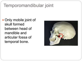
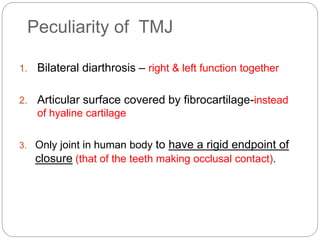
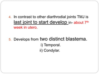
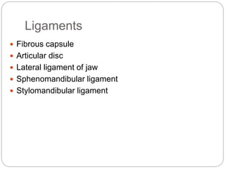
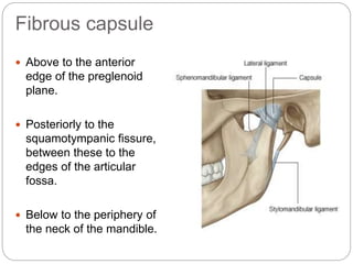
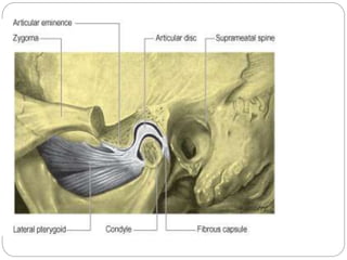
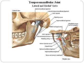
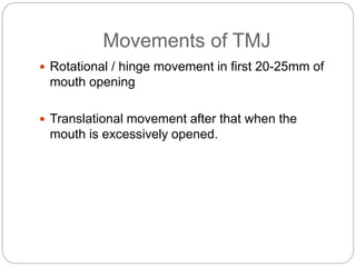
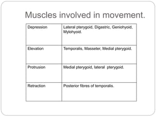
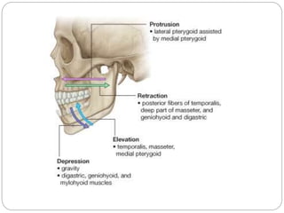
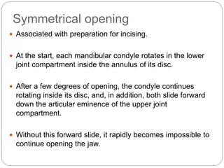
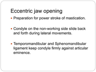
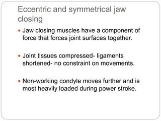
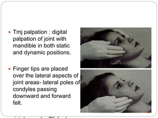
TEMPOROMANDIBULAR JOINT
Dr Bhaumik Thakkar
MDS-Part 1.
Dept. Of Periodontology and
Implantology.
INTRODUCTION
 The most important functions of the
temporomandibular joint (TMJ) are mastication and
speech and are of great interest to dentists,
orthodontists, clinicians, and radiologists.
 The TMJ is a ginglymoarthrodial joint, a term that is
derived from ginglymus, meaning a hinge joint,
allowing motion only backward and forward in one
plane, and arthrodia, meaning a joint of which
permits a gliding motion of the surfaces.
Temporomandibular joint
 Only mobile joint of
skull formed
between head of
mandible and
articular fossa of
temporal bone.
Peculiarity of TMJ
1. Bilateral diarthrosis – right & left function together
2. Articular surface covered by fibrocartilage-instead
of hyaline cartilage
3. Only joint in human body to have a rigid endpoint of
closure (that of the teeth making occlusal contact).
4. In contrast to other diarthrodial joints TMJ is
last joint to start develop in- about 7th
week in utero.
5. Develops from two distinct blastema.
i) Temporal.
ii) Condylar.
Ligaments
 Fibrous capsule
 Articular disc
 Lateral ligament of jaw
 Sphenomandibular ligament
 Stylomandibular ligament
Fibrous capsule
 Above to the anterior
edge of the preglenoid
plane.
 Posteriorly to the
squamotympanic fissure,
between these to the
edges of the articular
fossa.
 Below to the periphery of
the neck of the mandible.
Articular disc
 Fibrocartilaginous disc dividing joint cavity in upper and
lower compartment.
 Shape: Oval
 Makes articular surface congruent.
 In sagittal section- a thin intermediate zone and thickened
anterior and posterior bands, and its upper surface appears
concavo-convex.
 Posteriorly- Attached to a region of loose vascular and
nervous tissue which splits into two laminae, the bilaminar
region.
Functions of Articular disc
 Stabilize the TMJ.
 Makes articular surfaces congruent.
 Reduce wear of TMJ.
 Aid lubrication of the joint.
Lateral ligament of Jaw
 Attached above to the articular tubercle on the root
of the zygomatic process of the temporal bone.
 It extends downwards and backwards at an angle
of 45° to the horizontal, to attach to the lateral
surface and posterior border of the neck of the
condyle, deep to the parotid gland.
 Function: To prevent posterior displacement of the
resting condyle.
Sphenomandibular ligament
 Medial to, and normally separate from, the
capsule. It is a flat, thin band that descends from
the spine of the sphenoid.
 Widens at the lingula of the mandibular foramen.
This part is a vestige of the dorsal end of
Meckel's cartilage.
 It is separated from the pharynx by fat and a
pharyngeal vein.
Stylomandibular ligament
 A thickened band of deep cervical fascia that stretches
from the apex and adjacent anterior aspect of the
styloid process to the angle and posterior border of the
mandible.
 Along with sphenomandibular ligament it is
responsible for limitation of mandibular movement.
Blood supply & Innervation
Auriculotemporal nerve
maxillary artery
Masseteric nerve
Superficial
temporal
artery
Arterial supply : Superficial
temporal artery laterally and the
maxillary artery medially.
Nerve supply:
Masseteric and
auriculotemporal nerves
VASCULARIZATION
 Predominant vessals supplying tmj are:
Superficial temporal artery from the posterior
Middle meningeal artery from the anterior
Internal maxillary artery from the inferior
HISTOLOGY
OF
ARTICULAR SURFACE OF TMJ
1. The Articular zone
 Dense fibrous
connective tissue
 Poor blood supply
 Better ability to
repair
• Good adaption to sliding movement
• Shock absorber
• Less susceptible to the effect of
aging time & breakdown over time.
2. The proliferative
zone
 Mainly cellular zone
 Undifferentiated
mesenchymal cells
 Proliferation &
regeneration
throughout life
3. The cartilagenous
zone
 Collagen fibers
arranged in criss -
cross pattern of
bundles
 Fibrocartilage
appears in a random
orientation, providing
a three-dimensional
network that offers
resistance against
compressive and
lateral forces.
• Offers considerable resistance against
compressive & lateral forces
• But becomes thinner with age.
4. The calcified zone
 Deepest zone
 Chondrocytes,
chondroblasts &
osteoblasts
• Active site for remodeling activity as
bone growth proceeds.
Movements of TMJ
 Rotational / hinge movement in first 20-25mm of
mouth opening
 Translational movement after that when the
mouth is excessively opened.
Muscles involved in movement.
Depression Lateral pterygoid, Digastric, Geniohyoid,
Mylohyoid.
Elevation Temporalis, Masseter, Medial pterygoid.
Protrusion Medial pterygoid, lateral pterygoid.
Retraction Posterior fibres of temporalis.
Age changes of the TMJ:
 Condyle:
 Becomes more flattened
 Fibrous capsule becomes thicker.
 Osteoporosis of underlying bone.
 Thinning or absence of cartilaginous zone.
 Disk:
 Becomes thinner.
 Shows hyalinization and chondroid changes.
 Synovial fold:
 Become fibrotic with thick basement membrane.
 Blood vessels and nerves:
 Walls of blood vessels thickened.
 Nerves decrease in number
Age changes lead to:
 Decrease in the synovial fluid formation
 Impairment of motion due to decrease in the
disc and capsule extensibility
 Decrease the resilience during mastication
due to chondroid changes into collagenous
elements
 Dysfunction in older people
Symmetrical opening
 Associated with preparation for incising.
 At the start, each mandibular condyle rotates in the lower
joint compartment inside the annulus of its disc.
 After a few degrees of opening, the condyle continues
rotating inside its disc, and, in addition, both slide forward
down the articular eminence of the upper joint
compartment.
 Without this forward slide, it rapidly becomes impossible to
continue opening the jaw.
Eccentric jaw opening
 Preparation for power stroke of mastication.
 Condyle on the non-working side slide back
and forth during lateral movements.
 Temporomandibular and Sphenomandibular
ligament keep condyle firmly against articular
eminence.
Eccentric and symmetrical jaw
closing
 Jaw closing muscles have a component of
force that forces joint surfaces together.
 Joint tissues compressed- ligaments
shortened- no constraint on movements.
 Non-working condyle moves further and is
most heavily loaded during power stroke.
Temporomandibular Joint Disorder
 Various terms have been used to describe
disturbances of the masticatory system.
 1934 James costen described group of symptoms
centering around ear and TMJ- Costen syndrome.
 1959 Shore introduced TMJ Dysfunction
 Ash & Ramfjord- functional TMJ disturbances
Terminology
 Limited nature of these terms lead to a broader
term- Craniomandibular disorders.
 Bell coined the term Temporomandibular
disorders.
 Describes both problems associated with the joint
& disturbances associated with function of
masticatory system.
Event
 Events can be either local or systemic.
 Local: a change in proprioceptive input e.g
improper crown.
 Can be secondary to trauma- too wide opening of
mouth; unaccustomed use- bruxism.
 Also deep pain input- alters muscle function by
way of central excitatory effects.
 Systemic events; that alter normal function occurs
at a systemic level; emotional stress.
Physiologic tolerance
 Influenced by local and systemic factors.
 Local: orthopedic stability-relation between
mandible and maxilla- good stability; mandible
closes with the condyles in their most superior
and anterior position against posterior slopes of
articular eminence- even contact of all possible
teeth.
 Here masticatory system is best able to tolerate
local and systemic events.
 Poor stability: one way by which occlusal
condition influences symptoms associated with
TMD.
 Instability can be in occlusion/joints/both.
 Can be genetic/developmental/iatrogenic. Also
related to alterations in normal anatomic form.
 Systemic: multiple systemic factors influence
physiologic tolerance; genetic/gender diet/chronic
& acute diseases.
Etiologic considerations for TMD.
 5 major factors associated with TMD
1. Occlusal condition:
2. Trauma
3. Emotional stress
4. Deep pain input
5. Para-functional activities.
Occlusal condition
 Excessive load on the system due to orthopedic
instability may lead to intracapsular disorders.
 2 factors that determine it are: degree of
instability and amount of loading.
 Changes can be acute/ sudden or chronic.
Trauma
 Macro and micro
 Macro: sudden face that can result in structural
alterations. Eg blow to face.
 Micro : small force applied repeatedly to
structures over a long periods. Bruxism/
clenching.
Deep pain input.
 Centrally excites a brainstem – produces muscle
response- protective co-contraction.
 Functional disorders of masticatory system
 2 symptoms: Pain and dysfunction.
 In case of presence of pain. Evaluated based on
chief complaint
 Questions asked
1. Chief complaint.
A. Location of pain.
B. Onset of pain.
• Associated factors.
• Progression.
C. Characterstics of pain
1. Quality
2. Behavior
 Temporal
 Duration
 Localization.
3. Intensity of pain
4. Concomitant symptoms.
5. Flow of pain.
D. Aggrevating and alleviating factors
1. Function and parafunction
2. Physical modalities
3. Medications
4. Emotional stress
5. Sleep disturbances
E. Past consulations or treatment
II Medical history
III. Review of systems
 Interpretation;
0- no pain or tenderness
1- uncomfortable on palpation
2- definite discomfort
3- eye tearing/ extreme discomfort
 Tmj palpation : digital
palpation of joint with
mandible in both static
and dynamic positions.
 Finger tips are placed
over the lateral aspects of
joint areas- lateral poles of
condyles passing
downward and forward
felt.
 Click is a single sound of short duration; if
relatively loud referred to as POP.
 Crepitation – multiple gravel like sound – grating
and complicated
 Can be done using digits / stethoscope.
TMJ DISLOCATION
 The mandible can dislocate in the anterior,
posterior, lateral, or superior position.
 Anterior dislocations are the most common These
dislocations are classified as acute, chronic
recurrent, or chronic
 TMJ dislocation may occur with trauma, extreme
opening of the mouth during yawning, laughing,
singing, vomiting, or dental treatment .
 Symmetric mandibular dislocation is most common,
but unilateral dislocation with the jaw deviating to
the opposite side also can occur.
 TMJ dislocation is painful and frightening for the
patient.
TMJ DISLOCATION….
TMJ ANKYLOSIS
 Ankylosis of the TMJ
most often results from
trauma or infection.
 True bilateral congenital
ankylosis of the TMJ
leads to micrognathia or
“bird face”.
 If ankylosis affects only
one side, it produces a
lateral deviation of the
jaw to the non-affected
side, due to the fact that
this side continues its
growth normally.
LAB INVESTIGATIONS
1.Blood tests: ESR, CRP for inflammation.
2.Plain radiographs - show gross bony pathology
such as degeneration or trauma.
3.CT or MRI scan of the joint. MRI scan shows the
soft tissues and intra-articular disc well.
4.Ultrasound - this is a useful alternative imaging
technique for monitoring TMJ disorders.
5.Diagnostic nerve block.
6.Arthroscopy.
Management.
 Conservative
reversible therapy.
 Counsel
 Exercise
 Physical therapy
 Medications
 Appliances
 Selective Grinding
 Non conservative
irreversible therapy.
 High Condylectomy
 Meniscectomy
 Disectomy
 Orthodontic surgery
 High Condylotomy
 Hyaluronic acid
 Reconstruction
 Arthrocentisis
Long term studies for TMD treatment have
given 2 kinds of approaches ;
Definitive treatment
 Occlusal conditions- occlusal therapy(occlusal
splint)
 Reversible-stabilization appliance
 Irreversible – selective grinding, restorative
procedures.
 Emotional Stress: Restrictive use, Voluntary
avoidance, Relaxation therapy.
Supportive therapy
 Directed toward the reduction of pain and
dysfunction.
 Pharmacologic or Physical therapy.
 Pharmacologic:
1. Analgesics
2. NSAIDs
3. Corticosteroids
4. Muscle relaxants
5. Antidepressants.
 Physical therapy. Group of supportive actions, usually
instituted as an adjunct to definitive treatment. 2
types- Modalities & Manual techniques
 Modalities: Thermotherapy, ultrasound,
phonophoresis, iontophoresis, laser
 Manual techniques: provided by physical therapist; 3
types- soft tissue & joint mobilization, muscle
conditioning.
Temporomandibular joint surgery: what
does it mean to the dental practitioner
 In March 2011, G Dimitroulis in vincents hospital
melbourne assesed why dental practioners
should be aware of benefits and risks of TMJ
surgeries.
 They concluded that all dental practitioners
should be aware of the benefits of TMJ surgery
so that patients do not suffer unnecessarily from
ongoing non-surgical treatments that ultimately
prove to be ineffective in the management of their
condition.
Temporomandibular joint problems and
periodontal condition in rheumatoid arthritis
patients
 In December 2011, Garib BT1 and Qaradaxi SS in
College of Dentistry, University of Sulaimani,
Kurdistan assesedTemporomandibular joint problems
and periodontal condition in rheumatoid arthritis
patients in relation to their rheumatologic status.
 They took plaque index, bleeding index, clinical
attachment loss, radiographic bone loss, tooth loss,
and TMJ problems were assessed in the 2 groups.
 They concluded that Patients with advanced RA are
more likely to develop more significant periodontal
and TMJ problems compared with patients with PD
and without RA. There is a great need to instruct
patients with RA to consult a dentist to at least
decrease PD severity.
CONCLUSION
 It is impossible to comprehend the fine points of occlusion without
an in depth awareness of anatomy ,physiology ,and biomechanics
of the TMJ.
 The first requirement for successful occlusal treatment is stable,
comfortable TMJ.
 The jaw joints must be able to accept maximum loading by the
elevator muscles with no signs of discomfort.
 It is only through an understanding of how the normal, healthy TMJ
functions that we can make sense out of what is wrong when it isn't
functioning comfortably.
 This understanding of TMJ is foundational to diagnosis and
treatment.
THANK YOU

Temporomandibular joint

  • 1.
    TEMPOROMANDIBULAR JOINT Dr BhaumikThakkar MDS-Part 1. Dept. Of Periodontology and Implantology.
  • 2.
    INTRODUCTION  The mostimportant functions of the temporomandibular joint (TMJ) are mastication and speech and are of great interest to dentists, orthodontists, clinicians, and radiologists.  The TMJ is a ginglymoarthrodial joint, a term that is derived from ginglymus, meaning a hinge joint, allowing motion only backward and forward in one plane, and arthrodia, meaning a joint of which permits a gliding motion of the surfaces.
  • 3.
    Temporomandibular joint  Onlymobile joint of skull formed between head of mandible and articular fossa of temporal bone.
  • 4.
    Peculiarity of TMJ 1.Bilateral diarthrosis – right & left function together 2. Articular surface covered by fibrocartilage-instead of hyaline cartilage 3. Only joint in human body to have a rigid endpoint of closure (that of the teeth making occlusal contact).
  • 5.
    4. In contrastto other diarthrodial joints TMJ is last joint to start develop in- about 7th week in utero. 5. Develops from two distinct blastema. i) Temporal. ii) Condylar.
  • 6.
    Ligaments  Fibrous capsule Articular disc  Lateral ligament of jaw  Sphenomandibular ligament  Stylomandibular ligament
  • 7.
    Fibrous capsule  Aboveto the anterior edge of the preglenoid plane.  Posteriorly to the squamotympanic fissure, between these to the edges of the articular fossa.  Below to the periphery of the neck of the mandible.
  • 8.
    Articular disc  Fibrocartilaginousdisc dividing joint cavity in upper and lower compartment.  Shape: Oval  Makes articular surface congruent.  In sagittal section- a thin intermediate zone and thickened anterior and posterior bands, and its upper surface appears concavo-convex.  Posteriorly- Attached to a region of loose vascular and nervous tissue which splits into two laminae, the bilaminar region.
  • 10.
    Functions of Articulardisc  Stabilize the TMJ.  Makes articular surfaces congruent.  Reduce wear of TMJ.  Aid lubrication of the joint.
  • 11.
    Lateral ligament ofJaw  Attached above to the articular tubercle on the root of the zygomatic process of the temporal bone.  It extends downwards and backwards at an angle of 45° to the horizontal, to attach to the lateral surface and posterior border of the neck of the condyle, deep to the parotid gland.  Function: To prevent posterior displacement of the resting condyle.
  • 12.
    Sphenomandibular ligament  Medialto, and normally separate from, the capsule. It is a flat, thin band that descends from the spine of the sphenoid.  Widens at the lingula of the mandibular foramen. This part is a vestige of the dorsal end of Meckel's cartilage.  It is separated from the pharynx by fat and a pharyngeal vein.
  • 14.
    Stylomandibular ligament  Athickened band of deep cervical fascia that stretches from the apex and adjacent anterior aspect of the styloid process to the angle and posterior border of the mandible.  Along with sphenomandibular ligament it is responsible for limitation of mandibular movement.
  • 15.
    Blood supply &Innervation Auriculotemporal nerve maxillary artery Masseteric nerve Superficial temporal artery Arterial supply : Superficial temporal artery laterally and the maxillary artery medially. Nerve supply: Masseteric and auriculotemporal nerves
  • 16.
    VASCULARIZATION  Predominant vessalssupplying tmj are: Superficial temporal artery from the posterior Middle meningeal artery from the anterior Internal maxillary artery from the inferior
  • 17.
  • 18.
    1. The Articularzone  Dense fibrous connective tissue  Poor blood supply  Better ability to repair • Good adaption to sliding movement • Shock absorber • Less susceptible to the effect of aging time & breakdown over time.
  • 19.
    2. The proliferative zone Mainly cellular zone  Undifferentiated mesenchymal cells  Proliferation & regeneration throughout life
  • 20.
    3. The cartilagenous zone Collagen fibers arranged in criss - cross pattern of bundles  Fibrocartilage appears in a random orientation, providing a three-dimensional network that offers resistance against compressive and lateral forces. • Offers considerable resistance against compressive & lateral forces • But becomes thinner with age.
  • 21.
    4. The calcifiedzone  Deepest zone  Chondrocytes, chondroblasts & osteoblasts • Active site for remodeling activity as bone growth proceeds.
  • 22.
    Movements of TMJ Rotational / hinge movement in first 20-25mm of mouth opening  Translational movement after that when the mouth is excessively opened.
  • 23.
    Muscles involved inmovement. Depression Lateral pterygoid, Digastric, Geniohyoid, Mylohyoid. Elevation Temporalis, Masseter, Medial pterygoid. Protrusion Medial pterygoid, lateral pterygoid. Retraction Posterior fibres of temporalis.
  • 25.
    Age changes ofthe TMJ:  Condyle:  Becomes more flattened  Fibrous capsule becomes thicker.  Osteoporosis of underlying bone.  Thinning or absence of cartilaginous zone.  Disk:  Becomes thinner.  Shows hyalinization and chondroid changes.  Synovial fold:  Become fibrotic with thick basement membrane.  Blood vessels and nerves:  Walls of blood vessels thickened.  Nerves decrease in number
  • 26.
    Age changes leadto:  Decrease in the synovial fluid formation  Impairment of motion due to decrease in the disc and capsule extensibility  Decrease the resilience during mastication due to chondroid changes into collagenous elements  Dysfunction in older people
  • 27.
    Symmetrical opening  Associatedwith preparation for incising.  At the start, each mandibular condyle rotates in the lower joint compartment inside the annulus of its disc.  After a few degrees of opening, the condyle continues rotating inside its disc, and, in addition, both slide forward down the articular eminence of the upper joint compartment.  Without this forward slide, it rapidly becomes impossible to continue opening the jaw.
  • 28.
    Eccentric jaw opening Preparation for power stroke of mastication.  Condyle on the non-working side slide back and forth during lateral movements.  Temporomandibular and Sphenomandibular ligament keep condyle firmly against articular eminence.
  • 29.
    Eccentric and symmetricaljaw closing  Jaw closing muscles have a component of force that forces joint surfaces together.  Joint tissues compressed- ligaments shortened- no constraint on movements.  Non-working condyle moves further and is most heavily loaded during power stroke.
  • 30.
  • 31.
     Various termshave been used to describe disturbances of the masticatory system.  1934 James costen described group of symptoms centering around ear and TMJ- Costen syndrome.  1959 Shore introduced TMJ Dysfunction  Ash & Ramfjord- functional TMJ disturbances Terminology
  • 32.
     Limited natureof these terms lead to a broader term- Craniomandibular disorders.  Bell coined the term Temporomandibular disorders.  Describes both problems associated with the joint & disturbances associated with function of masticatory system.
  • 33.
    Event  Events canbe either local or systemic.  Local: a change in proprioceptive input e.g improper crown.  Can be secondary to trauma- too wide opening of mouth; unaccustomed use- bruxism.
  • 34.
     Also deeppain input- alters muscle function by way of central excitatory effects.  Systemic events; that alter normal function occurs at a systemic level; emotional stress.
  • 35.
    Physiologic tolerance  Influencedby local and systemic factors.  Local: orthopedic stability-relation between mandible and maxilla- good stability; mandible closes with the condyles in their most superior and anterior position against posterior slopes of articular eminence- even contact of all possible teeth.  Here masticatory system is best able to tolerate local and systemic events.
  • 36.
     Poor stability:one way by which occlusal condition influences symptoms associated with TMD.  Instability can be in occlusion/joints/both.  Can be genetic/developmental/iatrogenic. Also related to alterations in normal anatomic form.
  • 37.
     Systemic: multiplesystemic factors influence physiologic tolerance; genetic/gender diet/chronic & acute diseases.
  • 38.
    Etiologic considerations forTMD.  5 major factors associated with TMD 1. Occlusal condition: 2. Trauma 3. Emotional stress 4. Deep pain input 5. Para-functional activities.
  • 39.
    Occlusal condition  Excessiveload on the system due to orthopedic instability may lead to intracapsular disorders.  2 factors that determine it are: degree of instability and amount of loading.  Changes can be acute/ sudden or chronic.
  • 40.
    Trauma  Macro andmicro  Macro: sudden face that can result in structural alterations. Eg blow to face.  Micro : small force applied repeatedly to structures over a long periods. Bruxism/ clenching.
  • 41.
    Deep pain input. Centrally excites a brainstem – produces muscle response- protective co-contraction.  Functional disorders of masticatory system  2 symptoms: Pain and dysfunction.
  • 42.
     In caseof presence of pain. Evaluated based on chief complaint  Questions asked 1. Chief complaint. A. Location of pain. B. Onset of pain. • Associated factors. • Progression.
  • 43.
    C. Characterstics ofpain 1. Quality 2. Behavior  Temporal  Duration  Localization. 3. Intensity of pain 4. Concomitant symptoms. 5. Flow of pain.
  • 44.
    D. Aggrevating andalleviating factors 1. Function and parafunction 2. Physical modalities 3. Medications 4. Emotional stress 5. Sleep disturbances E. Past consulations or treatment II Medical history III. Review of systems
  • 45.
     Interpretation; 0- nopain or tenderness 1- uncomfortable on palpation 2- definite discomfort 3- eye tearing/ extreme discomfort
  • 46.
     Tmj palpation: digital palpation of joint with mandible in both static and dynamic positions.  Finger tips are placed over the lateral aspects of joint areas- lateral poles of condyles passing downward and forward felt.
  • 47.
     Click isa single sound of short duration; if relatively loud referred to as POP.  Crepitation – multiple gravel like sound – grating and complicated  Can be done using digits / stethoscope.
  • 48.
    TMJ DISLOCATION  Themandible can dislocate in the anterior, posterior, lateral, or superior position.  Anterior dislocations are the most common These dislocations are classified as acute, chronic recurrent, or chronic  TMJ dislocation may occur with trauma, extreme opening of the mouth during yawning, laughing, singing, vomiting, or dental treatment .  Symmetric mandibular dislocation is most common, but unilateral dislocation with the jaw deviating to the opposite side also can occur.  TMJ dislocation is painful and frightening for the patient.
  • 49.
  • 50.
    TMJ ANKYLOSIS  Ankylosisof the TMJ most often results from trauma or infection.  True bilateral congenital ankylosis of the TMJ leads to micrognathia or “bird face”.  If ankylosis affects only one side, it produces a lateral deviation of the jaw to the non-affected side, due to the fact that this side continues its growth normally.
  • 51.
    LAB INVESTIGATIONS 1.Blood tests:ESR, CRP for inflammation. 2.Plain radiographs - show gross bony pathology such as degeneration or trauma. 3.CT or MRI scan of the joint. MRI scan shows the soft tissues and intra-articular disc well. 4.Ultrasound - this is a useful alternative imaging technique for monitoring TMJ disorders. 5.Diagnostic nerve block. 6.Arthroscopy.
  • 52.
  • 53.
     Conservative reversible therapy. Counsel  Exercise  Physical therapy  Medications  Appliances  Selective Grinding  Non conservative irreversible therapy.  High Condylectomy  Meniscectomy  Disectomy  Orthodontic surgery  High Condylotomy  Hyaluronic acid  Reconstruction  Arthrocentisis Long term studies for TMD treatment have given 2 kinds of approaches ;
  • 54.
    Definitive treatment  Occlusalconditions- occlusal therapy(occlusal splint)  Reversible-stabilization appliance  Irreversible – selective grinding, restorative procedures.  Emotional Stress: Restrictive use, Voluntary avoidance, Relaxation therapy.
  • 55.
    Supportive therapy  Directedtoward the reduction of pain and dysfunction.  Pharmacologic or Physical therapy.  Pharmacologic: 1. Analgesics 2. NSAIDs 3. Corticosteroids 4. Muscle relaxants 5. Antidepressants.
  • 56.
     Physical therapy.Group of supportive actions, usually instituted as an adjunct to definitive treatment. 2 types- Modalities & Manual techniques  Modalities: Thermotherapy, ultrasound, phonophoresis, iontophoresis, laser  Manual techniques: provided by physical therapist; 3 types- soft tissue & joint mobilization, muscle conditioning.
  • 57.
    Temporomandibular joint surgery:what does it mean to the dental practitioner  In March 2011, G Dimitroulis in vincents hospital melbourne assesed why dental practioners should be aware of benefits and risks of TMJ surgeries.  They concluded that all dental practitioners should be aware of the benefits of TMJ surgery so that patients do not suffer unnecessarily from ongoing non-surgical treatments that ultimately prove to be ineffective in the management of their condition.
  • 58.
    Temporomandibular joint problemsand periodontal condition in rheumatoid arthritis patients  In December 2011, Garib BT1 and Qaradaxi SS in College of Dentistry, University of Sulaimani, Kurdistan assesedTemporomandibular joint problems and periodontal condition in rheumatoid arthritis patients in relation to their rheumatologic status.  They took plaque index, bleeding index, clinical attachment loss, radiographic bone loss, tooth loss, and TMJ problems were assessed in the 2 groups.  They concluded that Patients with advanced RA are more likely to develop more significant periodontal and TMJ problems compared with patients with PD and without RA. There is a great need to instruct patients with RA to consult a dentist to at least decrease PD severity.
  • 59.
    CONCLUSION  It isimpossible to comprehend the fine points of occlusion without an in depth awareness of anatomy ,physiology ,and biomechanics of the TMJ.  The first requirement for successful occlusal treatment is stable, comfortable TMJ.  The jaw joints must be able to accept maximum loading by the elevator muscles with no signs of discomfort.  It is only through an understanding of how the normal, healthy TMJ functions that we can make sense out of what is wrong when it isn't functioning comfortably.  This understanding of TMJ is foundational to diagnosis and treatment.
  • 60.

Editor's Notes

  • #6 T- 1st to develop C-rapid growth in dorsolateral dirctn. Blastema is mass of cells for grwtn n regnrtn of body parts.
  • #8 Squamotympanic-seperates tympanic part or temporal from squamous part.
  • #9 Congruent is same shape n size.
  • #13 Meckels cartilage-cartilaginous bar of mand. arch
  • #15 Limits excessive opening.
  • #16 STA- arises from ext carotid art. Supllies blood to scalp. Masseteric from mand.nerve innervates masseter and tmj. Auriculo from mand nerve innervates side of head.
  • #19 Collagen fibers are less susceptible than hyaline cartilage to the effect of ageing and therefore less likely to breakdown over time.
  • #28 Annulus-ring like anatomical parts
  • #30 Jaw close-masseter temporalis medial ptery.
  • #32 Costen synd- loss of hearing tinnitis dizziness headache etc. Mandibular joint neuralgia.
  • #33 Problems in chewing muscles and associated struc.
  • #34 Proprio- realted to stimuli.
  • #35 Central excitatory- exciatatory and depressant effect of pilocarpine due to direct central acion of drug.changes in pH,plasma co2 etc.
  • #47 Crepitation- crackling sound.
  • #52 Esr is erythrocyte sedimen rate reveals inflammatory act. Crp is c reactive protein for inflammation.
  • #54 Meniscectomy- removal of torn meniscus(thin fibre cartilage betwn joints) arthrocentisis-joint aspiration. For arthritis gout etc
  • #56 Analgesic-Aspirin ,tylenol. Nsaid-ibuprofen, naproxen corticosteriod-omnacortil muscle rela-diazepam antidepp-anxit .25mg