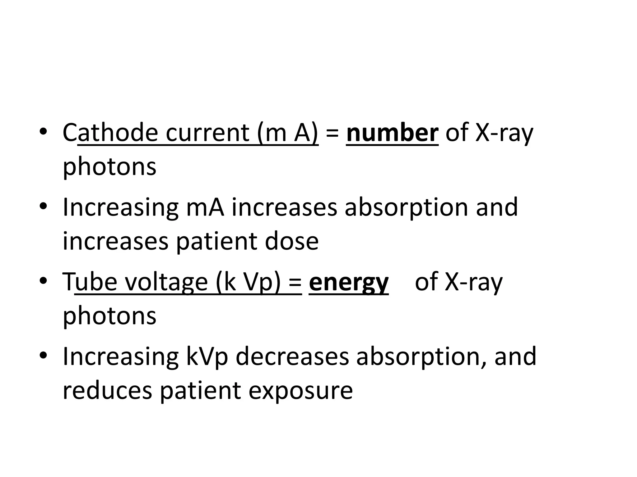
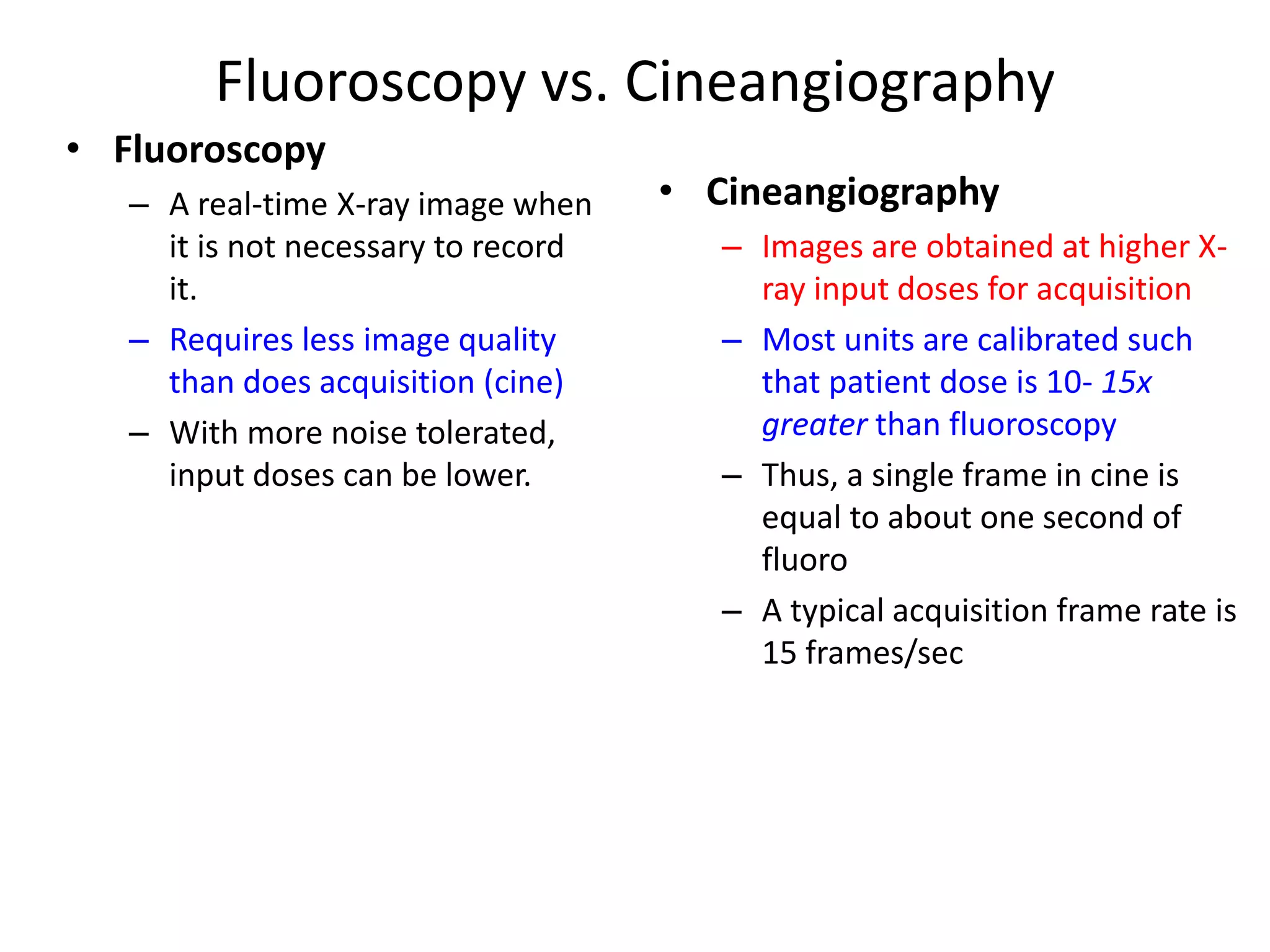
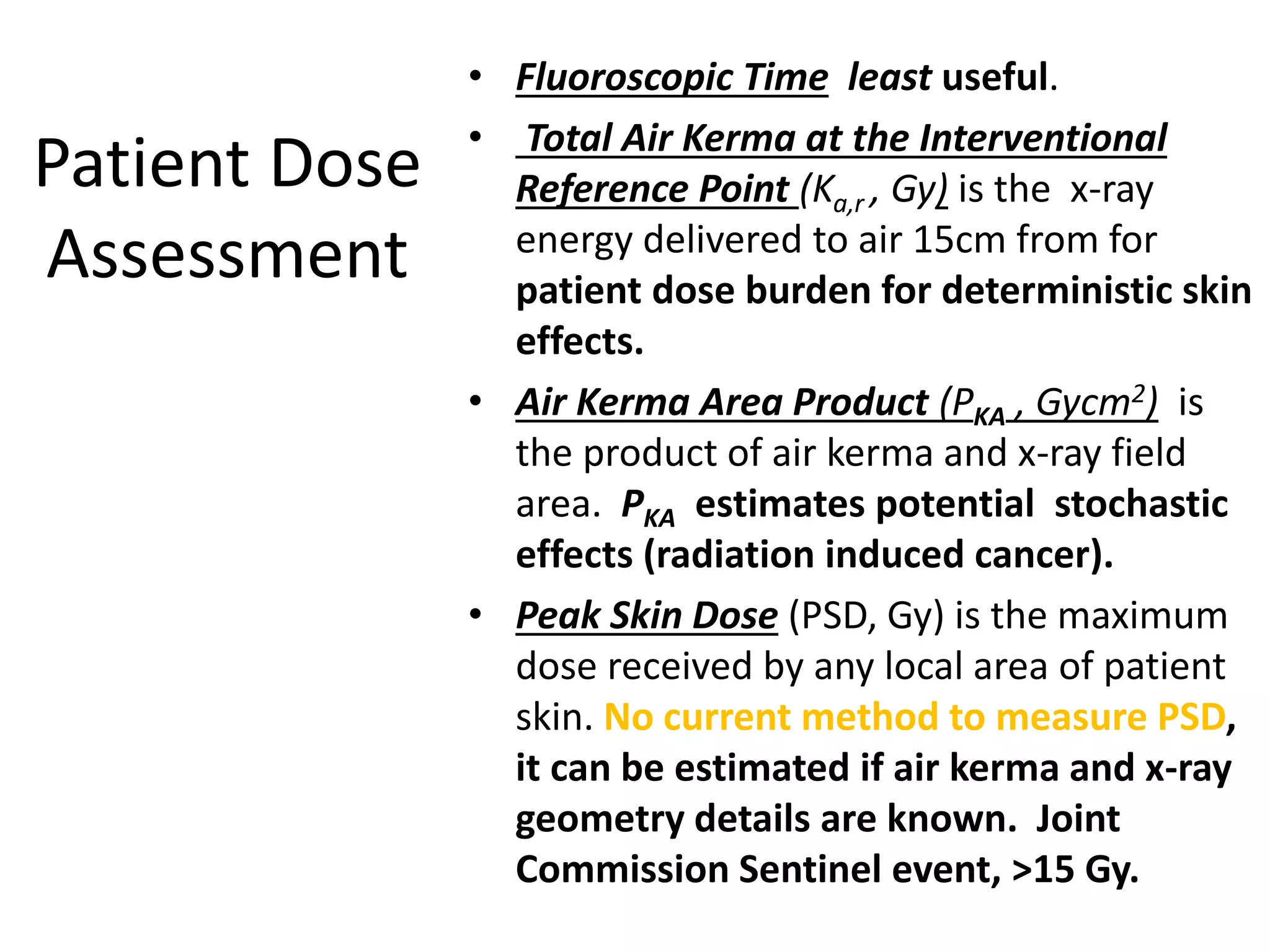
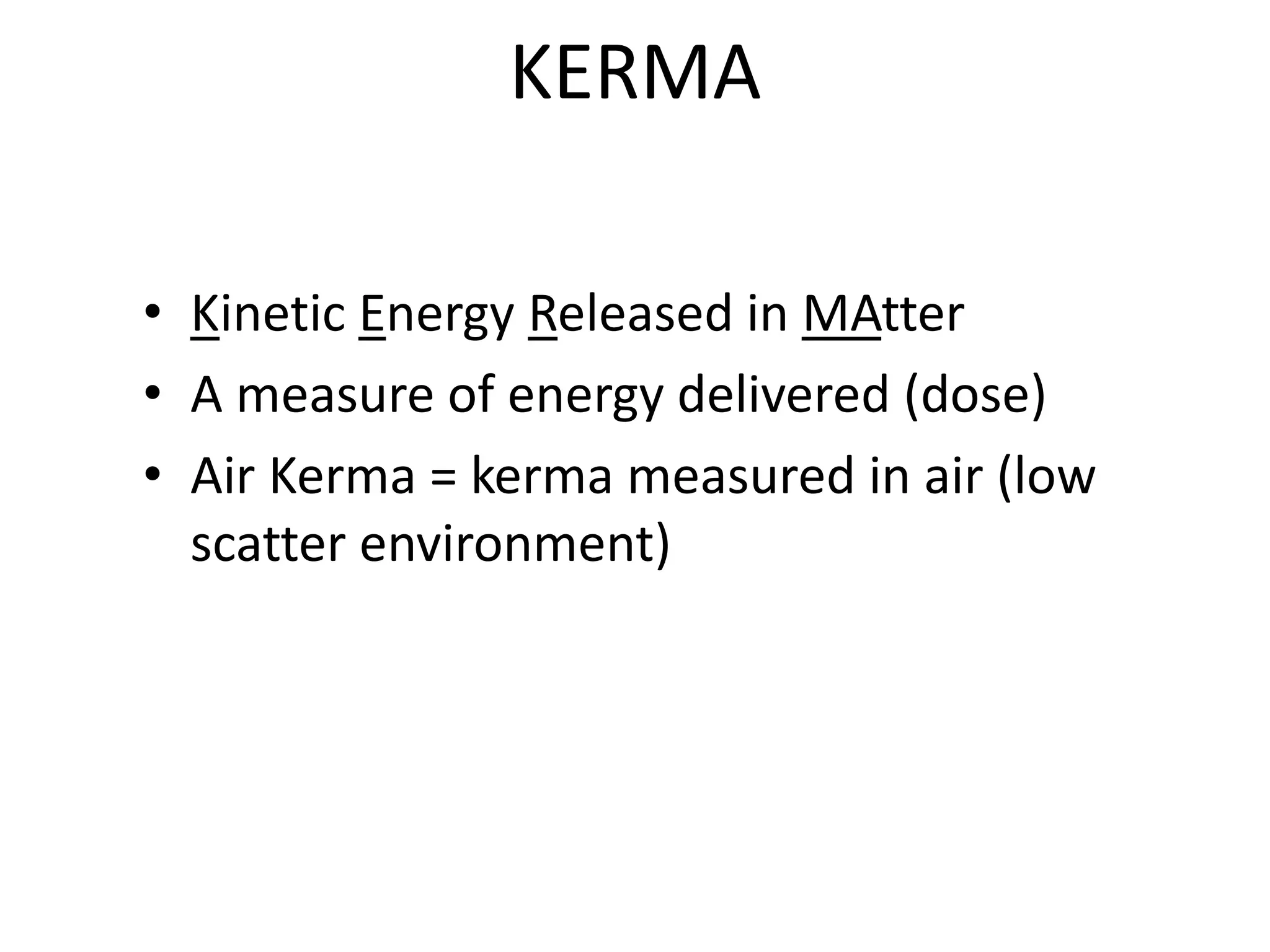
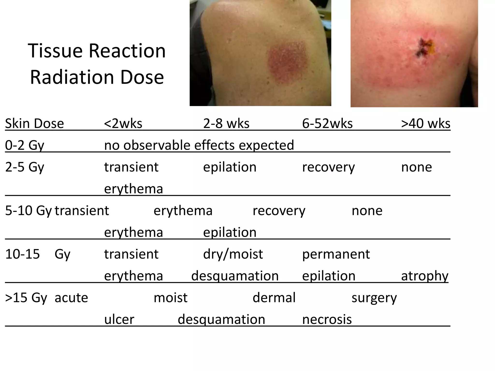
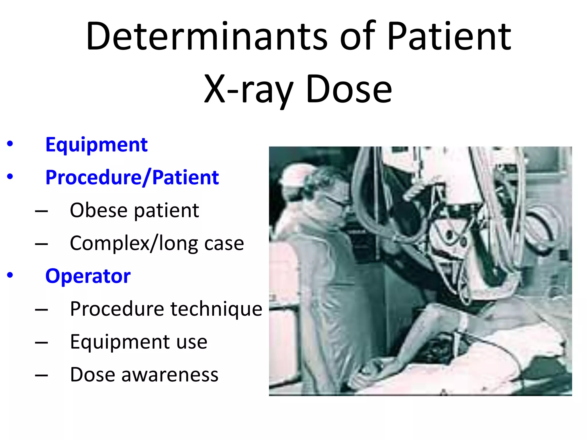
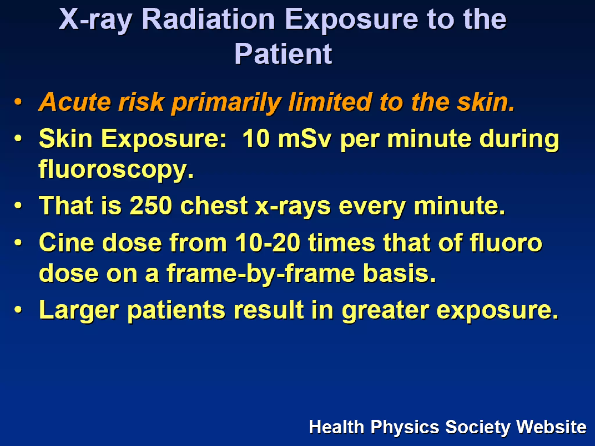
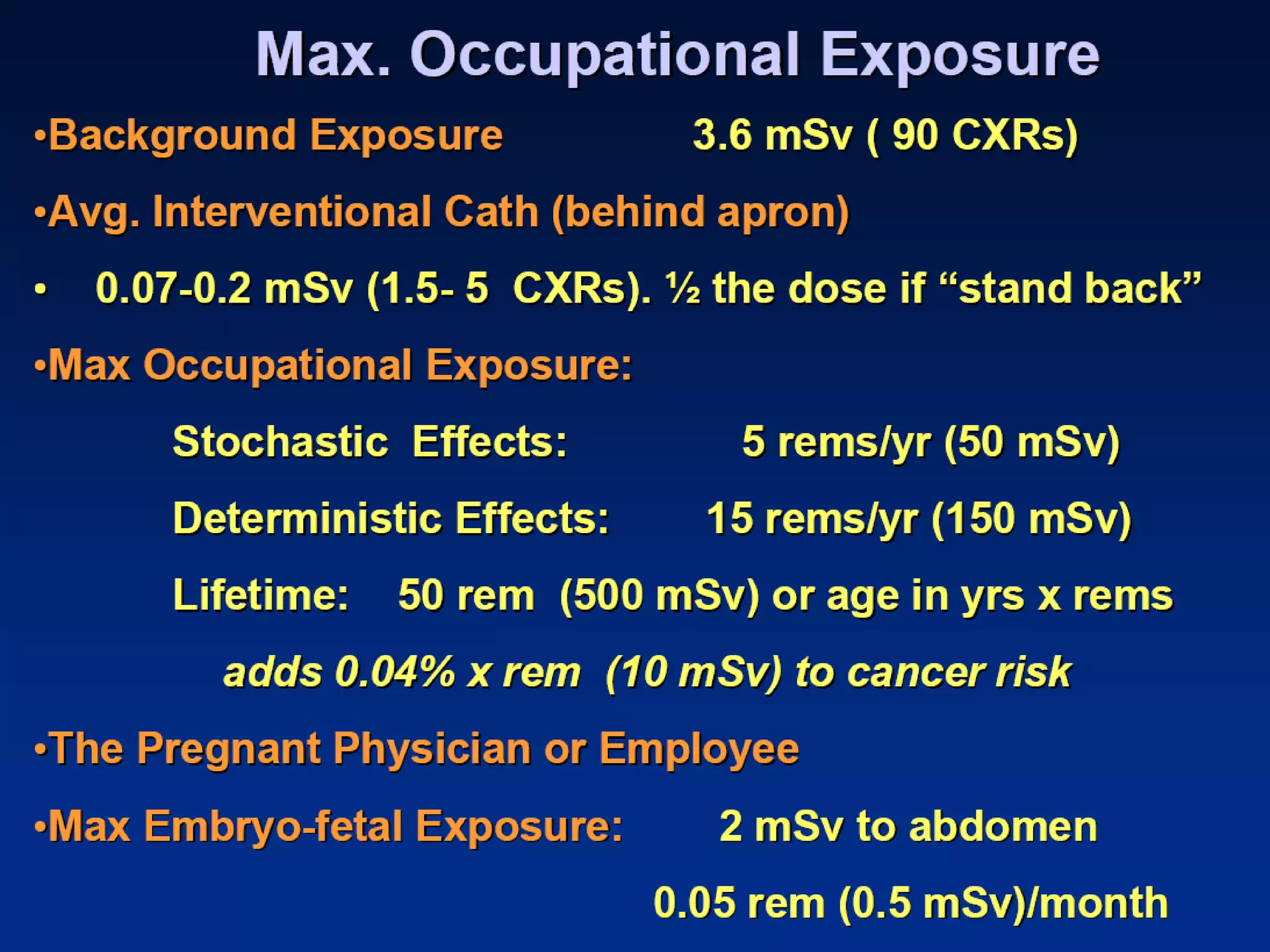
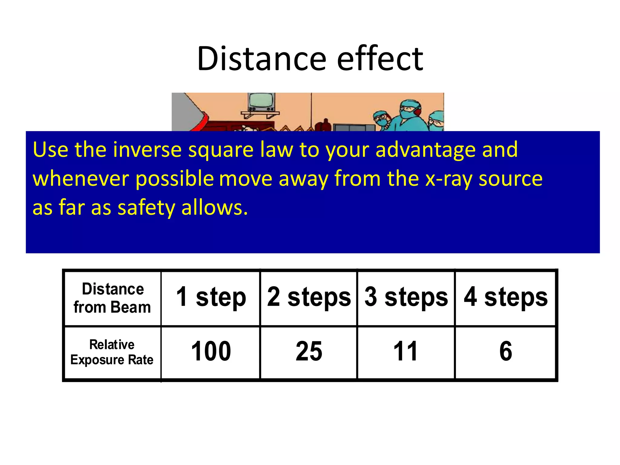
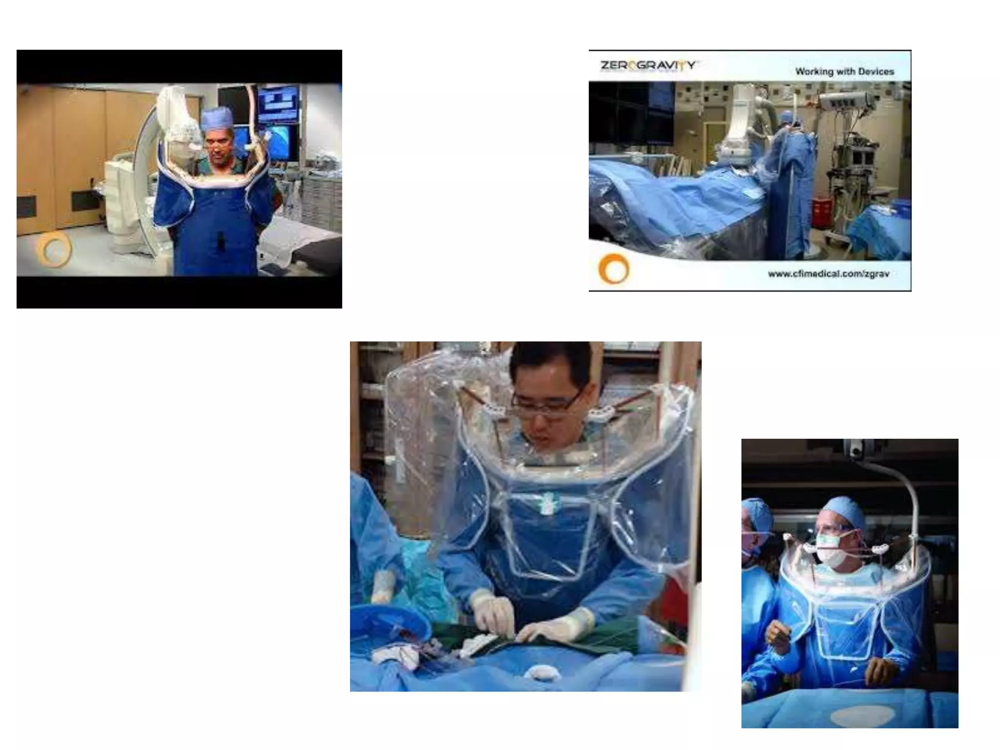
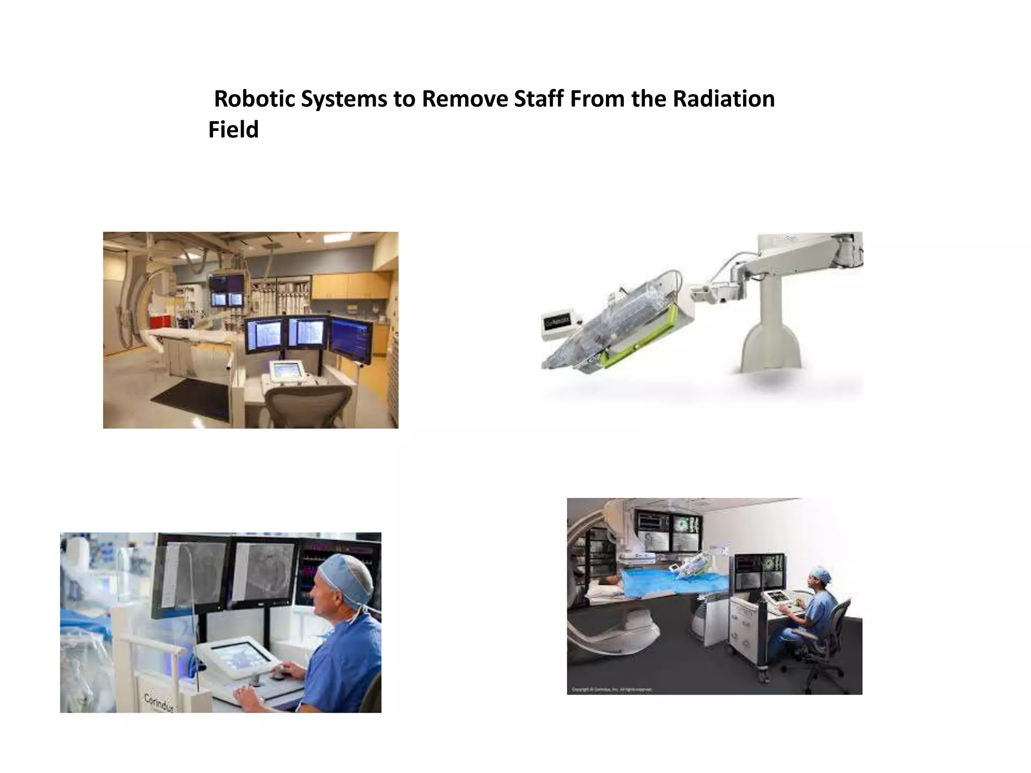
RADIATION SAFETY IN CATHLAB
• Cathode current (m A) = number of X-ray
photons
• Increasing mA increases absorption and
increases patient dose
• Tube voltage (k Vp) = energy of X-ray
photons
• Increasing kVp decreases absorption, and
reduces patient exposure
Fluoroscopy vs. Cineangiography
• Fluoroscopy
– A real-time X-ray image when
it is not necessary to record
it.
– Requires less image quality
than does acquisition (cine)
– With more noise tolerated,
input doses can be lower.
• Cineangiography
– Images are obtained at higher X-
ray input doses for acquisition
– Most units are calibrated such
that patient dose is 10- 15x
greater than fluoroscopy
– Thus, a single frame in cine is
equal to about one second of
fluoro
– A typical acquisition frame rate is
15 frames/sec
Patient Dose
Assessment
• Fluoroscopic Time least useful.
• Total Air Kerma at the Interventional
Reference Point (Ka,r , Gy) is the x-ray
energy delivered to air 15cm from for
patient dose burden for deterministic skin
effects.
• Air Kerma Area Product (PKA , Gycm2) is
the product of air kerma and x-ray field
area. PKA estimates potential stochastic
effects (radiation induced cancer).
• Peak Skin Dose (PSD, Gy) is the maximum
dose received by any local area of patient
skin. No current method to measure PSD,
it can be estimated if air kerma and x-ray
geometry details are known. Joint
Commission Sentinel event, >15 Gy.
KERMA
• Kinetic Energy Released in MAtter
• A measure of energy delivered (dose)
• Air Kerma = kerma measured in air (low
scatter environment)
Biologic Factors for Skin
Reaction (Deterministic)
• Patient-related factors
– hyperthyroidism , diabetes mellitus, connective tissue disorders, obesity,
smoking, and compromised skin integrity
– Medications: actinomycin D, doxorubicin, bleomycin, 5-
fluorouracil, and methotrexate mitoxantrone, 5-fluorourcil,
cyclophosphamide, paclitaxel, docetaxel, and possibly
tamoxifen
• Ethnic differences
– individuals with light-colored hair/ skin are most sensitive
• Defects in DNA repair genes
– autosomal recessive ATM gene, 1% population
Tissue Reaction
Radiation Dose
Skin Dose <2wks 2-8 wks 6-52wks >40 wks
0-2 Gy no observable effects expected
2-5 Gy transient epilation recovery none
erythema
5-10 Gy transient erythema recovery none
erythema epilation
10-15 Gy transient dry/moist permanent
erythema desquamation epilation atrophy
>15 Gy acute moist dermal surgery
ulcer desquamation necrosis
Patient Effects of X-ray Exposure
Stochastic Effects
• Induced Neoplasm
– Epidemiologic data suggest a linear dose-response
relationship, not a threshold, between ionizing radiation
exposure and induction of solid tumors.
– Fatal cancer risk of 0.04% to 0.12% for whole body exposure of
10 mSv (1 rem).
– Less for people>50 yrs; latent period > 5yrs.
– <1% for DAP of 200 Gycm2, typical PCI
• Heritable Abnormalities
– 0.01%/ 10cGy (1 rad) absorbed does to gonads
Determinants of Patient
X-ray Dose
• Equipment
• Procedure/Patient
– Obese patient
– Complex/long case
• Operator
– Procedure technique
– Equipment use
– Dose awareness
Distance effect
Distance
from Beam 1 step 2 steps 3 steps 4 steps
Relative
Exposure Rate 100 25 11 6
Use the inverse square law to your advantage and
whenever possible move away from the x-ray source
as far as safety allows.
Radiation Safety Principle
• Use the least amount of magnification
consistent with seeing the object adequately.
• BIGGER IS NOT ALWAYS BETTER!!
• A larger image means more radiation
– If it is necessary for adequate visualization, fine
– If it does not improve procedure safety or
performance, reduce the magnification
2011 PCI Guidelines
3.1 Radiation Safety Recommendation
Class I
Cardiac catheterization laboratories should routinely record
relevant patient procedural radiation dose data (e.g.., total
air kerma at the interventional reference point (Ka,r), air
kerma area product (PKA), fluoroscopy time, number of cine
images), and should define thresholds with corresponding
follow-up protocols for patients who receive a high
procedural radiation dose. (Level of Evidence: C)
Radiation Dose
Management in PCI
1. Pre-Procedure
• Radiation safety program for cath lab
• Dosimeter use, shielding,
training/education
• Equipment and operator knowledge
• On screen dose assessment (Ka,r , PKA)
• Dose saving: store fluoro, adj. pulse
and frame rate, and last image hold
• Pre-procedure dose planning
– assess patient and procedure including
– patient’s size and lesion(s) complexity
• Informed patient with appropriate
consent
2. Procedure
• Limit fluoro: use petal only when looking at screen
• Limit cine: store fluoro if image quality not key
• Limit magnification, frame rate, and steep angles
• Use collimation and filters to fullest extent possible
• Vary tube angle if possible to change skin exposed
• Position table & image receptor: x-ray tube close to
pt increases dose; high image receptor incr. scatter
• Keep pt & operator body parts out of field of view
• Maximize shielding and distance from x-ray source
for all personnel
• Manage and monitor dose in real time from the
beginning of the case
Procedure Related Issues to Minimize Exposure
to Patient and Operator
• Utilize radiation only when
imaging is necessary
• Minimize use of cine
• Minimize steep angle X-ray
beam
• Minimize use of magnification
modes
• Minimize frame rate of
fluoroscopy and cine
• Keep the image receptor close
to the patient
• Utilize collimation to the fullest
extent possible
• Monitor real time radiation dose
DRAPED:
• D-distance: inverse square law
• R-receptor: keep image receptor
close to patient and collimate
• A-angles: avoid steep angles
• P-pedal: keep foot off pedal
except when looking at the
monitor
• E: extremities-keep
patient/operator extremities out
of the beam
• D-dose: limit cine, adjust frame
rate, where personal dosimeter
Staff Radiation Protection
• Shielding
– Lead>90%; Proper care of aprons
– Thyroid shielding; most important <40
– Glasses-0.25 mm; must fit
– Portable: above/below table shielding
– Drapes (Bismuth-barium)
• Personnel Dosimeters:
Take Proper Care
of Your Apron
New Technologies to Reduce Cath Lab Radiation
Exposure
Increased Shielding Without the Weight
Zero-Gravity Radiation Protection System
Real Time Monitoring of Staff Dose in the
Cath Lab
Robotic Systems to Remove Staff From the Radiation
Field
BloXR Corp
lightweight Aprons and Anti-X-ray Hand Cream

RADIATION SAFETY IN CATHLAB

  • 1.
  • 2.
    • Cathode current(m A) = number of X-ray photons • Increasing mA increases absorption and increases patient dose • Tube voltage (k Vp) = energy of X-ray photons • Increasing kVp decreases absorption, and reduces patient exposure
  • 3.
    Fluoroscopy vs. Cineangiography •Fluoroscopy – A real-time X-ray image when it is not necessary to record it. – Requires less image quality than does acquisition (cine) – With more noise tolerated, input doses can be lower. • Cineangiography – Images are obtained at higher X- ray input doses for acquisition – Most units are calibrated such that patient dose is 10- 15x greater than fluoroscopy – Thus, a single frame in cine is equal to about one second of fluoro – A typical acquisition frame rate is 15 frames/sec
  • 4.
    Patient Dose Assessment • FluoroscopicTime least useful. • Total Air Kerma at the Interventional Reference Point (Ka,r , Gy) is the x-ray energy delivered to air 15cm from for patient dose burden for deterministic skin effects. • Air Kerma Area Product (PKA , Gycm2) is the product of air kerma and x-ray field area. PKA estimates potential stochastic effects (radiation induced cancer). • Peak Skin Dose (PSD, Gy) is the maximum dose received by any local area of patient skin. No current method to measure PSD, it can be estimated if air kerma and x-ray geometry details are known. Joint Commission Sentinel event, >15 Gy.
  • 5.
    KERMA • Kinetic EnergyReleased in MAtter • A measure of energy delivered (dose) • Air Kerma = kerma measured in air (low scatter environment)
  • 7.
    Biologic Factors forSkin Reaction (Deterministic) • Patient-related factors – hyperthyroidism , diabetes mellitus, connective tissue disorders, obesity, smoking, and compromised skin integrity – Medications: actinomycin D, doxorubicin, bleomycin, 5- fluorouracil, and methotrexate mitoxantrone, 5-fluorourcil, cyclophosphamide, paclitaxel, docetaxel, and possibly tamoxifen • Ethnic differences – individuals with light-colored hair/ skin are most sensitive • Defects in DNA repair genes – autosomal recessive ATM gene, 1% population
  • 8.
    Tissue Reaction Radiation Dose SkinDose <2wks 2-8 wks 6-52wks >40 wks 0-2 Gy no observable effects expected 2-5 Gy transient epilation recovery none erythema 5-10 Gy transient erythema recovery none erythema epilation 10-15 Gy transient dry/moist permanent erythema desquamation epilation atrophy >15 Gy acute moist dermal surgery ulcer desquamation necrosis
  • 9.
    Patient Effects ofX-ray Exposure Stochastic Effects • Induced Neoplasm – Epidemiologic data suggest a linear dose-response relationship, not a threshold, between ionizing radiation exposure and induction of solid tumors. – Fatal cancer risk of 0.04% to 0.12% for whole body exposure of 10 mSv (1 rem). – Less for people>50 yrs; latent period > 5yrs. – <1% for DAP of 200 Gycm2, typical PCI • Heritable Abnormalities – 0.01%/ 10cGy (1 rad) absorbed does to gonads
  • 10.
    Determinants of Patient X-rayDose • Equipment • Procedure/Patient – Obese patient – Complex/long case • Operator – Procedure technique – Equipment use – Dose awareness
  • 15.
    Distance effect Distance from Beam1 step 2 steps 3 steps 4 steps Relative Exposure Rate 100 25 11 6 Use the inverse square law to your advantage and whenever possible move away from the x-ray source as far as safety allows.
  • 16.
    Radiation Safety Principle •Use the least amount of magnification consistent with seeing the object adequately. • BIGGER IS NOT ALWAYS BETTER!! • A larger image means more radiation – If it is necessary for adequate visualization, fine – If it does not improve procedure safety or performance, reduce the magnification
  • 17.
    2011 PCI Guidelines 3.1Radiation Safety Recommendation Class I Cardiac catheterization laboratories should routinely record relevant patient procedural radiation dose data (e.g.., total air kerma at the interventional reference point (Ka,r), air kerma area product (PKA), fluoroscopy time, number of cine images), and should define thresholds with corresponding follow-up protocols for patients who receive a high procedural radiation dose. (Level of Evidence: C)
  • 18.
    Radiation Dose Management inPCI 1. Pre-Procedure • Radiation safety program for cath lab • Dosimeter use, shielding, training/education • Equipment and operator knowledge • On screen dose assessment (Ka,r , PKA) • Dose saving: store fluoro, adj. pulse and frame rate, and last image hold • Pre-procedure dose planning – assess patient and procedure including – patient’s size and lesion(s) complexity • Informed patient with appropriate consent 2. Procedure • Limit fluoro: use petal only when looking at screen • Limit cine: store fluoro if image quality not key • Limit magnification, frame rate, and steep angles • Use collimation and filters to fullest extent possible • Vary tube angle if possible to change skin exposed • Position table & image receptor: x-ray tube close to pt increases dose; high image receptor incr. scatter • Keep pt & operator body parts out of field of view • Maximize shielding and distance from x-ray source for all personnel • Manage and monitor dose in real time from the beginning of the case
  • 19.
    Procedure Related Issuesto Minimize Exposure to Patient and Operator • Utilize radiation only when imaging is necessary • Minimize use of cine • Minimize steep angle X-ray beam • Minimize use of magnification modes • Minimize frame rate of fluoroscopy and cine • Keep the image receptor close to the patient • Utilize collimation to the fullest extent possible • Monitor real time radiation dose DRAPED: • D-distance: inverse square law • R-receptor: keep image receptor close to patient and collimate • A-angles: avoid steep angles • P-pedal: keep foot off pedal except when looking at the monitor • E: extremities-keep patient/operator extremities out of the beam • D-dose: limit cine, adjust frame rate, where personal dosimeter
  • 20.
    Staff Radiation Protection •Shielding – Lead>90%; Proper care of aprons – Thyroid shielding; most important <40 – Glasses-0.25 mm; must fit – Portable: above/below table shielding – Drapes (Bismuth-barium) • Personnel Dosimeters: Take Proper Care of Your Apron
  • 21.
    New Technologies toReduce Cath Lab Radiation Exposure Increased Shielding Without the Weight Zero-Gravity Radiation Protection System Real Time Monitoring of Staff Dose in the Cath Lab
  • 23.
    Robotic Systems toRemove Staff From the Radiation Field
  • 24.
    BloXR Corp lightweight Apronsand Anti-X-ray Hand Cream