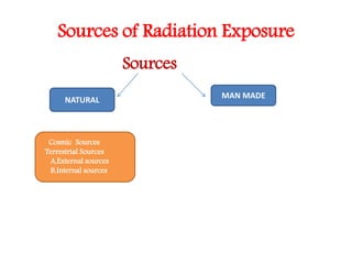
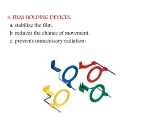
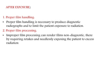
Radiation safety and protection measures are important to minimize radiation exposure for patients and dental staff. Early pioneers in dental radiography suffered health effects from excessive radiation doses. Radiation can come from natural sources like cosmic rays and radioactive elements in the ground, or man-made sources like medical imaging. Protection techniques include using thyroid collars and lead aprons for patients, fast film, rectangular collimation, and selecting proper exposure factors. Proper handling and processing of films is also needed to avoid retakes and reduce unnecessary radiation exposure.
This power-point presentation is very important for radiology resident radiologist and radiographers and technician. this includes principles, technique , biological effects of radiation and how to protect, whats should normal radiation dose with latest update. This slide also includes ALARA PRINCIPLE thanks.
Radiation protection and personnel monitoring devicesRubiSapkota
This document discusses radiation protection and personnel monitoring devices. It begins with an introduction to radiation and the electromagnetic spectrum. It then covers the effects of radiation, principles of radiation protection including justification of practice, optimization of protection, and dose limits. The document discusses various personnel monitoring devices including film badges, thermoluminescence dosimeters (TLD), and optically stimulated luminescence dosimeters. It provides details on how each device works and their advantages and disadvantages.
The document discusses principles of radiation protection including justification, optimization, and dose limits. It describes how justification requires that the benefits of any radiation exposure outweigh the risks, and optimization aims to keep radiation exposure as low as reasonably achievable. Various personnel protective devices are described such as lead aprons, gloves, glasses, and shields that can reduce radiation exposure.
This document discusses dosimetry and radiation protection. It begins with a brief history of radiation discovery and the first radiograph. It then discusses key concepts in radiation protection including dose limits for different exposure groups, the ALARA (As Low As Reasonably Achievable) philosophy, and the basic methods of reducing radiation exposure through time, distance and shielding. The document also covers external and internal radiation sources, dose and exposure concepts, and methods for reducing radiation exposure in dental radiography through proper equipment selection, technique choice, processing and interpreting images. Protection of patients, personnel and the public is emphasized throughout.
Dental radiography involves taking images of the teeth, bones, and soft tissues in the mouth to aid in diagnosis and treatment planning. There are several types of dental radiography procedures, including intraoral radiographs like bitewings and periapicals, as well as panoramic and cephalometric images. Radiographs are useful for detecting issues like dental caries, abnormalities, and monitoring treatment. Proper radiation safety protocols must be followed when performing dental radiography to minimize risk to patients and staff.
Thermoluminescent dosimeters (TLDs) are used to monitor radiation exposure. TLDs use crystals that emit light when heated, with the amount of light proportional to radiation exposure. Key aspects include:
1) TLDs measure exposure to ionizing radiation like x-rays and gamma rays by heating the crystal and detecting the light emitted using a photomultiplier tube.
2) Common TLD materials include lithium fluoride, calcium fluoride, lithium borate, and calcium sulfate. In India, calcium sulfate doped with dysprosium is commonly used.
3) TLDs are reusable, have good sensitivity and linearity, and allow quick readout of accumulated
Radiation protection, also known as radiological protection, is defined by the International Atomic Energy Agency (IAEA) as "The protection of people from harmful effects of exposure to ionizing radiation, and the means for achieving this". Exposure can be from a source of radiation external to the human body or due to internal irradiation caused by the ingestion of radioactive contamination.
Ionizing radiation is widely used in industry and medicine, and can present a significant health hazard by causing microscopic damage to living tissue. There are two main categories of ionizing radiation health effects. At high exposures, it can cause "tissue" effects, also called "deterministic" effects due to the certainty of them happening, conventionally indicated by the unit gray and resulting in acute radiation syndrome. For low level exposures there can be statistically elevated risks of radiation-induced cancer, called "stochastic effects" due to the uncertainty of them happening, conventionally indicated by the unit sievert.
Fundamental to radiation protection is the avoidance or reduction of dose using the simple protective measures of time, distance and shielding. The duration of exposure should be limited to that necessary, the distance from the source of radiation should be maxi mised, and the source shielded wherever possible. To measure personal dose uptake in occupational or emergency exposure, for external radiation personal dosimeters are used, and for internal dose to due to ingestion of radioactive contamination, bioassay techniques are applied.
This document discusses panoramic radiography, including its history, advantages, procedure details, and principles of image formation. Panoramic radiography uses a rotating x-ray beam and receptor to create a single image of the facial structures, including teeth and supporting bones. It provides broad anatomic coverage with a low radiation dose compared to full-mouth intraoral x-rays. Proper patient positioning is needed to place the dental arches within the "focal trough" where structures will be reasonably defined.
The document discusses different types of digital radiography technologies including computed radiography which uses photostimulable phosphor plates, indirect digital radiography using a scintillator and photodiode array, and direct digital radiography using photoconductive materials. It covers the processes of image acquisition, processing, display, and archiving for digital radiography systems. Key differences between direct and indirect digital radiography technologies are also outlined.
The document discusses filtration and collimation in x-ray beams. Filtration removes low-energy photons that do not contribute to the image but increase patient exposure. Filters are typically made of aluminum and selectively allow high-energy photons to pass. Collimation uses a lead plate with a central hole to restrict the beam to only the area being imaged, reducing patient exposure and preventing scatter that degrades image quality. Both filtration and collimation aim to improve image quality while lowering radiation dose.
This document discusses radiation protection and reducing occupational radiation exposure. It provides guidelines for permissible occupational radiation doses, including annual and cumulative limits. It discusses ways to reduce exposure through applying the principles of time, distance, and shielding. Specific techniques mentioned include using protective apparel like lead aprons and thyroid shields during exams, collimating the x-ray beam, and positioning patients to avoid exposing sensitive areas. The document also addresses monitoring occupational exposure through the use of film badges, pocket dosimeters, and thermoluminescent dosimeters.
The document discusses the general requirements and characteristics of dosimeters. A dosimeter is a device that measures radiation quantities like exposure, absorbed dose, and equivalent dose. It should be accurate, precise, and have a linear response over a wide dose range with little dependence on factors like energy, dose rate, or direction. Common types of personal dosimeters mentioned are film badges, pocket ion chambers, thermoluminescent dosimeters (TLD), optically stimulated luminescence (OSL) dosimeters, and solid state dosimeters. The document also discusses appropriate uses of personal dosimeters and factors to consider for different work environments.
This document describes the procedures for sialography and dacrocystography. Sialography involves cannulating the ducts of the parotid and submandibular salivary glands and injecting contrast medium to visualize the glands and ducts under fluoroscopy. Dacrocystography involves cannulating the lacrimal puncta and injecting contrast into the nasolacrimal duct system to identify any obstructions. Both procedures provide anatomical imaging of the relevant duct systems to evaluate conditions like stones, strictures, masses or trauma. The document outlines the anatomy, indications, contraindications, technique and expected imaging findings for each procedure.
Radiographic grids are devices placed between the patient and image receptor to reduce scattered radiation and improve image contrast. Invented in 1913 by Dr. Gustav Bucky, grids work by blocking scattered radiation while allowing primary radiation to pass through. The amount of scatter reduction depends on factors like grid ratio, line frequency, and focal distance. While grids improve image quality, they also increase patient radiation dose. Proper grid selection and positioning are important to maximize benefits and minimize patient exposure.
The document summarizes the process of radiographic film processing and the darkroom equipment used. It discusses:
1. How a latent image is formed on the film when exposed to x-rays and the chemical components involved.
2. The steps of film processing - developing the latent image into a visible one using developer solutions, fixing the image using fixer, and washing the film.
3. The components and purpose of developer and fixer solutions, and factors like temperature, time and replenishment that are important in processing.
4. Darkroom requirements like safelighting, manual processing tanks, timers and drying racks used to process films.
Sialography is an x-ray examination of the salivary glands that involves injecting contrast media into the ducts to evaluate any abnormalities. It can detect issues like stones, lesions, or masses that may be obstructing the ducts and causing pain or inflammation. There are three major pairs of salivary glands - parotid, submandibular, and sublingual - which produce saliva. Sialography can be used to evaluate masses, stones, pain, functional disorders, and suspected obstructions or strictures of the salivary glands. The procedure involves injecting contrast media under fluoroscopy and taking x-ray images to view the flow of saliva and identify any ob
The document discusses different types of radiography technologies, including computed radiography (CR), direct digital radiography (DR), and the components and layers of imaging plates (IPs) used in CR. It also covers image processing techniques for CR/DR such as histogram generation, exposure compensation, and potential artifacts that can occur during acquisition, post-acquisition or display.
Radiation can damage DNA and cause cell death or transformation. Exposure is measured in Sieverts or rem, with effects depending on dose and type of radiation. Protective measures include minimizing exposure time, maximizing distance from the source, and using shielding like lead aprons. Procedures should be justified medically and optimize radiation dose levels to as low as reasonably achievable.
This document discusses the history and development of radiation protection. Some key points:
- The harmful effects of radiation were initially not well understood after X-rays were discovered in the late 19th century. Several early researchers and technicians suffered health effects.
- Over time, concepts like tolerance doses, maximum permissible doses, and the "as low as reasonably achievable" principle were developed to set safe radiation exposure limits.
- International organizations like the ICRP and IAEA were formed to make recommendations on radiation safety standards and regulation. National bodies like AERB regulate radiation protection in India.
- The principles of justification, optimization and dose limitation form the foundation of modern radiation protection practices and regulation. Exposure
Radiation is energy that is given off by particular materials and devices.
Radiation protection, also known as radiological protection, is defined by the International Atomic Energy Agency (IAEA) as "The protection of people from harmful effects of exposure to ionizing radiation, and the means for achieving this". Exposure can be from a source of radiation external to the human body or due to internal irradiation caused by the ingestion of radioactive contamination
The objectives of radiation protection according to the ICRP and NCRP are to prevent serious radiation effects and reduce stochastic effects to acceptable levels while allowing beneficial practices involving radiation exposure. This is achieved through principles of justification, optimization and dose limitation. Justification requires that practices only be adopted if benefits outweigh radiation risks. Optimization aims to keep exposures as low as reasonably achievable. Dose limitation sets defined exposure limits for workers and the public.
Latent Image formation & Dark room Chemistry.pptxssuser71d7b1
This document discusses latent image formation on dental radiographic film and the darkroom chemistry involved in processing the film. It begins by explaining how x-ray photons interact with silver halide crystals in the film emulsion to form a latent image. It then describes the key components and functions of the darkroom, including safe lighting, temperature and humidity control. Finally, it provides details on manual and automatic film processing, the chemical compositions and purposes of developer and fixer solutions, and some additional darkroom techniques.
Principle of Radiation Protection- Avinesh ShresthaAvinesh Shrestha
Radiation protection is the science whose aim is to minimize the risks generated by the use of ionizing radiation. Briefly discusses The ICRP System of Radiological Protection, STRUCTURAL SHIELDING OF
IMAGING FACILITIES, APPLICATION OF INDIVIDUAL DOSE LIMTS, RADIATION EXPOSURE IN PREGNANCY, Diagnostic reference level, Personnel Protection in
Medical X-ray Imaging, Dose Optimization in CT, Radiation Protection in Nuclear Medicine.
This document discusses radiation protection and provides definitions, types of radiation effects, sources of radiation exposure, units of measurement, dose limits, and techniques to reduce radiation exposure in medical imaging. It defines radiation protection as protecting people from harmful effects of ionizing radiation. It describes stochastic and deterministic effects and lists examples of radiation anomalies. It also outlines regulatory bodies, dose limits for occupational workers and the public, and principles of radiation safety including time, distance, shielding and reducing exposure.
This document summarizes the biological effects of radiation at various levels of organization. It discusses:
1. The interaction of radiation with DNA and cells, including direct and indirect effects on DNA.
2. The cellular response to radiation damage, including stochastic and deterministic effects, DNA repair mechanisms, and factors influencing radiosensitivity.
3. Tissue and organ responses like acute radiation syndrome and late effects like fibrosis and osteoradionecrosis.
4. Genetic and carcinogenic risks of radiation exposure, especially for children and developing embryos.
The document discusses various radiation protection measures for patients, operators, and the environment during dental radiography. It outlines techniques to minimize radiation exposure before, during, and after x-ray procedures for patients such as proper prescribing, use of protective equipment like aprons and collars, and fast film. Operator protection includes guidelines on distance, positioning, shielding, and monitoring. The environment is protected by shielding walls, doors, and limiting the primary beam. Regulations establish safe exposure limits.
This document discusses radiation protection in dentistry. It describes sources of radiation exposure including natural sources like cosmic and terrestrial radiation as well as artificial sources from medical and consumer products. It emphasizes following the ALARA principle to keep radiation exposure as low as reasonably achievable. Specific techniques to protect patients include restricting radiographs to necessary views, using proper equipment, filtration, collimation, lead shielding, and film handling. Operator protection involves maintaining distance from the x-ray unit, using barriers, and monitoring radiation exposure with dosimeters.
Dose reduction in Conventional Radiography and FluoroscopyTarun Goyal
This document summarizes techniques to reduce radiation dose in diagnostic radiology, including fluoroscopy. It discusses using the smallest x-ray field, increasing distance between the patient and x-ray source, using filters, grids, and intensifying screens. It also covers automatic processing, avoiding unnecessary repeat images, and techniques to reduce dose in fluoroscopy like intermittent exposure, removal of grids, and last image hold. The document emphasizes training operators and using radiation only when necessary to obtain diagnostic information.
Radiation safety and protection for dental radiographyNitin Sharma
1) Licensed dentists must maintain radiation exposures as low as reasonably achievable and understand the health risks of radiation.
2) Dental radiographic equipment must be registered and follow safety protocols to protect patients and staff, such as using protective gear and collimation.
3) Dentists are responsible for quality assurance programs to ensure proper functioning and calibration of dental X-ray machines and processing of films. Guidelines help prescribe radiographs appropriately.
Operator protection when using radiation in dentistry includes following guidelines on distance, position, and shielding from the primary x-ray beam. Distance recommendations state the radiographer should stand at least 6 feet away, while position recommends being perpendicular or at a 90-135 degree angle to the beam. Shielding like protective barriers should also be used when possible. Radiation exposure is monitored through equipment monitoring for leakage radiation and personnel monitoring using film badges, thermoluminescent dosimeters, or ionization chambers. Radiation safety legislation establishes exposure limits for public and occupational exposure to ensure protection of patients, operators, and the environment.
Occupational radiation safety in Radiological imaging, Dr. Roshan S Livingstoneohscmcvellore
Occupational radiation safety in Radiological imaging
1) There is increased use of radiation-based medical imaging globally, but many staff lack proper training in radiation safety techniques.
2) Workers in cardiology cath labs receive the highest radiation doses, followed by radiology cath labs and other interventional procedures. Prolonged fluoroscopic screening can lead to hair loss and cataracts in interventionalists.
3) Basic principles of radiation safety include minimizing time, maximizing distance, and using shielding. Monitoring staff doses with dosimeters and following safety protocols helps ensure doses are as low as reasonably achievable.
This document provides an overview of x-ray machines and their components and uses. It discusses the history of x-rays and their discovery in 1895. The main components of an x-ray machine are described, including the high voltage generator, control panel, x-ray tube, collimator, grid, and film or digital sensor. Different types of x-ray machines are examined, such as conventional, computed radiography, and digital radiography systems. Factors that affect image quality like kilovoltage, milliamperes, and distance are outlined. The document also reviews exposure dose limits and protective procedures for radiation workers.
Personal dosimeters are used to measure the amount of radiation workers are exposed to. The three main types discussed are film badges, optically stimulated luminescent dosimeters, and thermoluminescent dosimeters. Film badges contain radiosensitive film that darkens with exposure. OSL dosimeters use aluminum oxide detectors that luminesce when struck by laser light, in proportion to exposure. TLDs contain lithium fluoride crystals that absorb radiation and emit light through thermoluminescence, allowing measurement of exposure. Dosimeters should be worn at collar level outside protective equipment to monitor head exposure.
The document discusses guidelines for orthodontic radiographs, including the damaging effects of radiation on human tissue and legislation regarding medical radiation exposure in the UK. It covers justifying the need for exposures, optimizing techniques to minimize radiation dose, and estimating effective radiation doses received from common orthodontic radiographs. Guidelines are provided on indication for taking radiographs at different treatment stages and ages based on clinical need. Techniques to reduce patient radiation dose include using faster film/receptors, appropriate collimation and filtration, and digital radiography.
This document discusses radiation protection for patients and operators during dental x-ray procedures. It covers key concepts like total filtration, collimation, protective equipment like lead aprons and thyroid collars, proper techniques to minimize exposure, and guidelines for radiation safety. The document emphasizes that while dental x-rays provide benefits, it is important to use all available methods to minimize the amount of radiation received by patients and operators, in accordance with legislation and the ALARA principle of keeping exposures as low as reasonably achievable.
X-rays are a form of ionizing radiation that produces positively and negatively charged particles when passing through matter. The goals of radiation protection are to protect persons from both short-term and long-term effects of radiation by adhering to an established radiation protection program. Effective radiation protection measures are employed by radiation workers to safeguard patients, personnel, and the general public from unnecessary exposure to ionizing radiation.
Brief introduction to the latest innovations that are used at dentistry, where equipment used are fully digitized and computerized, with the differences between using conventional methods and digital equipment in dentistry.
Main equipment to be discussed are dental imaging systems and CAD/CAM systems
1) A 22-year-old female presented with a keloid on her left ear lobe that developed after ear piercing. The keloid was excised and she received superficial radiotherapy (SXT) within 24-48 hours post-excision.
2) SXT is a non-invasive treatment that uses low-energy x-rays to destroy unhealthy cells just beneath the skin's surface. It has shown success in reducing keloid recurrence when used after excision.
3) Strict safety procedures were followed during the patient's SXT treatment, including immobilization, shielding of sensitive areas, real-time monitoring, and a single fraction dose of 12Gy delivered over a treatment time determined through calculation.
Radiation Safety - Dr Hafeesh Fazulu -Pushpagiri - June 2020Hafeesh Fazulu
This document summarizes radiation safety procedures in the cathlab. It discusses x-ray physics, imaging modes, and measures of radiation used to monitor patient irradiation. It describes the biological effects of radiation, including both stochastic and deterministic mechanisms. It provides recommendations to reduce radiation exposure through principles of justification, optimization and dose limitation. Specific techniques discussed include minimizing fluoroscopy time, increasing distance from the patient, using protective shielding, and new technologies to monitor and reduce radiation dose in the cathlab.
Those who administer ionizing radiation must become familiar with the magnitude of exposure encountered in medicine, dentistry and every day life; the possible risks associated with such exposure; and the methods used to affect exposure.
Practitioners should remain informed about safety updates to further improve diagnostic quality of radiographs and decrease radiation exposure.
This document discusses radiation safety in vascular and endovascular procedures. It defines key terms like absorbed dose, equivalent dose, and effective dose. It describes the biological effects of radiation as either deterministic or stochastic. It provides guidance on minimizing radiation exposure for patients and operators during diagnostic imaging and endovascular interventions through techniques like collimation, distance, barriers, and monitoring dose. The goal is to justify and optimize radiation use to provide benefit while limiting harm.
X-rays are a form of ionizing radiation that produces charged particles when passing through matter. The goals of radiation protection are to protect persons from both short-term and long-term effects of radiation by adhering to an established radiation protection program. Effective radiation protection employs measures to safeguard patients, personnel, and the public from unnecessary radiation exposure.
Radiation protection principles aim to safeguard patients, personnel, and the public from unnecessary exposure to ionizing radiation during medical procedures. Key concepts include justifying procedures, applying ALARA principles to minimize dose, monitoring personnel dose, and protecting sensitive populations like pregnant workers and children. Radiation safety is an ongoing responsibility requiring adherence to protection programs and guidelines.
This document summarizes a radiographic system, which uses x-rays to perform diagnostic medical imaging. It describes the main components of an x-ray system including the table, Bucky film tray, grid system, x-ray tube, and collimators. It also discusses different types of x-ray imaging techniques like conventional radiography, CT, angiography, and mammography. Digital radiography is highlighted as an advancement over traditional film radiography. Considerations for purchasing and maintaining a radiographic system are provided.
This document discusses image receptors used in dental radiography. It defines image receptors, provides a brief history, and classifies receptors as analog film or digital sensors. It describes common intra-oral and extra-oral radiographs and films used, and compares analog film speeds and packaging types. Digital receptors like CCD, CMOS, and PSP sensors are also outlined. The document reviews pathogens that can survive on receptors and strategies to reduce cross-contamination risk. Overall, the document provides a comprehensive overview of analog and digital dental image receptors.
This document provides an overview of fibro-osseous lesions of the jaw. It begins by defining fibro-osseous lesions as a diverse group of processes characterized by replacement of normal bone by fibrous tissue containing newly formed mineralized product. It then discusses the classifications, common lesions including fibrous dysplasia, cemento-osseous dysplasia, and ossifying fibroma. The importance of specific diagnosis is highlighted. Radiological features and differential diagnosis of fibro-osseous lesions are also summarized.
This document discusses sialography, a radiographic technique used to visualize the major salivary glands. Sialography involves introducing a radiopaque contrast medium into the ductal system of the glands. It is effective for diagnosing obstructions caused by stones or strictures. The procedure involves preoperative imaging, filling the gland with contrast medium using one of three techniques, and imaging the emptying phase. Sialography can reveal pathological findings like stones, ductal dilatation, contrast retention, and underfilling related to tumors or Sjogren's syndrome.
dental management patients with cardiovascular disorders.pptxPooja461465
This document discusses cardiovascular diseases and their relevance to dentistry. It describes common conditions like hypertension, coronary heart disease, myocardial infarction, and infective endocarditis. For hypertension, it covers classification, signs/symptoms, diagnosis and dental management considerations like stress reduction and cautious use of vasoconstrictors. For coronary heart disease, it explains angina, myocardial infarction, and emphasizes stress reduction during dental treatment. It provides guidance on managing patients who are taking antiplatelet drugs or anticoagulants. The document concludes by discussing infective endocarditis and recommendations for antibiotic prophylaxis during certain dental procedures to prevent bacteremia.
This document discusses various entities that can produce pericoronal radiolucencies, including both those without and with opacities. It describes normal follicular spaces and several pathologies that appear as radiolucencies surrounding unerupted teeth, such as dentigerous cysts, odontogenic keratocysts, and ameloblastic fibromas. It also provides details on inflammatory paradental cysts and pericoronitis. Differential diagnoses and typical management approaches are provided for each condition.
This document discusses radiotherapy (radiation therapy) and its use in treating cancer. It covers the origins and physics of radiotherapy, how radiation affects cells, and methods to improve radiation therapy outcomes. These methods include using radiosensitizers and radioprotectors, hyperbaric oxygen therapy, altered fractionation schedules, and combining radiation with chemotherapy or hyperthermia. The document also addresses complications and dental care considerations related to radiotherapy.
DIFFERENTIAL DIAGNOSIS FOR PERIAPICAL RADIOLUCENCY.pptxPooja461465
This document discusses the differential diagnosis of periapical radiolucencies. It describes true periapical radiolucencies such as pulpo-periapical radiolucencies, dentigerous cysts, periapical cemento-osseous dysplasias and malignant tumors. It also discusses pseudo-periapical radiolucencies caused by anatomical variations. Different pathological conditions are described including their etiology, clinical features, radiographic features, differential diagnosis and management.
This document discusses projection geometry principles for radiography. It describes how focal spot size and position affect image clarity and distortion. Smaller focal spots and increasing the distance between the focal spot, object, and film can improve sharpness and resolution. Two common techniques for localizing foreign objects are the right-angle technique using two perpendicular projections, and tube shift techniques which examine how object positions change with projection angle.
How to Setup Warehouse & Location in Odoo 17 InventoryCeline George
In this slide, we'll explore how to set up warehouses and locations in Odoo 17 Inventory. This will help us manage our stock effectively, track inventory levels, and streamline warehouse operations.
it describes the bony anatomy including the femoral head , acetabulum, labrum . also discusses the capsule , ligaments . muscle that act on the hip joint and the range of motion are outlined. factors affecting hip joint stability and weight transmission through the joint are summarized.
LAND USE LAND COVER AND NDVI OF MIRZAPUR DISTRICT, UPRAHUL
This Dissertation explores the particular circumstances of Mirzapur, a region located in the
core of India. Mirzapur, with its varied terrains and abundant biodiversity, offers an optimal
environment for investigating the changes in vegetation cover dynamics. Our study utilizes
advanced technologies such as GIS (Geographic Information Systems) and Remote sensing to
analyze the transformations that have taken place over the course of a decade.
The complex relationship between human activities and the environment has been the focus
of extensive research and worry. As the global community grapples with swift urbanization,
population expansion, and economic progress, the effects on natural ecosystems are becoming
more evident. A crucial element of this impact is the alteration of vegetation cover, which plays a
significant role in maintaining the ecological equilibrium of our planet.Land serves as the foundation for all human activities and provides the necessary materials for
these activities. As the most crucial natural resource, its utilization by humans results in different
'Land uses,' which are determined by both human activities and the physical characteristics of the
land.
The utilization of land is impacted by human needs and environmental factors. In countries
like India, rapid population growth and the emphasis on extensive resource exploitation can lead
to significant land degradation, adversely affecting the region's land cover.
Therefore, human intervention has significantly influenced land use patterns over many
centuries, evolving its structure over time and space. In the present era, these changes have
accelerated due to factors such as agriculture and urbanization. Information regarding land use and
cover is essential for various planning and management tasks related to the Earth's surface,
providing crucial environmental data for scientific, resource management, policy purposes, and
diverse human activities.
Accurate understanding of land use and cover is imperative for the development planning
of any area. Consequently, a wide range of professionals, including earth system scientists, land
and water managers, and urban planners, are interested in obtaining data on land use and cover
changes, conversion trends, and other related patterns. The spatial dimensions of land use and
cover support policymakers and scientists in making well-informed decisions, as alterations in
these patterns indicate shifts in economic and social conditions. Monitoring such changes with the
help of Advanced technologies like Remote Sensing and Geographic Information Systems is
crucial for coordinated efforts across different administrative levels. Advanced technologies like
Remote Sensing and Geographic Information Systems
9
Changes in vegetation cover refer to variations in the distribution, composition, and overall
structure of plant communities across different temporal and spatial scales. These changes can
occur natural.
Gender and Mental Health - Counselling and Family Therapy Applications and In...PsychoTech Services
A proprietary approach developed by bringing together the best of learning theories from Psychology, design principles from the world of visualization, and pedagogical methods from over a decade of training experience, that enables you to: Learn better, faster!
Philippine Edukasyong Pantahanan at Pangkabuhayan (EPP) CurriculumMJDuyan
(𝐓𝐋𝐄 𝟏𝟎𝟎) (𝐋𝐞𝐬𝐬𝐨𝐧 𝟏)-𝐏𝐫𝐞𝐥𝐢𝐦𝐬
𝐃𝐢𝐬𝐜𝐮𝐬𝐬 𝐭𝐡𝐞 𝐄𝐏𝐏 𝐂𝐮𝐫𝐫𝐢𝐜𝐮𝐥𝐮𝐦 𝐢𝐧 𝐭𝐡𝐞 𝐏𝐡𝐢𝐥𝐢𝐩𝐩𝐢𝐧𝐞𝐬:
- Understand the goals and objectives of the Edukasyong Pantahanan at Pangkabuhayan (EPP) curriculum, recognizing its importance in fostering practical life skills and values among students. Students will also be able to identify the key components and subjects covered, such as agriculture, home economics, industrial arts, and information and communication technology.
𝐄𝐱𝐩𝐥𝐚𝐢𝐧 𝐭𝐡𝐞 𝐍𝐚𝐭𝐮𝐫𝐞 𝐚𝐧𝐝 𝐒𝐜𝐨𝐩𝐞 𝐨𝐟 𝐚𝐧 𝐄𝐧𝐭𝐫𝐞𝐩𝐫𝐞𝐧𝐞𝐮𝐫:
-Define entrepreneurship, distinguishing it from general business activities by emphasizing its focus on innovation, risk-taking, and value creation. Students will describe the characteristics and traits of successful entrepreneurs, including their roles and responsibilities, and discuss the broader economic and social impacts of entrepreneurial activities on both local and global scales.
Walmart Business+ and Spark Good for Nonprofits.pdfTechSoup
"Learn about all the ways Walmart supports nonprofit organizations.
You will hear from Liz Willett, the Head of Nonprofits, and hear about what Walmart is doing to help nonprofits, including Walmart Business and Spark Good. Walmart Business+ is a new offer for nonprofits that offers discounts and also streamlines nonprofits order and expense tracking, saving time and money.
The webinar may also give some examples on how nonprofits can best leverage Walmart Business+.
The event will cover the following::
Walmart Business + (https://business.walmart.com/plus) is a new shopping experience for nonprofits, schools, and local business customers that connects an exclusive online shopping experience to stores. Benefits include free delivery and shipping, a 'Spend Analytics” feature, special discounts, deals and tax-exempt shopping.
Special TechSoup offer for a free 180 days membership, and up to $150 in discounts on eligible orders.
Spark Good (walmart.com/sparkgood) is a charitable platform that enables nonprofits to receive donations directly from customers and associates.
Answers about how you can do more with Walmart!"
Main Java[All of the Base Concepts}.docxadhitya5119
This is part 1 of my Java Learning Journey. This Contains Custom methods, classes, constructors, packages, multithreading , try- catch block, finally block and more.
ISO/IEC 27001, ISO/IEC 42001, and GDPR: Best Practices for Implementation and...PECB
Denis is a dynamic and results-driven Chief Information Officer (CIO) with a distinguished career spanning information systems analysis and technical project management. With a proven track record of spearheading the design and delivery of cutting-edge Information Management solutions, he has consistently elevated business operations, streamlined reporting functions, and maximized process efficiency.
Certified as an ISO/IEC 27001: Information Security Management Systems (ISMS) Lead Implementer, Data Protection Officer, and Cyber Risks Analyst, Denis brings a heightened focus on data security, privacy, and cyber resilience to every endeavor.
His expertise extends across a diverse spectrum of reporting, database, and web development applications, underpinned by an exceptional grasp of data storage and virtualization technologies. His proficiency in application testing, database administration, and data cleansing ensures seamless execution of complex projects.
What sets Denis apart is his comprehensive understanding of Business and Systems Analysis technologies, honed through involvement in all phases of the Software Development Lifecycle (SDLC). From meticulous requirements gathering to precise analysis, innovative design, rigorous development, thorough testing, and successful implementation, he has consistently delivered exceptional results.
Throughout his career, he has taken on multifaceted roles, from leading technical project management teams to owning solutions that drive operational excellence. His conscientious and proactive approach is unwavering, whether he is working independently or collaboratively within a team. His ability to connect with colleagues on a personal level underscores his commitment to fostering a harmonious and productive workplace environment.
Date: May 29, 2024
Tags: Information Security, ISO/IEC 27001, ISO/IEC 42001, Artificial Intelligence, GDPR
-------------------------------------------------------------------------------
Find out more about ISO training and certification services
Training: ISO/IEC 27001 Information Security Management System - EN | PECB
ISO/IEC 42001 Artificial Intelligence Management System - EN | PECB
General Data Protection Regulation (GDPR) - Training Courses - EN | PECB
Webinars: https://pecb.com/webinars
Article: https://pecb.com/article
-------------------------------------------------------------------------------
For more information about PECB:
Website: https://pecb.com/
LinkedIn: https://www.linkedin.com/company/pecb/
Facebook: https://www.facebook.com/PECBInternational/
Slideshare: http://www.slideshare.net/PECBCERTIFICATION
2. INTRODUCTION
• Many of the early pioneers in dental radiography
suffered from adverse effects of radiations.
• Resulting into loss of fingers , limbs and ultimately
their lives due excessive doses of radiation.
• Hence radiation protection measures need to be
taken to minimize radiation exposure to both
patients and dental radiographer before during and
after exposure to X-rays
3. Sources of Radiation Exposure
Sources
NATURAL
MAN MADE
Cosmic Sources
Terrestrial Sources
A,External sources
B,Internal sources
4. NATURAL RADIATION
• Background radiation from cosmic and terrestrial
sources yields an average annual effective dose of about
2.4 millisieverts (mSv) worldwide.
5. Cosmic Sources
Cosmic radiation includes
• Energetic subatomic particles,
• Photons from the sun and supernova and
• To a lesser extent, the particles and photons (Secondary cosmic
radiation) generated by the interactions of primary cosmic
radiation with atoms and molecules of the earth’s atmosphere.
Exposure from cosmic radiation
• At sea level - 0.24 mSv per year
• At an elevation of 1600 m - 0.50 mSv per year.
7. External Radiation:
• Soil - Radioactive nuclides like potassium 40 Radioactive
decay products of uranium 238 and thorium 232.
• The average terrestrial exposure rate is about 0.5 mSv per
year
8. INTERNAL SOURCES:
Radionuclides that are taken up from the external environment by
ingestion.
Radon:
• A decay product in the uranium series,
• It is the largest single contributor to natural radiation (1.2 mSv ).
9. MAN-MADE RADIATION
3 major groups:
• Medical diagnosis and treatment,
• Consumer and industrial products and sources,
• other minor sources.
It is estimated that the average doses from medical exposures are
comparable to natural background exposure.
10. • The hazards of radiation are now well documented and the
radiation protection measures can be used to minimize the
dental exposure of both the patient and the dental
radiographer.
11. Radiation Protection can be
• 1.patient protection
• 2.operator protection
• 3.environment protection
12. PATIENT PROTECTION
• Patient protection techniques can be used before, during and after exposure.
• BEFORE EXPOSURE :
1.Prescribing dental radiographs.
2.Proper equipment.
• DURING EXPOSURE :
1. Thyroid collar
2. Lead apron,
3. Fast film,
4. Film holding devices
5.Exposure factor selection
6. Proper technique
7.source to skin distance
• AFTER EXPOSURE :
• 1.Proper film handling
2.Proper film processing:
13. 1. Prescribing dental radiographs:
The first important step in limiting the amount of x-radiation
received by the dental patient is proper prescribing or ordering
of dental radiographs.
14. • The dentist uses professional judgment to make decisions about
the number, Type & frequency of dental radiographs.
• Every patient’s dental condition is different & consequently every
patient should be evaluated for dental radiographs on an
individual basis.
• A radiographic examination should never include a
predetermined number of radiographs nor should radiographs
should be taken at predetermined time intervals.
15. 2.PROPER EQUIPMENT
• The dental x-ray tube must be equipped with appropriate
aluminum filters, lead collimators & position indicating devices.
16. FILTRATION:
• The purpose of aluminum disks is to filter out longer wavelength,
low energy x-rays from x-ray beam.
• Filtration of the x-ray beam results in a higher energy & more
penetrating useful beam.
1. Inherent filtration: filtration resulting from absorption of X-ray
as they pass through
a. Glass wall of the x-ray tube
b. Insulating oil and
c. Barrier Material - usable beam outside the tube enclosure
• Inherent filtration of dental x-ray machine is approximately 0.5
to 1.0 millimeter of aluminum.
17. • 2. Added filtration:
• Refers to placement of Al disk in the path of X-ray beam between
collimator and tube head seal which is added externally.
• It should try to absorb maximum low energy photon.
• Aluminum disks can be added to the tube head in 0.5 mm
increments
3.Total filtration:
• Inherent filtration + Added filtration.
• According to state and fedral law regulation the required thickness
of total filtration should be
• 1.5mm of aluminum to 70KVp
• 2.5mm for all higher voltages.
18. • WEDGE FILTER
• This filter is like sledge
• Occasionally used in diagnostic radiology to obtain film of
more uniform density
• Used when the part being examine is thinner than other side
within the field.
• Less radiation is absorbed by thinner part of filter so more is
available for thicker part.
19. COLLIMATION:
• Collimator is used to restrict the size & shape of
x-ray beam & to reduce patient exposure.
• Scattered radiation minimized by collimating the beam.
• It reduces Pt exposure and improves image quality
• Types : 1.tubular
2.slit type
3.round
4.rectangular
21. • The collimator may have either a round or rectangular opening.
• When using a circular collimator, federal regulations require that
the x-ray beam be collimated to a diameter of no more than 2 3/4
inches as it exits from position indicating device & reaches the
skin of the patient.
• These have larger diameter then that of size 2 films-increased
exposure to pt
22. • Since a rectangular collimator decreases the radiation dose by
up to fivefold as compared with a circular one, radiographic
equipment should provide rectangular collimation for
exposure of periapical and bitewing radiographs. (ADA,
2006).
23. POSITION INDICATING DEVICE:
appears as an extension of the x-ray tube head & is used to direct the
x-ray beam
There are three basic types of PID’s
1. conical
2. rectangular
3. round.
• Of the three types of PID the rectangular type is most effective in
reducing patient exposure.
24. To limit of the size of the x-ray beam.
A rectangular position-indicating device (PID) –
• Has an exit opening of 3.5 × 4.4 cm (1.38 × 1.34 inches)
reduces the area of the patient’s skin surface exposed by 60%
over that of a round (7 cm) PID.
• Make aiming the beam difficult (cone cutting), a film-
holding instrument is recommended
25. • Alternatively, film and sensor positioning devices with
rectangular collimators may be used with round aiming
cylinders .
26.
27. DURING EXPOSURE:
• A thyroid collar, lead apron, fast film, film holding devices
are all used to limit the amount of radiation received by
patient.
28. 1. THYROID COLLAR: the thyroid collar is a flexible lead shield
that is placed securely around the patient’s neck to protect the
thyroid gland from scatter radiation.
• it is recommended for all intra oral films , however it is not
recommended for extra oral films.
29. 2. LEAD APRON: it is a flexible shield placed over the patients
chest & lap to protect the reproductive & blood-forming tissues
from scatter radiation;
the lead prevents the radiation to reach the radiosensitive
organs.
30. 3. FAST FILM:
Film of a speed slower than E-speed should not be used for
dental radiographs. (ADA, 2006)
Currently different film's which are used are
• F-speed or in-sight.,
• E-speed film or ektaspeed, &
• D-speed film or ultra speed.
Clinically, film of speed group E is almost twice as fast (sensitive)
as film of group D and about 50 times as fast as regular dental
x-ray film.
31. • The current F-speed films require about 75% the exposure of
E-speed film and only about 40% that of D-speed.
• Multiple studies have found that F-speed film has the same
useful density range, latitude, contrast, and image quality as D-
and E-speed films and can be used in routine intraoral
radiographic examinations without sacrifice of diagnostic
information.
32. • Current digital sensors offer equal or greater dose savings
than F-speed film and comparable diagnostic utility.
33. Extraoral radiography :
• Rare-earth intensifying screens are recommended...
combined with high-speed film of 400 or greater. (ADA,
2006)
• Compared with the older calcium tungstate screens, rare
earth screens decrease patient exposure by as much as 55%
in panoramic and cephalometric radiography.
34. • Unlike digital intraoral imaging, there is no significant dose
reduction to be gained by replacing extraoral screen-film systems
with digital imaging.
• Image resolution with digital systems is comparable to that
obtained with rare earth screens matched with appropriate film.
35. 4. FILM HOLDING DEVICES:
a. stabilize the film.
b. reduces the chance of movement.
c. prevents unnecessary radiation.
36. 5.Exposure factor selection:
• Exposure factor selection also limits the amount of x-
radiation exposure a patient receives.
• The dental radiographer can control the exposure factors by
adjusting the kilo voltage peak, mill amperage, & time settings
on the control panel of the dental x-ray machine.
• Operating potential of dental x-ray machine should range
between 70 to 90kvp, keeps patient exposure to minimum.
37. 6. Proper technique:
Proper technique helps to ensure the diagnostic quality of films
& to reduce the amount of exposure a patient receives.
ALL RETAKES MUST BE AVOIDED…
38. 7.Source-to-Skin Distance:
• Use of long source-to-skin distances of 40 cm, rather than short
distances of 20 cm, decreases exposure by 10 to 25 percent.
• Distances between 20 cm and 40 cm are appropriate, but the
longer distances are optimal. (ADA, 2006).
• As the x-ray beam will be less divergent.
• The use of a longer source-to-object distance also results in a
smaller apparent focal spot size and thereby theoretically
increases the resolution of the radiograph
39. AFTER EXPOSURE:
1. Proper film handling:
• Proper film handling is necessary to produce diagnostic
radiographs and to limit the patient exposure to radiation.
2. Proper film processing:
• Improper film processing can render films non-diagnostic, there
by requiring retakes and needlessly exposing the patient to excess
radiation
Exposure from cosmic radiation is primarily a function of altitude, almost doubling with each 2000-meter (m) increase in elevation, because less atmosphere is present to attenuate the radiation.
Most of the γ radiation from these sources comes from the top 20 cm of soil.
Indoor exposure from radionuclides is very close to that occurring outdoors because the shielding provided by structural materials balances the exposure from radioactive nuclides contained within these shielding materials. or approximately 20% of the average annual background exposure
is estimated to be responsible for approximately 52% of the radiation exposure of the world’s population.
Humans have contributed many additional sources of radiation to the environment .
Recent estimates suggest that medical exposure in the developed countries has grown rapidly in recent decades, particularly computed tomography (CT) of the chest and abdomen and increased use of cardiac nuclear medicine studies. Dental exposure constitutes about less den 1% of annual exposure from man made sources.
Uterine dose Full mouth iopa-without lead apron-1mrem
American dental association in conjunction with US Food and Drug Administration(FDA) has adapted guide lines for prescribing numb. Type & frequency of dental radiographs.
State and fedral law regulates the required thickness of total filtration.
3.5 × 4.4 cm
or cone,.
Rectangular collimation. An alternative means of limiting the size of an x-ray beam to a rectangle is to insert the device shown here into the end of a circular aiming cylinder that restricts the beam field to a rectangle.
Contemporary intensifying screens used in extraoral radiography use the rare earth elements gadolinium and lanthanum.
These rare earth phosphors emit green light on interaction with x rays . Unlike digital intraoral imaging, there is no significant dose reduction
to be gained by replacing extraoral screen-film systems with digital imaging. Image resolution with digital systems is comparable
to that obtained with rare earth screens matched with appropriate
fi lm.