SlideShare a Scribd company logo
1 of 67
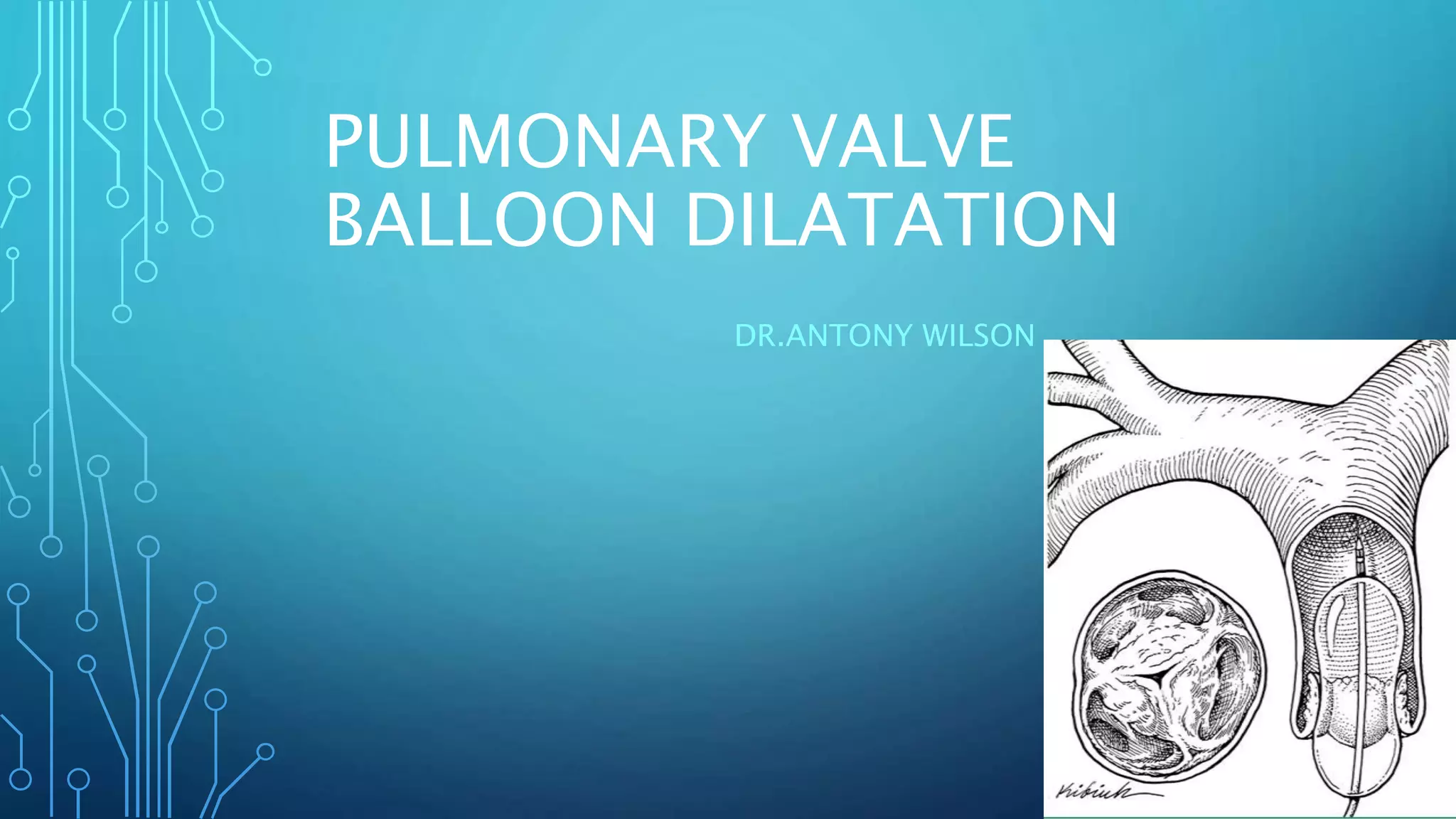
PULMONARY VALVE
BALLOON DILATATION
DR.ANTONY WILSON
• Percutaneous pulmonary valvuloplasty in children was first
reported by Semb et al in 1979.
• However, the static balloon technique, reported by Kan et al. in
1 982, was the first to be applied widely.
• Results have demonstrated the safety and effectiveness of this
technique and have established it as the treatment of choice for
children and adults with isolated pulmonary valve stenosis
CAUSES OF PULMONARY VALVE DISEASE
• Acquired pulmonary valve disease in the adult population is unusual
and mostly relates to rarities such as carcinoid disease.
• Even though rheumatic heart disease is common, pulmonary valve is
the least affected one(rare).
• Infective endocarditis is on uprise as there are increased cases of IV
drug abuse and CKD patients on dialysis.
• Congenital heart diseases mostly assosciated with dysfunction of
pulmonary valve either as a primary component or post repair.
PULMONARY VALVE STENOSIS
• First described by John Baptist Morgagni(1761).
• 8-10% of all congenital heart diseases.
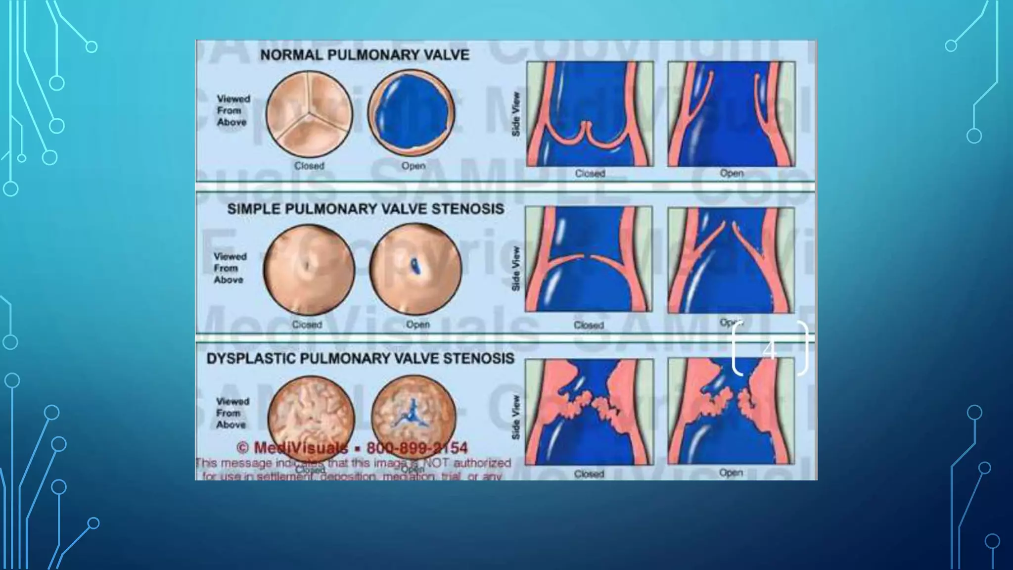
• MC form – valve is dome shaped,2-4 raphes present but no separation into valve leaflets.
• Trileaflet dysplastic myxomatous valve stenosis (15%) , they have no commissural fusion.
• Valve annulus hypoplasia
• Commonly seen in patients with Noonan’s syndrome.
• Secondary changes are – RVH,TR and RV failure.
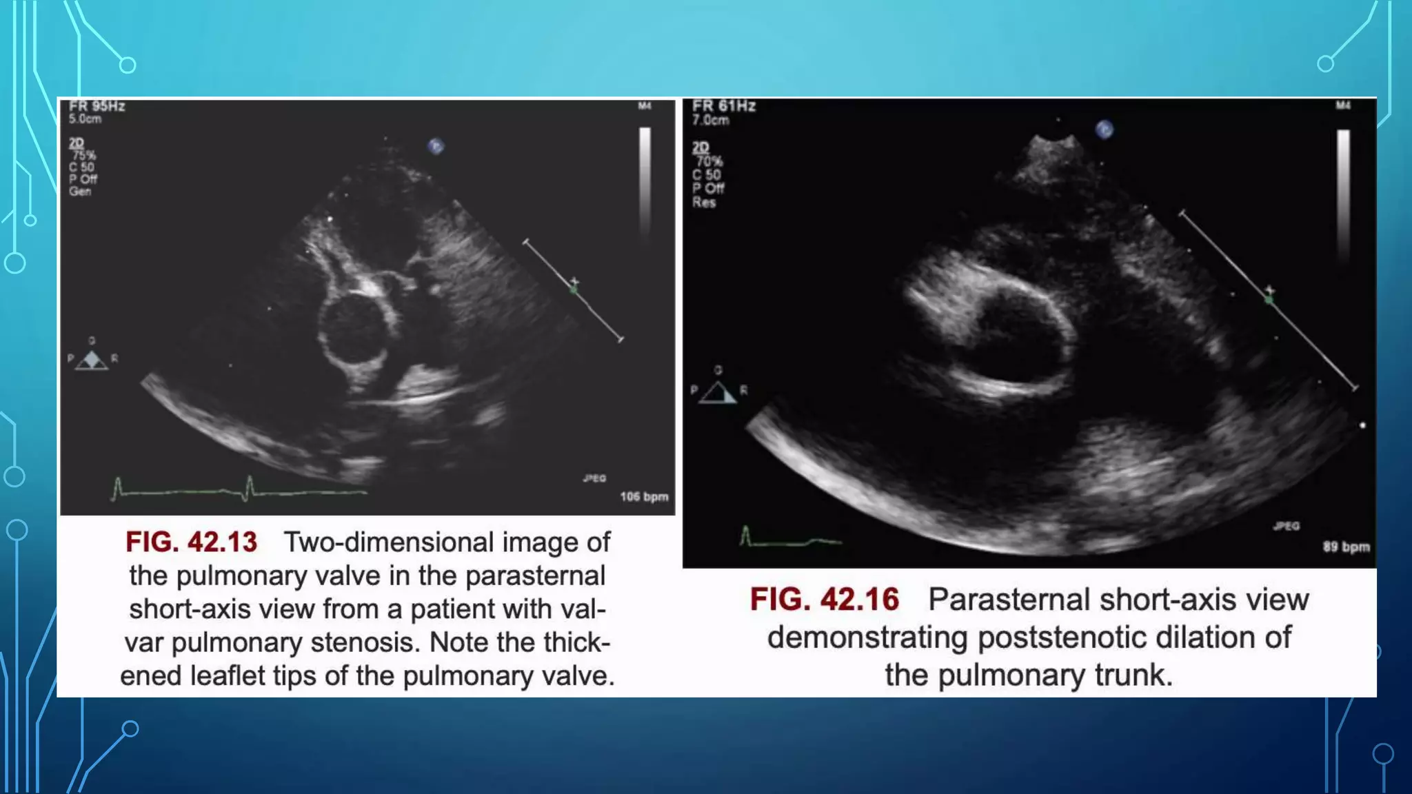
• Post stentotic dilation is usually seen in MPA –can extend into LPA.
• Usually asymptomatic – exercise intolerance MC symptom.
• Systolic ejection click and Ejection systolic murmur heard at left upper sternal border.
• Confrimation of severity – Echo – gradient,RVH,RV pressures.
• Except for neonates with critical pulmonary stenosis, patients
with untreated pulmonary valve stenosis often survive well into
adulthood
• Left untreated, significant pulmonary valve stenosis eventually
produces clinical symptoms such as fatigue, dyspnea, and
exercise intolerance.
• Nevertheless, treatment is indicated at any age if
hemodynamically significant pulmonary stenosis is documented
• Balloon pulmonary valvuloplasty can be performed safely and is
minimally invasive.
• It is therefore regarded as the treatment of choice for patients
with moderate to severe isolated pulmonary valve stenosis
• In isolated pulmonary valve stenosis, balloon dilation reduces
the degree of valvular obstruction by separating fused
commissures or by tearing the valve leaflets themselves.
INDICATIONS FOR BALLOON PULMONARY
VALVULOPLASTY
• Critical pulmonary stenosis, defined as pulmonary stenosis in a cyanotic
infant requiring a PDA to provide adequate pulmonary blood flow
• Resting catheterization peak systolic ejection gradient or echocardiographic
peak instantaneous pressure gradient 40 mm Hg or greater
• Resting catheterization or echocardiographic gradient less than 40 mm Hg
in the setting of RV dysfunction or symptoms
• Other indications for balloon pulmonary valvuloplasty, pulmonary atresia
with an intact ventricular septum but without RV-dependent coronary
circulation and as a palliative procedure for patients with cyanotic CHD
associated with pulmonary stenosis (e.g., tetralogy of Fallot [TOF]).
ACC/AHA’18
PREPROCEDURAL EVALUATION
ECG
• The electrocardiogram (ECG) findings are normal in mild cases.
• Right-axis deviation (RAD) and right ventricular hypertrophy (RVH)
are present in moderate PS.
• Right atrial hypertrophy (RAH) and RVH with “strain” may be seen in
severe PS.
• Neonates with critical PS may show left ventricular hypertrophy (LVH)
because of a hypoplastic RV and relatively large left ventricle (LV).
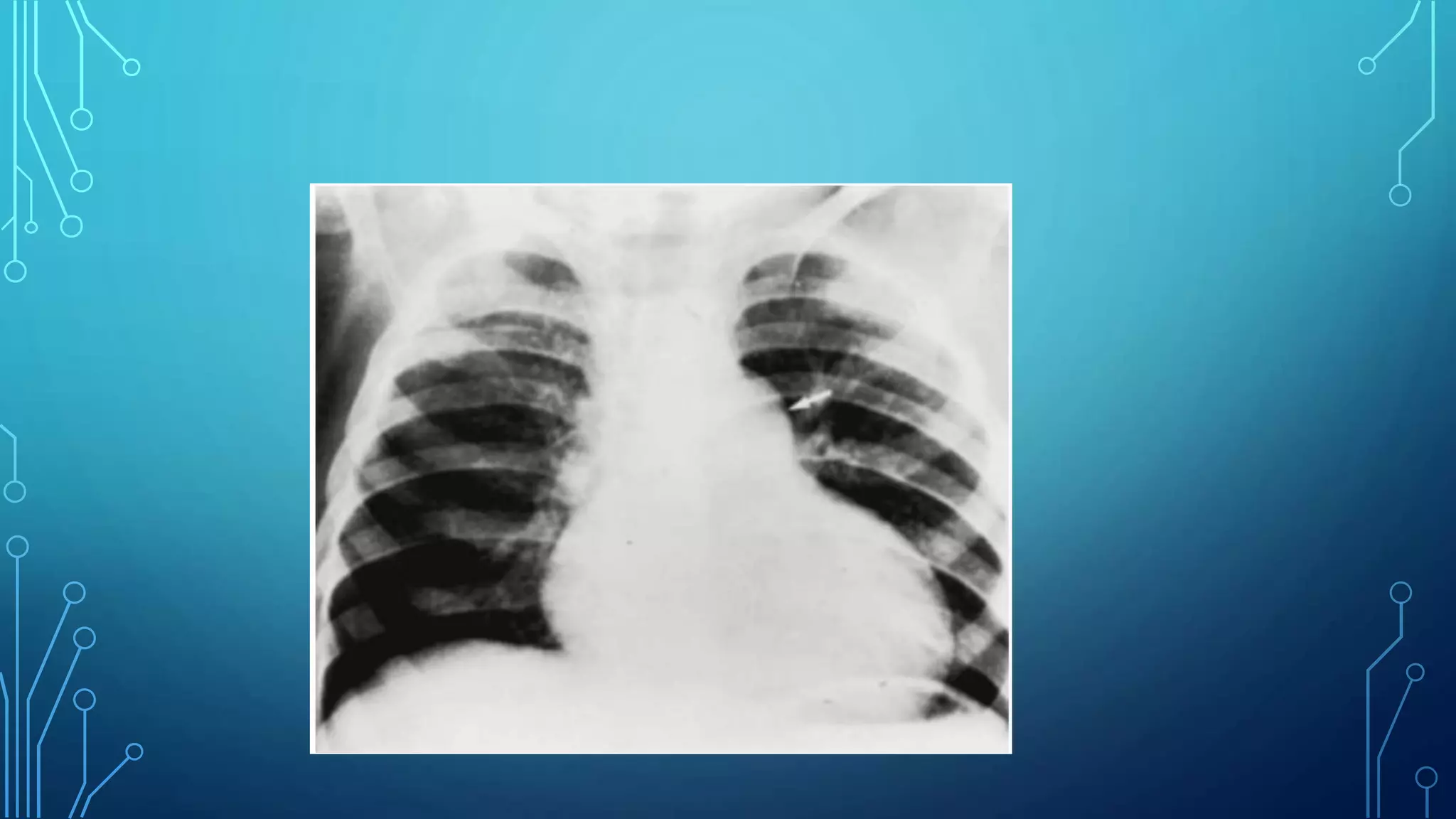
Radiography
• Heart size is usually normal, but the main PA segment may be
prominent with valvular stenosis (caused by poststenotic dilatation).
Cardiomegaly is present only if CHF develops.
• Pulmonary vascular markings are usually normal but may decrease
with severe PS.
• In neonates with critical PS, lung fields are oligemic with a varying
degree of cardiomegaly.
ECHO-DOPPLER STUDIES
• Two-dimensional echocardiographic precordial short-axis and
subcostal views are most useful in the evaluation of the
pulmonary valve leaflets
• Thickening and doming of the pulmonary valve leaflets can
often be visualized
• Markedly thickened, nodular, and immobile pulmonary valve
leaflets, suggestive of dysplastic pulmonary valves
• The pulmonary valve annulus can also be visualized and
measured
• Such measurements are also useful in the selection of balloon
diameter for balloon valvuloplasty
• Pulsed, continuous-wave, and color Doppler evaluation in
conjunction with two-dimensional echocardiography is most
useful in confirming the clinical diagnosis and in quantifying
the degree of obstruction
• Several studies have demonstrated the usefulness of peak Doppler
velocities in predicting the catheter-measured peak-to-peak
gradients across the pulmonary valve
• The peak instantaneous Doppler gradient may be calculated using a
modified Bernoulli equation:
ΔP = 4 V2
• where ΔP is the pressure gradient and V is the peak Doppler flow
velocity in the main pulmonary artery.
• Color Doppler and pulsed Doppler interrogation of the atrial
septum is useful and may reveal left-to-right or right-to-left
shunt
• Computed tomographic (CT) scans and magnetic resonance
imaging (MRI) may demonstrate pulmonary valve stenosis, but
the current state-of-the-art echo-Doppler studies are more
useful in diagnosing and quantifying pulmonary valve
obstruction.
SEDATION AND ANESTHESIA
• Fasting from the night before procedure
• Local anaesthesia is usually enough
• General anesthesia with endotracheal ventilation is used in
infants below the age of 3 months
VASCULAR ACCESS
• The percutaneous femoral venous route is the most preferred entry
site.
• However, other sites such as axillary or jugular venous or
transhepatic routes have been successfully used in the absence of
femoral venous access.
• A 5F to 7F sheath is inserted into the vein depending upon the age
and size of the patient as well as the anticipated size of the balloon
dilatation catheter.
• An arterial line is inserted percutaneously into the femoral artery to
continuously monitor the arterial blood pressure and to intermittently
HEMODYNAMIC ASSESSMENT
• The measurement of right
ventricular (RV) and pulmonary
artery pressures along with the
peak-to-peak gradient across the
pulmonary valve is performed
• A simultaneous recording of the
right ventricular and femoral artery
pressures is also undertaken. This
also helps assess the severity of
pulmonary valve obstruction; right
ventricular peak systolic pressure
≥75% of peak systolic systemic
pressure is considered significant.
ANGIOGRAPHY
• Biplane RV cineangiogram in a sitting-up (anterio-posterior
camera tilted to 15° LAO and 35° cranial) and lateral view are
performed to confirm the site of obstruction, to evaluate the
size and function of the right ventricle, and to measure
pulmonary valve annulus, preparatory to balloon pulmonary
valvuloplasty
• Berman angiographic catheter for the right ventriculogram, with
the inflated balloon positioned in the right ventricular apex.
• Selective left ventricular angiography and coronary
arteriography may be performed in patients older than 50
years
CATHETERS/WIRES PREPARATORY TO BALLOON
DILATATION
• Positioning a guide in the distal right or left pulmonary artery
or in the descending aorta in neonates is a necessary
prerequisite for undertaking balloon valvuloplasty
• In neonates and young infants, a 0.014-inch coronary guide
wire with a floppy end is used to cross the pulmonary valve
• The catheter is then advanced over the guide wire, again into
the distal right or left pulmonary artery or into the descending
aorta.
• The catheter is left in place and the guide wire is removed
slowly and replaced with a guide wire (extra-stiff, exchange
length Amplatz, Platinum Plus, or others) that is suited to
position the balloon dilatation catheter.
BALLOON DILATATION CATHETERS
• A variety of balloon angioplasty catheters have been used in the
past
• Subsequently, specifically designed catheters such as XXL,
Ultrathin, and Diamond, Marshal,Opta LP, Opta Pro,
PowerFlex,Tyshak I and II, Tyshak-mini, Z-Med, Z-Med II and
Mullins catheters and others have been used.
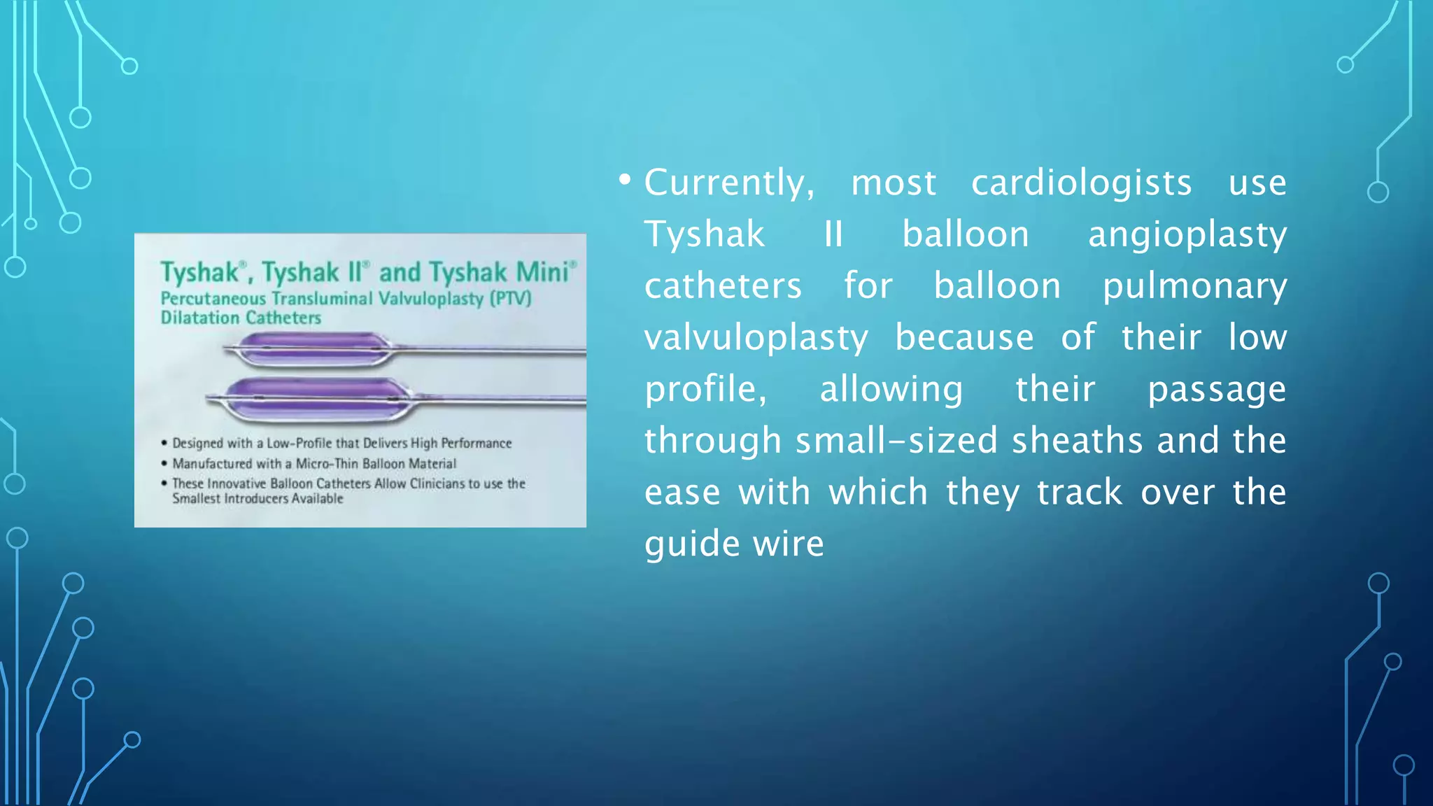
• Currently, most cardiologists use
Tyshak II balloon angioplasty
catheters for balloon pulmonary
valvuloplasty because of their low
profile, allowing their passage
through small-sized sheaths and the
ease with which they track over the
guide wire
• The Inoue balloon has been used in
adults with success.
• The major advantage of the Inoue
balloon over conventional balloons
is its adjustable diameter, which
makes stepwise dilation possible
BALLOON DIAMETER
• balloon/annulus ratio of 1.2–1.25 instead of the previously
recommended 1.2–1.4.
• Such smaller balloons are likely to result in good relief of
pulmonary valve obstruction while at the same time may help
to prevent significant pulmonary insufficiency at late follow-
up.
• Balloon size with >150% the diameter of the normal pulmonary
valve annulus will result in rupture.Ring JC et al,Am J Cardiol
1985;55(1):210-4
• The results of balloon pulmonary valvuloplasty for patients with
dysplastic pulmonary valves are generally poor with the use of
conventional balloon pulmonary valvuloplasty techniques.
• The use of large balloons, up to 150% of pulmonary valve
annulus, or high-pressure balloons may increase the
effectiveness of balloon therapy and avoid the need for
surgery.
BALLOON LENGTH
• Generally use 20-mm-long balloons in neonates and infants,
30-mm-long balloons in children and 40- or 50-mm-long
balloons in adolescents and adults.
• With shorter balloons, it is difficult to maintain the balloon
center across the pulmonary valve annulus during balloon
inflation.
• Longer balloons may impinge upon the tricuspid valve, causing
tricuspid insufficiency, or on the conduction system, causing
heart block.
NUMBER OF BALLOONS
• When the pulmonary valve annulus is too large to dilate with a
single balloon, valvuloplasty with simultaneous inflation of two
balloons across the pulmonary valve may be performed.
• where D1 and D2 are diameters of the balloons used. This
formula has been further simplified:Effective balloon diameter
= 0.82 (D1 + D2).
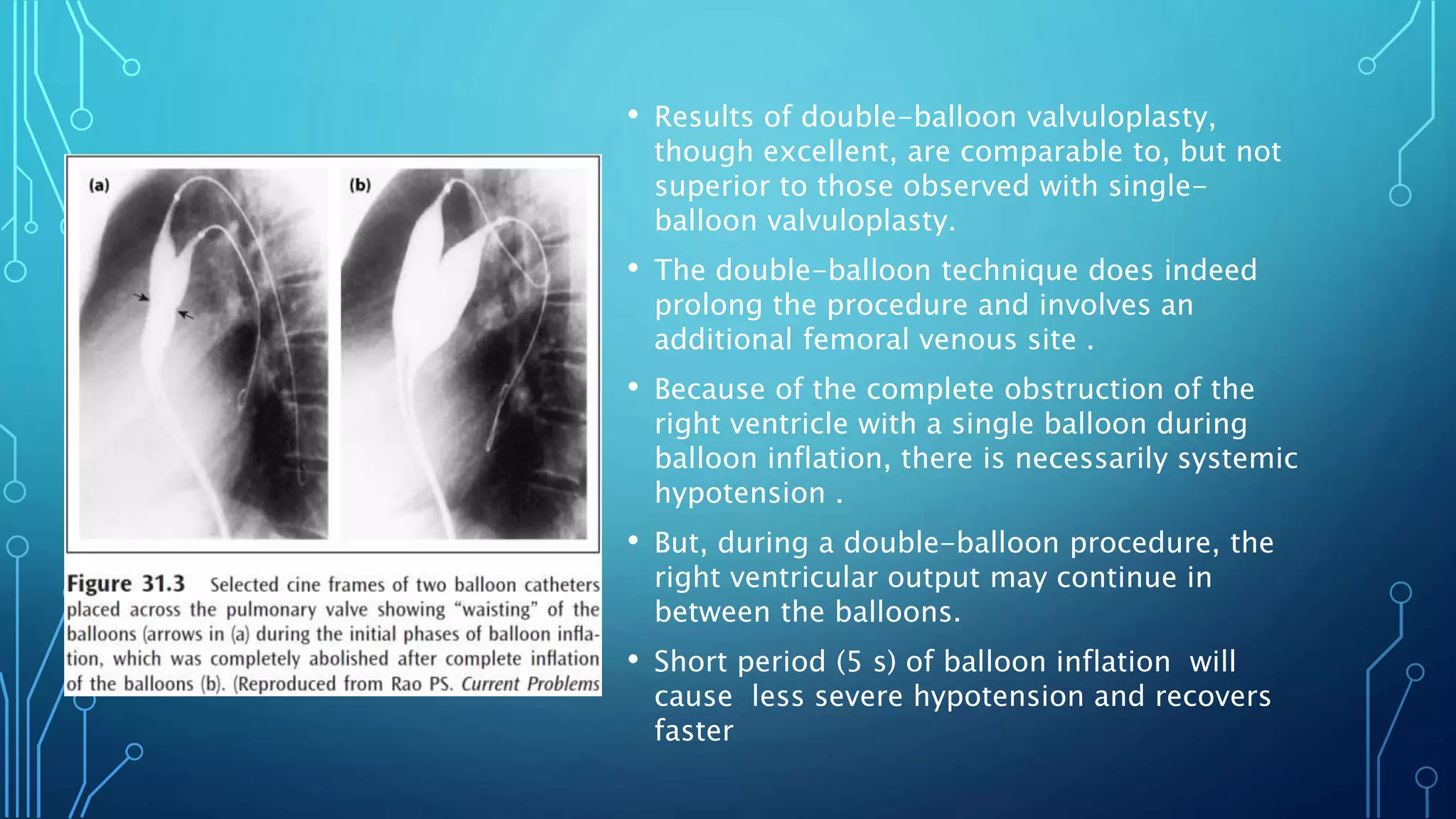
• Results of double-balloon valvuloplasty,
though excellent, are comparable to, but not
superior to those observed with single-
balloon valvuloplasty.
• The double-balloon technique does indeed
prolong the procedure and involves an
additional femoral venous site .
• Because of the complete obstruction of the
right ventricle with a single balloon during
balloon inflation, there is necessarily systemic
hypotension .
• But, during a double-balloon procedure, the
right ventricular output may continue in
between the balloons.
• Short period (5 s) of balloon inflation will
cause less severe hypotension and recovers
faster
PRESSURE, NUMBER, AND DURATION OF
BALLOON INFLATION• balloon inflation at or below the level of
balloon burst pressure stated by the
manufacturer, and will continue balloon
inflation until the waisting of the balloon
disappears .
• The duration of inflation is kept as short as
possible, usually just until after the waisting
disappears.
• Shorter balloon inflation cycles produce less
hypotension and more rapid return of
pressures toward normal
• No significant differences were found,
suggesting that the outcome of
valvuloplasty is not related to the balloon
inflation characteristics
BALLOON VALVULOPLASTY PROCEDURE (STEP-
BY-STEP)
1. Clinical and echocardiographic diagnosis of moderate to severe
valvar pulmonary stenosis.
2. Informed consent.
3. Confirmation of the severity of the stenosis by hemodynamic
measurements: gradients across the pulmonary valve and/or
comparison of the RV systolic pressure with systemic pressure.
4. RV angiography in sitting-up (15° LAO and 35° cranial) and straight
lateral views.
5. Measurement of pulmonary valve annulus is undertaken in
both views and an average of these is calculated. If the valve
annulus could not clearly be identified, the echocardiographic
measurement of the valve annulus may be used.
6. Selection of the balloon catheter to be used is made. An inflated
diameter is selected such that it is 1.2–1.25 times the pulmonary valve
annulus. The length of the balloon should be 20–40/50 mm depending
upon the patient’s age and size.
7. If a femoral arterial line is not in place, an arterial line is placed into
the femoral artery (3F in neonates and infants; 4F in children and 5F
in adolescents and adults) for monitoring of arterial pressure
continuously. Heart rate, blood pressure, respirations, and pulse
oximetry are also continuously monitored throughout the procedure.
8. No need to administer additional heparin for pulmonary valve
dilatations, but, if there is an intracardiac communication (patent
foramen ovale or an atrial septal defect), need to administer
heparin (100 units/ kg intravenously) and monitor activated
clotting times (ACTs) and maintain between 200 and 250 s.
9. A 4F to 6F multipurpose (multi A-2 [Cordis]) catheter is
introduced into the femoral venous sheath and advanced across
the pulmonary valve and the tip of the catheter is positioned in
the distal left (preferable) or right pulmonary artery. In neonates
and young infants, the catheter may be positioned in the
descending aorta via the ductus; this will increase the stability of
the wire and may make it easier to pass the balloon catheter
across the pulmonary valve
10. A 0.014–0.035 inch J-tipped, exchange-length, extra-stiff
guide wire is passed through the catheter already in place and
the catheter is removed.
11. The selected balloon angioplasty catheter is advanced over
the guide wire, but within the sheath and positioned across the
pulmonary valve. A frozen video frame of the RV cineangiogram
displayed on the screen is helpful in this regard.
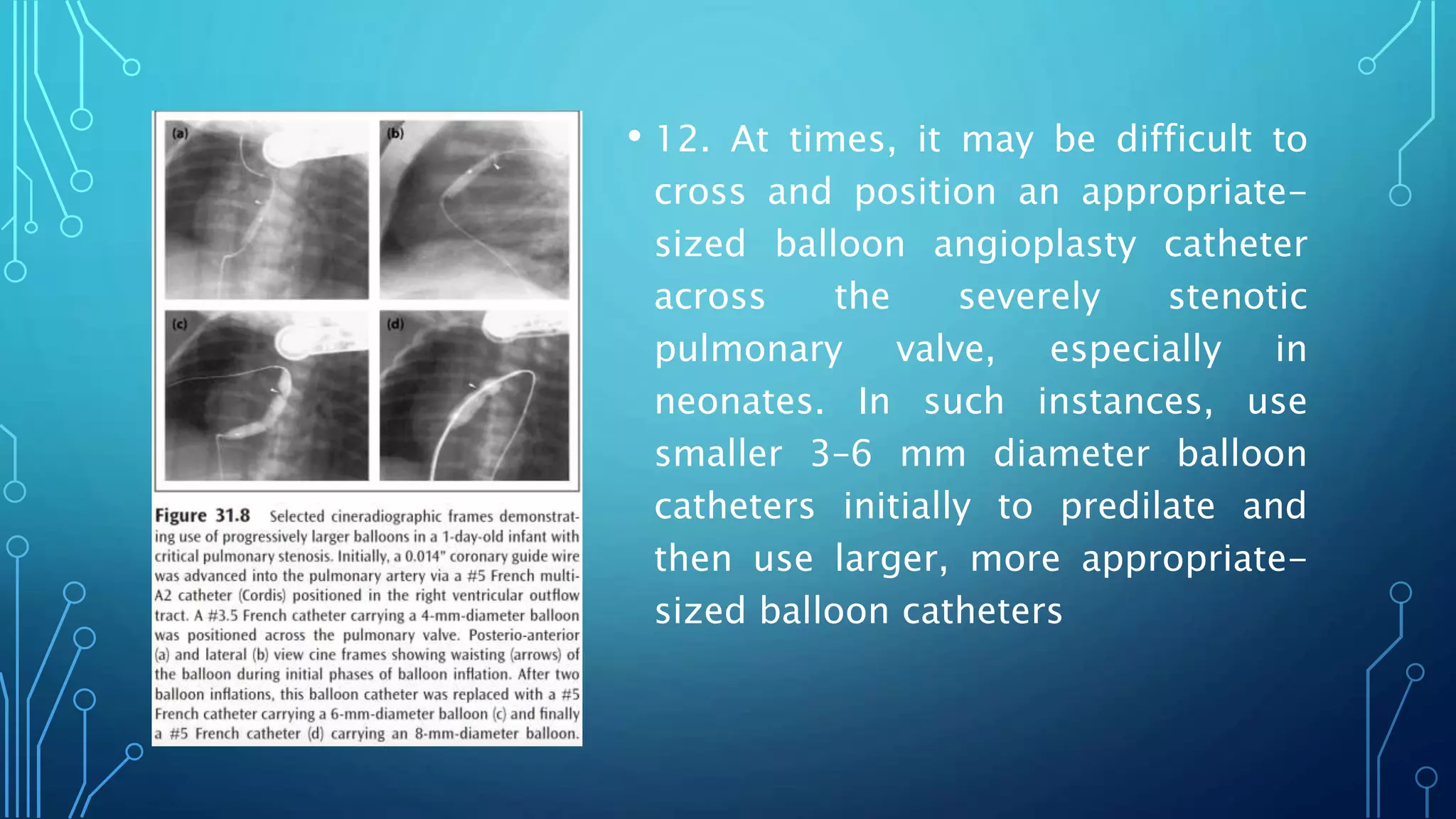
• 12. At times, it may be difficult to
cross and position an appropriate-
sized balloon angioplasty catheter
across the severely stenotic
pulmonary valve, especially in
neonates. In such instances, use
smaller 3–6 mm diameter balloon
catheters initially to predilate and
then use larger, more appropriate-
sized balloon catheters
13. The balloon is inflated with diluted contrast material (1 in 4) using
any of the commercially available inflators, while monitoring the
pressure of inflation. The inflation pressure is gradually increased up
to the manufacturer-recommended pressure or until disappearance of
the balloon waist.
If the balloon is not appropriately centered across the pulmonary valve,
the position of the catheter is readjusted and balloon inflation
repeated.
Once satisfactory balloon inflation is achieved, one more balloon
inflation may be performed as per the operator’s preference.
14. Sometimes, it may be difficult to maintain the position of the balloon
across the pulmonary valve during balloon inflation; the use of an
appropriate-length balloon, depending upon the age of the patient, and
holding the balloon catheter tight to prevent its movement is likely to resolve
this issue.
The use of the double-balloon technique, particularly in adolescents and
adults, has been useful. Others use adenosine-induced transient cardiac
standstill or rapid right ventricular pacing to achieve a stable position of the
balloon during valvuloplasty.
A nucleus balloon (NuMed) with a “barbell” configuration may keep the balloon
across the valve. However, there are no known reports of its use and these
catheters are bulky, requiring large sheaths.
POSTBALLOON PROTOCOL
• Following balloon valvuloplasty, the measurement of pressure
gradient across the pulmonary valve, pulmonary and femoral
arterial oxygen saturations, and simultaneous femoral artery
and right ventricular pressures are undertaken to assess the
result of valvuloplasty
• If the result is not satisfactory (peak-to-peak valvar gradient in
excess of 30 mmHg), a repeat dilatation with a larger balloon (2
mm larger than the first) is undertaken
• Finally, the catheter and guide wire are removed and Berman
angiography catheter is repositioned in the right ventricular
apex and an angiogram is performed to evaluate the mobility of
the pulmonary valve leaflets, to visualize the jet of contrast
across the dilated pulmonary valve to detect infundibular
stenosis, and to discern any complications such as tricuspid
insufficiency.
PITFALLS, PROBLEMS, AND COMPLICATIONS
• The balloon may not be truly across the pulmonary valve during
balloon inflation
• Waisting of the balloon may be produced by supravalvar
stenosis or infundibular constriction
• When in doubt, centering the balloon at various locations
across the right ventricular outflow region may become
necessary.
ACUTE COMPLICATIONS
• Complications during and immediately after balloon
valvuloplasty have been remarkably minimal; the VACA registry
reported a 0.24% death rate and 0.35% major complication rate
from the 822 balloon pulmonary valvuloplasty procedures from
26 institutions, attesting to the relative safety of the procedure.
• Transient bradycardia, premature beats, and a fall in systemic
pressure during balloon inflation have been uniformly noted by
all. These abnormalities rapidly return to normal following
balloon deflation
• Blood loss requiring transfusion has been reported, but with
better catheter/sheath systems that are currently available, the
blood loss is minimal
• Complete right bundle branch block, transient or permanent
heart block, cerebrovascular accident, loss of consciousness,
cardiac arrest, convulsions, balloon rupture at high balloon-
inflation pressures, rupture of tricuspid valve papillary muscle,
and pulmonary artery tears, though rare, have been reported.
• Transient severe right ventricular outflow tract obstruction ("suicidal
right ventricle") has been reported after the pulmonic valvular
obstruction has been relieved . This may be treated by volume
expansion and beta blocker therapy and tends to regress with time
• Infundibular gradients occur in nearly 30% of patients; the higher the
age and higher the severity of obstruction, the greater is the
prevalence of infundibular reaction.
• When the residual infundibular gradient is ≥50 mmHg, beta-
blockade therapy is generally recommended.
• Infundibular obstruction regresses to a great degree , with a rare
patient requiring surgical intervention
• Rare cases of ventricular arrhythmia have been reported
COMPLICATIONS AT FOLLOW-UP
• Seven to nineteen percent of the patients may develop femoral
venous obstruction, the femoral venous obstruction is more
likely in small infants.
• Recurrent pulmonary valve obstruction may occur in about 8%
of patients and repeat balloon valvuloplasty may help relieve
the residual or recurrent obstruction
• If the issues related to the technique are the reason for
recurrence, repeat balloon valvuloplasty is useful
• If the substrate (dysplastic valves without commissural fusion,
supravalvar pulmonary artery stenosis, or severe fixed
infundibular obstruction) is the problem, surgical intervention
may become necessary.
• Development of pulmonary insufficiency (PI); the frequency and
severity of PI increases with time.
• From one study group, 70 of 80 (88%) had PI at long-term
follow-up, while only 10% had PI prior to balloon valvuloplasty
POSTCATHETER MANAGEMENT
• Clinical, electrocardiographic, and echo-Doppler evaluation at 1, 6,
12, 24, and 60 months after the procedure, and every 5 years
thereafter is generally recommended.
• Regression of RV hypertrophy on the electrocardiogram following
balloon dilatation has been well documented6 and the
electrocardiogram is a useful adjunct in the evaluation of follow-up
results.
• Regression of RV hypertrophy on the electrocardiogram following
balloon dilatation has been well documented and the
electrocardiogram is a useful adjunct in the evaluation of follow-up
results
• However, electrocardiographic evidence for hemodynamic
improvement does not become apparent until 6 months after
valvuloplasty.
• Doppler gradient is generally reflective of the residual
obstruction and is a useful and reliable noninvasive monitoring
tool.
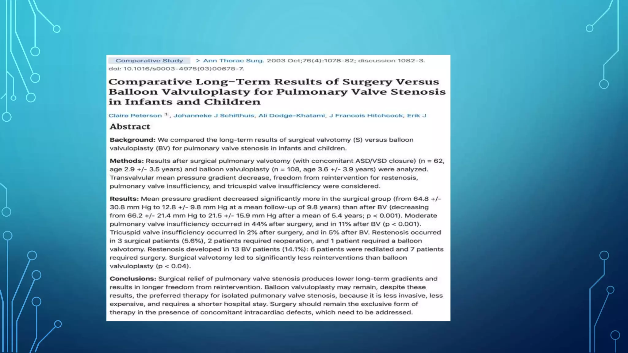
• 822 BPVs at the 26 member institutions of the
VACA Registry.
• Between 1981 to 1986
• The gradient decreased from 71±33 to
28±21mmHg
• Clinical success was achieved with balloon
valvotomy in 98 percent
• There were 5 major complications (0.6%),
including 2 deaths (0.2%), a cardiac perforation
with tamponade (0.1%) and 2 tricuspid
insufficiencies (0.2%) and11 minor
complications
THANK YOU
PVBD
PVBD
PVBD

More Related Content

What's hot

Collection of cath tracings by navin
Collection of cath tracings by navinCollection of cath tracings by navin
Collection of cath tracings by navinNavin Agrawal
 
Echocardiography of Mitral regurgitation
Echocardiography of Mitral regurgitationEchocardiography of Mitral regurgitation
Echocardiography of Mitral regurgitationDr. Muhammad AzAm Shah
 
Echo assessment of aortic stenosis
Echo assessment of aortic stenosisEcho assessment of aortic stenosis
Echo assessment of aortic stenosisNizam Uddin
 
MVP Mitral Valve Prolapse - Echocardiographic Evaluation
 MVP Mitral Valve  Prolapse - Echocardiographic Evaluation MVP Mitral Valve  Prolapse - Echocardiographic Evaluation
MVP Mitral Valve Prolapse - Echocardiographic EvaluationPraveen Nagula
 
Trans septal Puncture in Cardiology
Trans septal Puncture in CardiologyTrans septal Puncture in Cardiology
Trans septal Puncture in CardiologyRaghu Kishore Galla
 
Shunt quantification
Shunt quantificationShunt quantification
Shunt quantificationAnkur Gupta
 
BMV,PTMC,BALLOON MITRAL VALVOTOMY, BAL, VIRBHAN BALAI, DR VIRBHAN
BMV,PTMC,BALLOON MITRAL VALVOTOMY, BAL, VIRBHAN BALAI, DR VIRBHANBMV,PTMC,BALLOON MITRAL VALVOTOMY, BAL, VIRBHAN BALAI, DR VIRBHAN
BMV,PTMC,BALLOON MITRAL VALVOTOMY, BAL, VIRBHAN BALAI, DR VIRBHANDr Virbhan Balai
 
Guide catheters in coronary intervention
Guide catheters in coronary interventionGuide catheters in coronary intervention
Guide catheters in coronary interventionRohitWalse2
 
TAVI - Transcatheter Aortic Valve Implantation
TAVI - Transcatheter Aortic Valve ImplantationTAVI - Transcatheter Aortic Valve Implantation
TAVI - Transcatheter Aortic Valve ImplantationSrikanthK120
 
Right heart catheters
Right heart cathetersRight heart catheters
Right heart cathetersRohitWalse2
 
Echocardiography assessment of Aortic Regurgitation severity
Echocardiography assessment of Aortic Regurgitation severityEchocardiography assessment of Aortic Regurgitation severity
Echocardiography assessment of Aortic Regurgitation severityPRAVEEN GUPTA
 
Distal protection device
Distal protection deviceDistal protection device
Distal protection deviceAshish Golwara
 

What's hot (20)

Collection of cath tracings by navin
Collection of cath tracings by navinCollection of cath tracings by navin
Collection of cath tracings by navin
 
Echocardiography of Mitral regurgitation
Echocardiography of Mitral regurgitationEchocardiography of Mitral regurgitation
Echocardiography of Mitral regurgitation
 
cath Lab Hemoduhynamic
cath Lab Hemoduhynamiccath Lab Hemoduhynamic
cath Lab Hemoduhynamic
 
Echo assessment of aortic stenosis
Echo assessment of aortic stenosisEcho assessment of aortic stenosis
Echo assessment of aortic stenosis
 
Left ventricular angiogram (1)
Left ventricular angiogram (1)Left ventricular angiogram (1)
Left ventricular angiogram (1)
 
MVP Mitral Valve Prolapse - Echocardiographic Evaluation
 MVP Mitral Valve  Prolapse - Echocardiographic Evaluation MVP Mitral Valve  Prolapse - Echocardiographic Evaluation
MVP Mitral Valve Prolapse - Echocardiographic Evaluation
 
Trans septal Puncture in Cardiology
Trans septal Puncture in CardiologyTrans septal Puncture in Cardiology
Trans septal Puncture in Cardiology
 
Septal puncure ppt
Septal puncure pptSeptal puncure ppt
Septal puncure ppt
 
Trans septal puncture
Trans septal punctureTrans septal puncture
Trans septal puncture
 
Shunt quantification
Shunt quantificationShunt quantification
Shunt quantification
 
VSD devices
VSD devicesVSD devices
VSD devices
 
BMV,PTMC,BALLOON MITRAL VALVOTOMY, BAL, VIRBHAN BALAI, DR VIRBHAN
BMV,PTMC,BALLOON MITRAL VALVOTOMY, BAL, VIRBHAN BALAI, DR VIRBHANBMV,PTMC,BALLOON MITRAL VALVOTOMY, BAL, VIRBHAN BALAI, DR VIRBHAN
BMV,PTMC,BALLOON MITRAL VALVOTOMY, BAL, VIRBHAN BALAI, DR VIRBHAN
 
Guide catheters in coronary intervention
Guide catheters in coronary interventionGuide catheters in coronary intervention
Guide catheters in coronary intervention
 
ECHO ASSESSMENT OF ASD FOR DEVICE CLOSURE
ECHO ASSESSMENT OF ASD FOR DEVICE CLOSURE ECHO ASSESSMENT OF ASD FOR DEVICE CLOSURE
ECHO ASSESSMENT OF ASD FOR DEVICE CLOSURE
 
TAVI - Transcatheter Aortic Valve Implantation
TAVI - Transcatheter Aortic Valve ImplantationTAVI - Transcatheter Aortic Valve Implantation
TAVI - Transcatheter Aortic Valve Implantation
 
Right heart catheters
Right heart cathetersRight heart catheters
Right heart catheters
 
Echocardiography assessment of Aortic Regurgitation severity
Echocardiography assessment of Aortic Regurgitation severityEchocardiography assessment of Aortic Regurgitation severity
Echocardiography assessment of Aortic Regurgitation severity
 
Lbbb pacing
Lbbb pacingLbbb pacing
Lbbb pacing
 
Distal protection device
Distal protection deviceDistal protection device
Distal protection device
 
Vsd device closure
Vsd device closureVsd device closure
Vsd device closure
 

Similar to PVBD

congenital heart diseases.pptx
congenital heart diseases.pptxcongenital heart diseases.pptx
congenital heart diseases.pptxCHANDAN PADHAN
 
Transcatheter therapies for congenital heart disease
Transcatheter therapies for congenital heart diseaseTranscatheter therapies for congenital heart disease
Transcatheter therapies for congenital heart diseaseNAJEEB ULLAH SOFI
 
valvular heart disease LECT.pdf
valvular heart disease LECT.pdfvalvular heart disease LECT.pdf
valvular heart disease LECT.pdfisrashiekh
 
Central venous catheter complications
Central venous catheter complicationsCentral venous catheter complications
Central venous catheter complicationsRanjita Pallavi
 
Endocardial Cushion Defect / AVSD
Endocardial Cushion Defect / AVSDEndocardial Cushion Defect / AVSD
Endocardial Cushion Defect / AVSDHarshitha
 
Late complications in tof and redo surgeries
Late complications in tof and redo surgeriesLate complications in tof and redo surgeries
Late complications in tof and redo surgeriesbackstabber089
 
Transposition of great arteries
Transposition of great arteriesTransposition of great arteries
Transposition of great arteriesKuntal Surana
 
ANESTHESIA FOR TOF SURGERY
ANESTHESIA FOR TOF SURGERYANESTHESIA FOR TOF SURGERY
ANESTHESIA FOR TOF SURGERYRaju Jadhav
 
Hypoplastic Left heart dr Alister 3.pptx
Hypoplastic Left heart dr Alister 3.pptxHypoplastic Left heart dr Alister 3.pptx
Hypoplastic Left heart dr Alister 3.pptxGarvAmeta1
 
Cyanotic heart disease complete ppts
Cyanotic heart disease complete pptsCyanotic heart disease complete ppts
Cyanotic heart disease complete pptsDrMuddasarHussain
 
TETRALOGY OF FALLOT
TETRALOGY OF FALLOTTETRALOGY OF FALLOT
TETRALOGY OF FALLOTIndhu Reddy
 
Surgical management of tetralogy of fallot
Surgical management of tetralogy of fallotSurgical management of tetralogy of fallot
Surgical management of tetralogy of fallotrahul arora
 
Ultrasound in critically ill patients
Ultrasound in critically ill patients Ultrasound in critically ill patients
Ultrasound in critically ill patients Ahmed Bahnassy
 
emergency echo in critically ill patients.ppt
emergency echo in critically ill patients.pptemergency echo in critically ill patients.ppt
emergency echo in critically ill patients.pptShivani Rao
 
Ventricular septal defect DR NIKUNJ .R .SHRKHADA (MBBS,MS GEN SURG DNB CTS SR)
Ventricular septal defect  DR NIKUNJ .R .SHRKHADA (MBBS,MS GEN SURG DNB CTS SR)Ventricular septal defect  DR NIKUNJ .R .SHRKHADA (MBBS,MS GEN SURG DNB CTS SR)
Ventricular septal defect DR NIKUNJ .R .SHRKHADA (MBBS,MS GEN SURG DNB CTS SR)DR NIKUNJ SHEKHADA
 
Hypoplastic left heart syndrome
Hypoplastic left heart syndromeHypoplastic left heart syndrome
Hypoplastic left heart syndromeDrhunny88
 
Basic ultrasound in icu
Basic ultrasound in icuBasic ultrasound in icu
Basic ultrasound in icuAnor Abidin
 

Similar to PVBD (20)

congenital heart diseases.pptx
congenital heart diseases.pptxcongenital heart diseases.pptx
congenital heart diseases.pptx
 
Transcatheter therapies for congenital heart disease
Transcatheter therapies for congenital heart diseaseTranscatheter therapies for congenital heart disease
Transcatheter therapies for congenital heart disease
 
valvular heart disease LECT.pdf
valvular heart disease LECT.pdfvalvular heart disease LECT.pdf
valvular heart disease LECT.pdf
 
Central venous catheter complications
Central venous catheter complicationsCentral venous catheter complications
Central venous catheter complications
 
Endocardial Cushion Defect / AVSD
Endocardial Cushion Defect / AVSDEndocardial Cushion Defect / AVSD
Endocardial Cushion Defect / AVSD
 
Late complications in tof and redo surgeries
Late complications in tof and redo surgeriesLate complications in tof and redo surgeries
Late complications in tof and redo surgeries
 
Transposition of great arteries
Transposition of great arteriesTransposition of great arteries
Transposition of great arteries
 
ANESTHESIA FOR TOF SURGERY
ANESTHESIA FOR TOF SURGERYANESTHESIA FOR TOF SURGERY
ANESTHESIA FOR TOF SURGERY
 
Hypoplastic Left heart dr Alister 3.pptx
Hypoplastic Left heart dr Alister 3.pptxHypoplastic Left heart dr Alister 3.pptx
Hypoplastic Left heart dr Alister 3.pptx
 
Cyanotic heart disease complete ppts
Cyanotic heart disease complete pptsCyanotic heart disease complete ppts
Cyanotic heart disease complete ppts
 
Tricuspid atresia
Tricuspid atresiaTricuspid atresia
Tricuspid atresia
 
TETRALOGY OF FALLOT
TETRALOGY OF FALLOTTETRALOGY OF FALLOT
TETRALOGY OF FALLOT
 
Surgical management of tetralogy of fallot
Surgical management of tetralogy of fallotSurgical management of tetralogy of fallot
Surgical management of tetralogy of fallot
 
Ultrasound in critically ill patients
Ultrasound in critically ill patients Ultrasound in critically ill patients
Ultrasound in critically ill patients
 
emergency echo in critically ill patients.ppt
emergency echo in critically ill patients.pptemergency echo in critically ill patients.ppt
emergency echo in critically ill patients.ppt
 
VTE AND PE.pptx
VTE AND PE.pptxVTE AND PE.pptx
VTE AND PE.pptx
 
Adult Lines and Tubes in Radiology
Adult Lines and Tubes in RadiologyAdult Lines and Tubes in Radiology
Adult Lines and Tubes in Radiology
 
Ventricular septal defect DR NIKUNJ .R .SHRKHADA (MBBS,MS GEN SURG DNB CTS SR)
Ventricular septal defect  DR NIKUNJ .R .SHRKHADA (MBBS,MS GEN SURG DNB CTS SR)Ventricular septal defect  DR NIKUNJ .R .SHRKHADA (MBBS,MS GEN SURG DNB CTS SR)
Ventricular septal defect DR NIKUNJ .R .SHRKHADA (MBBS,MS GEN SURG DNB CTS SR)
 
Hypoplastic left heart syndrome
Hypoplastic left heart syndromeHypoplastic left heart syndrome
Hypoplastic left heart syndrome
 
Basic ultrasound in icu
Basic ultrasound in icuBasic ultrasound in icu
Basic ultrasound in icu
 

Recently uploaded

Crayon Activity Handout For the Crayon A
Crayon Activity Handout For the Crayon ACrayon Activity Handout For the Crayon A
Crayon Activity Handout For the Crayon AUnboundStockton
 
MARGINALIZATION (Different learners in Marginalized Group
MARGINALIZATION (Different learners in Marginalized GroupMARGINALIZATION (Different learners in Marginalized Group
MARGINALIZATION (Different learners in Marginalized GroupJonathanParaisoCruz
 
Earth Day Presentation wow hello nice great
Earth Day Presentation wow hello nice greatEarth Day Presentation wow hello nice great
Earth Day Presentation wow hello nice greatYousafMalik24
 
Blooming Together_ Growing a Community Garden Worksheet.docx
Blooming Together_ Growing a Community Garden Worksheet.docxBlooming Together_ Growing a Community Garden Worksheet.docx
Blooming Together_ Growing a Community Garden Worksheet.docxUnboundStockton
 
How to Make a Pirate ship Primary Education.pptx
How to Make a Pirate ship Primary Education.pptxHow to Make a Pirate ship Primary Education.pptx
How to Make a Pirate ship Primary Education.pptxmanuelaromero2013
 
Pharmacognosy Flower 3. Compositae 2023.pdf
Pharmacognosy Flower 3. Compositae 2023.pdfPharmacognosy Flower 3. Compositae 2023.pdf
Pharmacognosy Flower 3. Compositae 2023.pdfMahmoud M. Sallam
 
MICROBIOLOGY biochemical test detailed.pptx
MICROBIOLOGY biochemical test detailed.pptxMICROBIOLOGY biochemical test detailed.pptx
MICROBIOLOGY biochemical test detailed.pptxabhijeetpadhi001
 
Introduction to ArtificiaI Intelligence in Higher Education
Introduction to ArtificiaI Intelligence in Higher EducationIntroduction to ArtificiaI Intelligence in Higher Education
Introduction to ArtificiaI Intelligence in Higher Educationpboyjonauth
 
Like-prefer-love -hate+verb+ing & silent letters & citizenship text.pdf
Like-prefer-love -hate+verb+ing & silent letters & citizenship text.pdfLike-prefer-love -hate+verb+ing & silent letters & citizenship text.pdf
Like-prefer-love -hate+verb+ing & silent letters & citizenship text.pdfMr Bounab Samir
 
Historical philosophical, theoretical, and legal foundations of special and i...
Historical philosophical, theoretical, and legal foundations of special and i...Historical philosophical, theoretical, and legal foundations of special and i...
Historical philosophical, theoretical, and legal foundations of special and i...jaredbarbolino94
 
Types of Journalistic Writing Grade 8.pptx
Types of Journalistic Writing Grade 8.pptxTypes of Journalistic Writing Grade 8.pptx
Types of Journalistic Writing Grade 8.pptxEyham Joco
 
Meghan Sutherland In Media Res Media Component
Meghan Sutherland In Media Res Media ComponentMeghan Sutherland In Media Res Media Component
Meghan Sutherland In Media Res Media ComponentInMediaRes1
 
Computed Fields and api Depends in the Odoo 17
Computed Fields and api Depends in the Odoo 17Computed Fields and api Depends in the Odoo 17
Computed Fields and api Depends in the Odoo 17Celine George
 
Employee wellbeing at the workplace.pptx
Employee wellbeing at the workplace.pptxEmployee wellbeing at the workplace.pptx
Employee wellbeing at the workplace.pptxNirmalaLoungPoorunde1
 
ECONOMIC CONTEXT - LONG FORM TV DRAMA - PPT
ECONOMIC CONTEXT - LONG FORM TV DRAMA - PPTECONOMIC CONTEXT - LONG FORM TV DRAMA - PPT
ECONOMIC CONTEXT - LONG FORM TV DRAMA - PPTiammrhaywood
 
Enzyme, Pharmaceutical Aids, Miscellaneous Last Part of Chapter no 5th.pdf
Enzyme, Pharmaceutical Aids, Miscellaneous Last Part of Chapter no 5th.pdfEnzyme, Pharmaceutical Aids, Miscellaneous Last Part of Chapter no 5th.pdf
Enzyme, Pharmaceutical Aids, Miscellaneous Last Part of Chapter no 5th.pdfSumit Tiwari
 

Recently uploaded (20)

Crayon Activity Handout For the Crayon A
Crayon Activity Handout For the Crayon ACrayon Activity Handout For the Crayon A
Crayon Activity Handout For the Crayon A
 
MARGINALIZATION (Different learners in Marginalized Group
MARGINALIZATION (Different learners in Marginalized GroupMARGINALIZATION (Different learners in Marginalized Group
MARGINALIZATION (Different learners in Marginalized Group
 
Earth Day Presentation wow hello nice great
Earth Day Presentation wow hello nice greatEarth Day Presentation wow hello nice great
Earth Day Presentation wow hello nice great
 
Blooming Together_ Growing a Community Garden Worksheet.docx
Blooming Together_ Growing a Community Garden Worksheet.docxBlooming Together_ Growing a Community Garden Worksheet.docx
Blooming Together_ Growing a Community Garden Worksheet.docx
 
How to Make a Pirate ship Primary Education.pptx
How to Make a Pirate ship Primary Education.pptxHow to Make a Pirate ship Primary Education.pptx
How to Make a Pirate ship Primary Education.pptx
 
ESSENTIAL of (CS/IT/IS) class 06 (database)
ESSENTIAL of (CS/IT/IS) class 06 (database)ESSENTIAL of (CS/IT/IS) class 06 (database)
ESSENTIAL of (CS/IT/IS) class 06 (database)
 
Pharmacognosy Flower 3. Compositae 2023.pdf
Pharmacognosy Flower 3. Compositae 2023.pdfPharmacognosy Flower 3. Compositae 2023.pdf
Pharmacognosy Flower 3. Compositae 2023.pdf
 
MICROBIOLOGY biochemical test detailed.pptx
MICROBIOLOGY biochemical test detailed.pptxMICROBIOLOGY biochemical test detailed.pptx
MICROBIOLOGY biochemical test detailed.pptx
 
Introduction to ArtificiaI Intelligence in Higher Education
Introduction to ArtificiaI Intelligence in Higher EducationIntroduction to ArtificiaI Intelligence in Higher Education
Introduction to ArtificiaI Intelligence in Higher Education
 
TataKelola dan KamSiber Kecerdasan Buatan v022.pdf
TataKelola dan KamSiber Kecerdasan Buatan v022.pdfTataKelola dan KamSiber Kecerdasan Buatan v022.pdf
TataKelola dan KamSiber Kecerdasan Buatan v022.pdf
 
Like-prefer-love -hate+verb+ing & silent letters & citizenship text.pdf
Like-prefer-love -hate+verb+ing & silent letters & citizenship text.pdfLike-prefer-love -hate+verb+ing & silent letters & citizenship text.pdf
Like-prefer-love -hate+verb+ing & silent letters & citizenship text.pdf
 
Model Call Girl in Tilak Nagar Delhi reach out to us at 🔝9953056974🔝
Model Call Girl in Tilak Nagar Delhi reach out to us at 🔝9953056974🔝Model Call Girl in Tilak Nagar Delhi reach out to us at 🔝9953056974🔝
Model Call Girl in Tilak Nagar Delhi reach out to us at 🔝9953056974🔝
 
Historical philosophical, theoretical, and legal foundations of special and i...
Historical philosophical, theoretical, and legal foundations of special and i...Historical philosophical, theoretical, and legal foundations of special and i...
Historical philosophical, theoretical, and legal foundations of special and i...
 
Types of Journalistic Writing Grade 8.pptx
Types of Journalistic Writing Grade 8.pptxTypes of Journalistic Writing Grade 8.pptx
Types of Journalistic Writing Grade 8.pptx
 
Meghan Sutherland In Media Res Media Component
Meghan Sutherland In Media Res Media ComponentMeghan Sutherland In Media Res Media Component
Meghan Sutherland In Media Res Media Component
 
Computed Fields and api Depends in the Odoo 17
Computed Fields and api Depends in the Odoo 17Computed Fields and api Depends in the Odoo 17
Computed Fields and api Depends in the Odoo 17
 
9953330565 Low Rate Call Girls In Rohini Delhi NCR
9953330565 Low Rate Call Girls In Rohini  Delhi NCR9953330565 Low Rate Call Girls In Rohini  Delhi NCR
9953330565 Low Rate Call Girls In Rohini Delhi NCR
 
Employee wellbeing at the workplace.pptx
Employee wellbeing at the workplace.pptxEmployee wellbeing at the workplace.pptx
Employee wellbeing at the workplace.pptx
 
ECONOMIC CONTEXT - LONG FORM TV DRAMA - PPT
ECONOMIC CONTEXT - LONG FORM TV DRAMA - PPTECONOMIC CONTEXT - LONG FORM TV DRAMA - PPT
ECONOMIC CONTEXT - LONG FORM TV DRAMA - PPT
 
Enzyme, Pharmaceutical Aids, Miscellaneous Last Part of Chapter no 5th.pdf
Enzyme, Pharmaceutical Aids, Miscellaneous Last Part of Chapter no 5th.pdfEnzyme, Pharmaceutical Aids, Miscellaneous Last Part of Chapter no 5th.pdf
Enzyme, Pharmaceutical Aids, Miscellaneous Last Part of Chapter no 5th.pdf
 

PVBD

  • 2. • Percutaneous pulmonary valvuloplasty in children was first reported by Semb et al in 1979. • However, the static balloon technique, reported by Kan et al. in 1 982, was the first to be applied widely. • Results have demonstrated the safety and effectiveness of this technique and have established it as the treatment of choice for children and adults with isolated pulmonary valve stenosis
  • 3.
  • 4. CAUSES OF PULMONARY VALVE DISEASE • Acquired pulmonary valve disease in the adult population is unusual and mostly relates to rarities such as carcinoid disease. • Even though rheumatic heart disease is common, pulmonary valve is the least affected one(rare). • Infective endocarditis is on uprise as there are increased cases of IV drug abuse and CKD patients on dialysis. • Congenital heart diseases mostly assosciated with dysfunction of pulmonary valve either as a primary component or post repair.
  • 5. PULMONARY VALVE STENOSIS • First described by John Baptist Morgagni(1761). • 8-10% of all congenital heart diseases. • MC form – valve is dome shaped,2-4 raphes present but no separation into valve leaflets. • Trileaflet dysplastic myxomatous valve stenosis (15%) , they have no commissural fusion. • Valve annulus hypoplasia • Commonly seen in patients with Noonan’s syndrome. • Secondary changes are – RVH,TR and RV failure. • Post stentotic dilation is usually seen in MPA –can extend into LPA. • Usually asymptomatic – exercise intolerance MC symptom. • Systolic ejection click and Ejection systolic murmur heard at left upper sternal border. • Confrimation of severity – Echo – gradient,RVH,RV pressures.
  • 6. • Except for neonates with critical pulmonary stenosis, patients with untreated pulmonary valve stenosis often survive well into adulthood • Left untreated, significant pulmonary valve stenosis eventually produces clinical symptoms such as fatigue, dyspnea, and exercise intolerance. • Nevertheless, treatment is indicated at any age if hemodynamically significant pulmonary stenosis is documented
  • 7. • Balloon pulmonary valvuloplasty can be performed safely and is minimally invasive. • It is therefore regarded as the treatment of choice for patients with moderate to severe isolated pulmonary valve stenosis • In isolated pulmonary valve stenosis, balloon dilation reduces the degree of valvular obstruction by separating fused commissures or by tearing the valve leaflets themselves.
  • 8. INDICATIONS FOR BALLOON PULMONARY VALVULOPLASTY • Critical pulmonary stenosis, defined as pulmonary stenosis in a cyanotic infant requiring a PDA to provide adequate pulmonary blood flow • Resting catheterization peak systolic ejection gradient or echocardiographic peak instantaneous pressure gradient 40 mm Hg or greater • Resting catheterization or echocardiographic gradient less than 40 mm Hg in the setting of RV dysfunction or symptoms • Other indications for balloon pulmonary valvuloplasty, pulmonary atresia with an intact ventricular septum but without RV-dependent coronary circulation and as a palliative procedure for patients with cyanotic CHD associated with pulmonary stenosis (e.g., tetralogy of Fallot [TOF]).
  • 10. PREPROCEDURAL EVALUATION ECG • The electrocardiogram (ECG) findings are normal in mild cases. • Right-axis deviation (RAD) and right ventricular hypertrophy (RVH) are present in moderate PS. • Right atrial hypertrophy (RAH) and RVH with “strain” may be seen in severe PS. • Neonates with critical PS may show left ventricular hypertrophy (LVH) because of a hypoplastic RV and relatively large left ventricle (LV).
  • 11. Radiography • Heart size is usually normal, but the main PA segment may be prominent with valvular stenosis (caused by poststenotic dilatation). Cardiomegaly is present only if CHF develops. • Pulmonary vascular markings are usually normal but may decrease with severe PS. • In neonates with critical PS, lung fields are oligemic with a varying degree of cardiomegaly.
  • 12.
  • 13. ECHO-DOPPLER STUDIES • Two-dimensional echocardiographic precordial short-axis and subcostal views are most useful in the evaluation of the pulmonary valve leaflets • Thickening and doming of the pulmonary valve leaflets can often be visualized • Markedly thickened, nodular, and immobile pulmonary valve leaflets, suggestive of dysplastic pulmonary valves
  • 14.
  • 15.
  • 16. • The pulmonary valve annulus can also be visualized and measured • Such measurements are also useful in the selection of balloon diameter for balloon valvuloplasty • Pulsed, continuous-wave, and color Doppler evaluation in conjunction with two-dimensional echocardiography is most useful in confirming the clinical diagnosis and in quantifying the degree of obstruction
  • 17.
  • 18.
  • 19. • Several studies have demonstrated the usefulness of peak Doppler velocities in predicting the catheter-measured peak-to-peak gradients across the pulmonary valve • The peak instantaneous Doppler gradient may be calculated using a modified Bernoulli equation: ΔP = 4 V2 • where ΔP is the pressure gradient and V is the peak Doppler flow velocity in the main pulmonary artery.
  • 20. • Color Doppler and pulsed Doppler interrogation of the atrial septum is useful and may reveal left-to-right or right-to-left shunt • Computed tomographic (CT) scans and magnetic resonance imaging (MRI) may demonstrate pulmonary valve stenosis, but the current state-of-the-art echo-Doppler studies are more useful in diagnosing and quantifying pulmonary valve obstruction.
  • 21. SEDATION AND ANESTHESIA • Fasting from the night before procedure • Local anaesthesia is usually enough • General anesthesia with endotracheal ventilation is used in infants below the age of 3 months
  • 22. VASCULAR ACCESS • The percutaneous femoral venous route is the most preferred entry site. • However, other sites such as axillary or jugular venous or transhepatic routes have been successfully used in the absence of femoral venous access. • A 5F to 7F sheath is inserted into the vein depending upon the age and size of the patient as well as the anticipated size of the balloon dilatation catheter. • An arterial line is inserted percutaneously into the femoral artery to continuously monitor the arterial blood pressure and to intermittently
  • 23. HEMODYNAMIC ASSESSMENT • The measurement of right ventricular (RV) and pulmonary artery pressures along with the peak-to-peak gradient across the pulmonary valve is performed • A simultaneous recording of the right ventricular and femoral artery pressures is also undertaken. This also helps assess the severity of pulmonary valve obstruction; right ventricular peak systolic pressure ≥75% of peak systolic systemic pressure is considered significant.
  • 24. ANGIOGRAPHY • Biplane RV cineangiogram in a sitting-up (anterio-posterior camera tilted to 15° LAO and 35° cranial) and lateral view are performed to confirm the site of obstruction, to evaluate the size and function of the right ventricle, and to measure pulmonary valve annulus, preparatory to balloon pulmonary valvuloplasty • Berman angiographic catheter for the right ventriculogram, with the inflated balloon positioned in the right ventricular apex. • Selective left ventricular angiography and coronary arteriography may be performed in patients older than 50 years
  • 25.
  • 26. CATHETERS/WIRES PREPARATORY TO BALLOON DILATATION • Positioning a guide in the distal right or left pulmonary artery or in the descending aorta in neonates is a necessary prerequisite for undertaking balloon valvuloplasty • In neonates and young infants, a 0.014-inch coronary guide wire with a floppy end is used to cross the pulmonary valve
  • 27. • The catheter is then advanced over the guide wire, again into the distal right or left pulmonary artery or into the descending aorta. • The catheter is left in place and the guide wire is removed slowly and replaced with a guide wire (extra-stiff, exchange length Amplatz, Platinum Plus, or others) that is suited to position the balloon dilatation catheter.
  • 28. BALLOON DILATATION CATHETERS • A variety of balloon angioplasty catheters have been used in the past • Subsequently, specifically designed catheters such as XXL, Ultrathin, and Diamond, Marshal,Opta LP, Opta Pro, PowerFlex,Tyshak I and II, Tyshak-mini, Z-Med, Z-Med II and Mullins catheters and others have been used.
  • 29. • Currently, most cardiologists use Tyshak II balloon angioplasty catheters for balloon pulmonary valvuloplasty because of their low profile, allowing their passage through small-sized sheaths and the ease with which they track over the guide wire
  • 30. • The Inoue balloon has been used in adults with success. • The major advantage of the Inoue balloon over conventional balloons is its adjustable diameter, which makes stepwise dilation possible
  • 31. BALLOON DIAMETER • balloon/annulus ratio of 1.2–1.25 instead of the previously recommended 1.2–1.4. • Such smaller balloons are likely to result in good relief of pulmonary valve obstruction while at the same time may help to prevent significant pulmonary insufficiency at late follow- up. • Balloon size with >150% the diameter of the normal pulmonary valve annulus will result in rupture.Ring JC et al,Am J Cardiol 1985;55(1):210-4
  • 32. • The results of balloon pulmonary valvuloplasty for patients with dysplastic pulmonary valves are generally poor with the use of conventional balloon pulmonary valvuloplasty techniques. • The use of large balloons, up to 150% of pulmonary valve annulus, or high-pressure balloons may increase the effectiveness of balloon therapy and avoid the need for surgery.
  • 33. BALLOON LENGTH • Generally use 20-mm-long balloons in neonates and infants, 30-mm-long balloons in children and 40- or 50-mm-long balloons in adolescents and adults. • With shorter balloons, it is difficult to maintain the balloon center across the pulmonary valve annulus during balloon inflation. • Longer balloons may impinge upon the tricuspid valve, causing tricuspid insufficiency, or on the conduction system, causing heart block.
  • 34. NUMBER OF BALLOONS • When the pulmonary valve annulus is too large to dilate with a single balloon, valvuloplasty with simultaneous inflation of two balloons across the pulmonary valve may be performed. • where D1 and D2 are diameters of the balloons used. This formula has been further simplified:Effective balloon diameter = 0.82 (D1 + D2).
  • 35. • Results of double-balloon valvuloplasty, though excellent, are comparable to, but not superior to those observed with single- balloon valvuloplasty. • The double-balloon technique does indeed prolong the procedure and involves an additional femoral venous site . • Because of the complete obstruction of the right ventricle with a single balloon during balloon inflation, there is necessarily systemic hypotension . • But, during a double-balloon procedure, the right ventricular output may continue in between the balloons. • Short period (5 s) of balloon inflation will cause less severe hypotension and recovers faster
  • 36. PRESSURE, NUMBER, AND DURATION OF BALLOON INFLATION• balloon inflation at or below the level of balloon burst pressure stated by the manufacturer, and will continue balloon inflation until the waisting of the balloon disappears . • The duration of inflation is kept as short as possible, usually just until after the waisting disappears. • Shorter balloon inflation cycles produce less hypotension and more rapid return of pressures toward normal • No significant differences were found, suggesting that the outcome of valvuloplasty is not related to the balloon inflation characteristics
  • 37. BALLOON VALVULOPLASTY PROCEDURE (STEP- BY-STEP) 1. Clinical and echocardiographic diagnosis of moderate to severe valvar pulmonary stenosis. 2. Informed consent. 3. Confirmation of the severity of the stenosis by hemodynamic measurements: gradients across the pulmonary valve and/or comparison of the RV systolic pressure with systemic pressure. 4. RV angiography in sitting-up (15° LAO and 35° cranial) and straight lateral views.
  • 38. 5. Measurement of pulmonary valve annulus is undertaken in both views and an average of these is calculated. If the valve annulus could not clearly be identified, the echocardiographic measurement of the valve annulus may be used.
  • 39. 6. Selection of the balloon catheter to be used is made. An inflated diameter is selected such that it is 1.2–1.25 times the pulmonary valve annulus. The length of the balloon should be 20–40/50 mm depending upon the patient’s age and size. 7. If a femoral arterial line is not in place, an arterial line is placed into the femoral artery (3F in neonates and infants; 4F in children and 5F in adolescents and adults) for monitoring of arterial pressure continuously. Heart rate, blood pressure, respirations, and pulse oximetry are also continuously monitored throughout the procedure.
  • 40. 8. No need to administer additional heparin for pulmonary valve dilatations, but, if there is an intracardiac communication (patent foramen ovale or an atrial septal defect), need to administer heparin (100 units/ kg intravenously) and monitor activated clotting times (ACTs) and maintain between 200 and 250 s.
  • 41. 9. A 4F to 6F multipurpose (multi A-2 [Cordis]) catheter is introduced into the femoral venous sheath and advanced across the pulmonary valve and the tip of the catheter is positioned in the distal left (preferable) or right pulmonary artery. In neonates and young infants, the catheter may be positioned in the descending aorta via the ductus; this will increase the stability of the wire and may make it easier to pass the balloon catheter across the pulmonary valve
  • 42. 10. A 0.014–0.035 inch J-tipped, exchange-length, extra-stiff guide wire is passed through the catheter already in place and the catheter is removed. 11. The selected balloon angioplasty catheter is advanced over the guide wire, but within the sheath and positioned across the pulmonary valve. A frozen video frame of the RV cineangiogram displayed on the screen is helpful in this regard.
  • 43. • 12. At times, it may be difficult to cross and position an appropriate- sized balloon angioplasty catheter across the severely stenotic pulmonary valve, especially in neonates. In such instances, use smaller 3–6 mm diameter balloon catheters initially to predilate and then use larger, more appropriate- sized balloon catheters
  • 44. 13. The balloon is inflated with diluted contrast material (1 in 4) using any of the commercially available inflators, while monitoring the pressure of inflation. The inflation pressure is gradually increased up to the manufacturer-recommended pressure or until disappearance of the balloon waist. If the balloon is not appropriately centered across the pulmonary valve, the position of the catheter is readjusted and balloon inflation repeated. Once satisfactory balloon inflation is achieved, one more balloon inflation may be performed as per the operator’s preference.
  • 45. 14. Sometimes, it may be difficult to maintain the position of the balloon across the pulmonary valve during balloon inflation; the use of an appropriate-length balloon, depending upon the age of the patient, and holding the balloon catheter tight to prevent its movement is likely to resolve this issue. The use of the double-balloon technique, particularly in adolescents and adults, has been useful. Others use adenosine-induced transient cardiac standstill or rapid right ventricular pacing to achieve a stable position of the balloon during valvuloplasty. A nucleus balloon (NuMed) with a “barbell” configuration may keep the balloon across the valve. However, there are no known reports of its use and these catheters are bulky, requiring large sheaths.
  • 46.
  • 47. POSTBALLOON PROTOCOL • Following balloon valvuloplasty, the measurement of pressure gradient across the pulmonary valve, pulmonary and femoral arterial oxygen saturations, and simultaneous femoral artery and right ventricular pressures are undertaken to assess the result of valvuloplasty • If the result is not satisfactory (peak-to-peak valvar gradient in excess of 30 mmHg), a repeat dilatation with a larger balloon (2 mm larger than the first) is undertaken
  • 48. • Finally, the catheter and guide wire are removed and Berman angiography catheter is repositioned in the right ventricular apex and an angiogram is performed to evaluate the mobility of the pulmonary valve leaflets, to visualize the jet of contrast across the dilated pulmonary valve to detect infundibular stenosis, and to discern any complications such as tricuspid insufficiency.
  • 49. PITFALLS, PROBLEMS, AND COMPLICATIONS • The balloon may not be truly across the pulmonary valve during balloon inflation • Waisting of the balloon may be produced by supravalvar stenosis or infundibular constriction • When in doubt, centering the balloon at various locations across the right ventricular outflow region may become necessary.
  • 50. ACUTE COMPLICATIONS • Complications during and immediately after balloon valvuloplasty have been remarkably minimal; the VACA registry reported a 0.24% death rate and 0.35% major complication rate from the 822 balloon pulmonary valvuloplasty procedures from 26 institutions, attesting to the relative safety of the procedure. • Transient bradycardia, premature beats, and a fall in systemic pressure during balloon inflation have been uniformly noted by all. These abnormalities rapidly return to normal following balloon deflation
  • 51. • Blood loss requiring transfusion has been reported, but with better catheter/sheath systems that are currently available, the blood loss is minimal • Complete right bundle branch block, transient or permanent heart block, cerebrovascular accident, loss of consciousness, cardiac arrest, convulsions, balloon rupture at high balloon- inflation pressures, rupture of tricuspid valve papillary muscle, and pulmonary artery tears, though rare, have been reported.
  • 52. • Transient severe right ventricular outflow tract obstruction ("suicidal right ventricle") has been reported after the pulmonic valvular obstruction has been relieved . This may be treated by volume expansion and beta blocker therapy and tends to regress with time • Infundibular gradients occur in nearly 30% of patients; the higher the age and higher the severity of obstruction, the greater is the prevalence of infundibular reaction. • When the residual infundibular gradient is ≥50 mmHg, beta- blockade therapy is generally recommended. • Infundibular obstruction regresses to a great degree , with a rare patient requiring surgical intervention • Rare cases of ventricular arrhythmia have been reported
  • 53.
  • 54. COMPLICATIONS AT FOLLOW-UP • Seven to nineteen percent of the patients may develop femoral venous obstruction, the femoral venous obstruction is more likely in small infants. • Recurrent pulmonary valve obstruction may occur in about 8% of patients and repeat balloon valvuloplasty may help relieve the residual or recurrent obstruction • If the issues related to the technique are the reason for recurrence, repeat balloon valvuloplasty is useful
  • 55. • If the substrate (dysplastic valves without commissural fusion, supravalvar pulmonary artery stenosis, or severe fixed infundibular obstruction) is the problem, surgical intervention may become necessary. • Development of pulmonary insufficiency (PI); the frequency and severity of PI increases with time. • From one study group, 70 of 80 (88%) had PI at long-term follow-up, while only 10% had PI prior to balloon valvuloplasty
  • 56. POSTCATHETER MANAGEMENT • Clinical, electrocardiographic, and echo-Doppler evaluation at 1, 6, 12, 24, and 60 months after the procedure, and every 5 years thereafter is generally recommended. • Regression of RV hypertrophy on the electrocardiogram following balloon dilatation has been well documented6 and the electrocardiogram is a useful adjunct in the evaluation of follow-up results. • Regression of RV hypertrophy on the electrocardiogram following balloon dilatation has been well documented and the electrocardiogram is a useful adjunct in the evaluation of follow-up results
  • 57. • However, electrocardiographic evidence for hemodynamic improvement does not become apparent until 6 months after valvuloplasty. • Doppler gradient is generally reflective of the residual obstruction and is a useful and reliable noninvasive monitoring tool.
  • 58. • 822 BPVs at the 26 member institutions of the VACA Registry. • Between 1981 to 1986 • The gradient decreased from 71±33 to 28±21mmHg • Clinical success was achieved with balloon valvotomy in 98 percent • There were 5 major complications (0.6%), including 2 deaths (0.2%), a cardiac perforation with tamponade (0.1%) and 2 tricuspid insufficiencies (0.2%) and11 minor complications
  • 59.
  • 60.
  • 61.
  • 62.
  • 63.