Download as PDF, PPTX
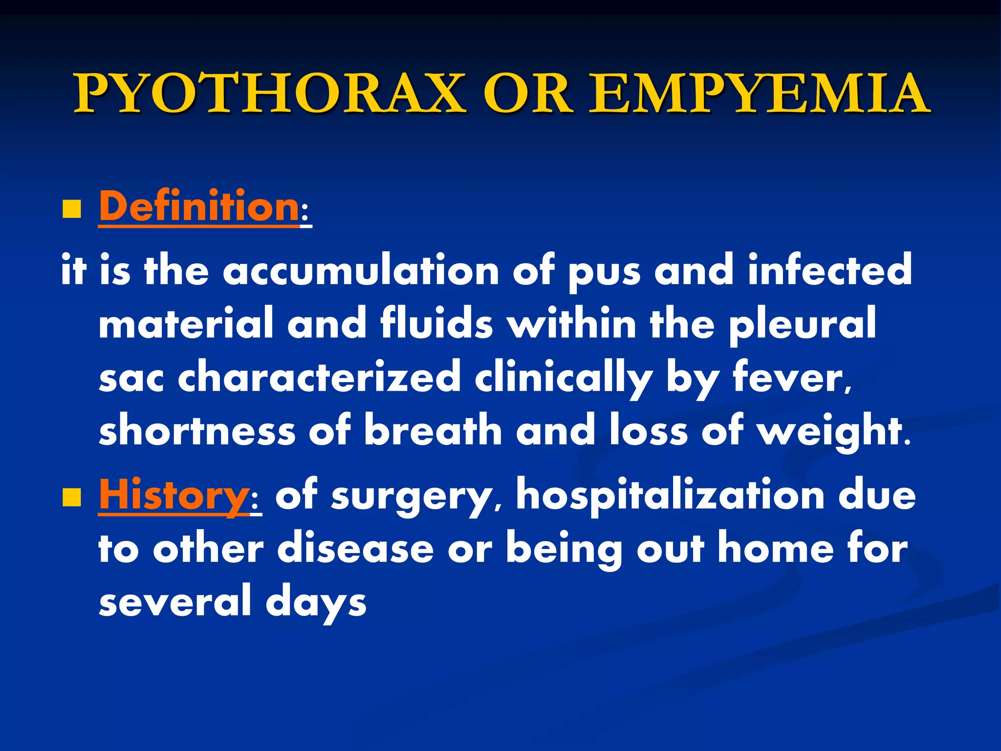

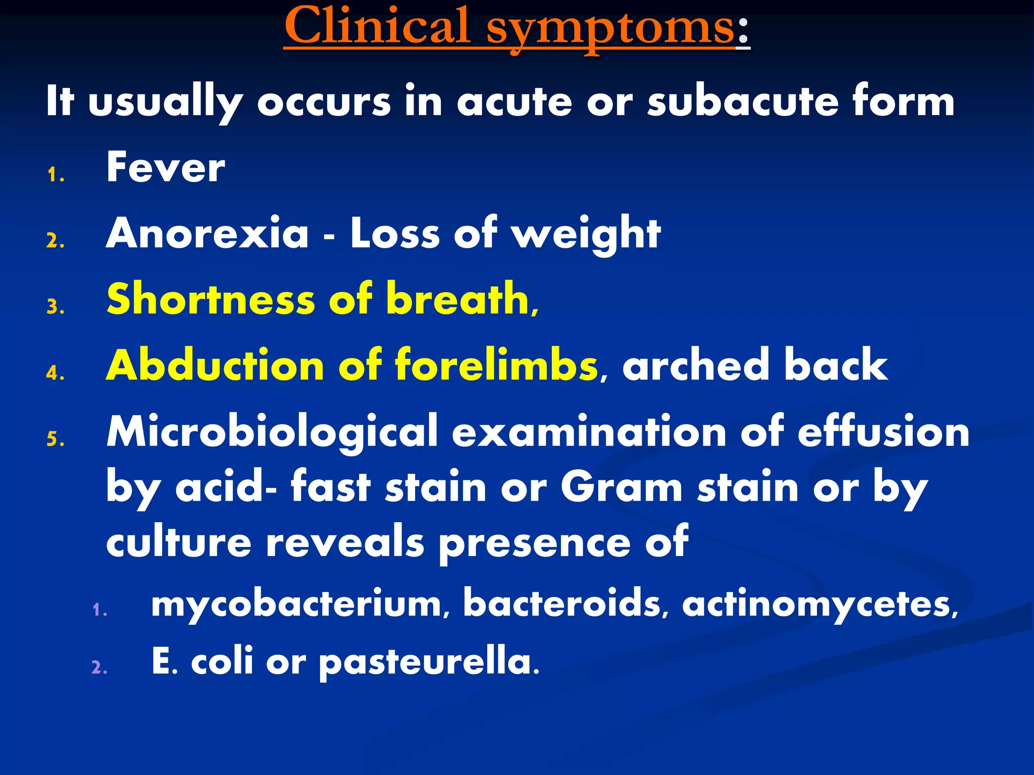
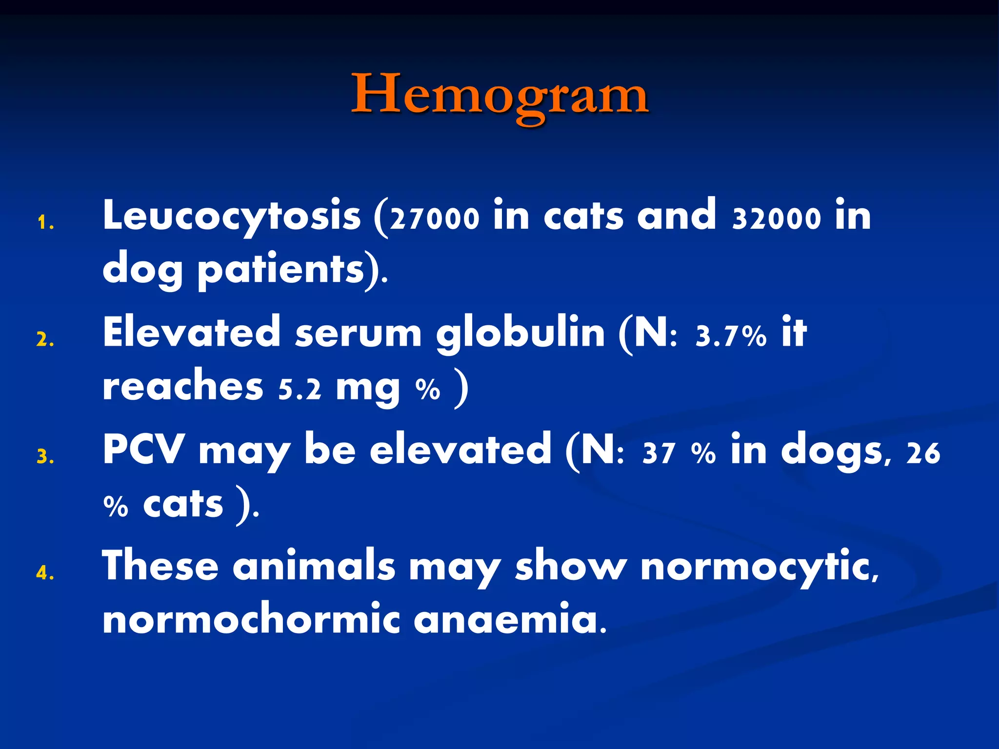
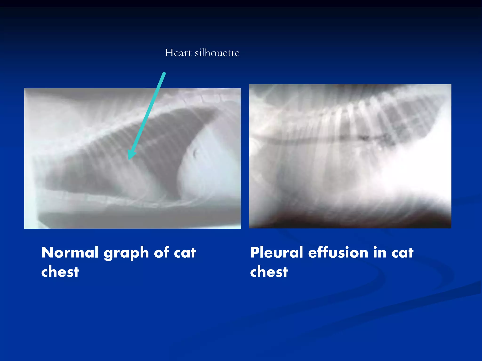
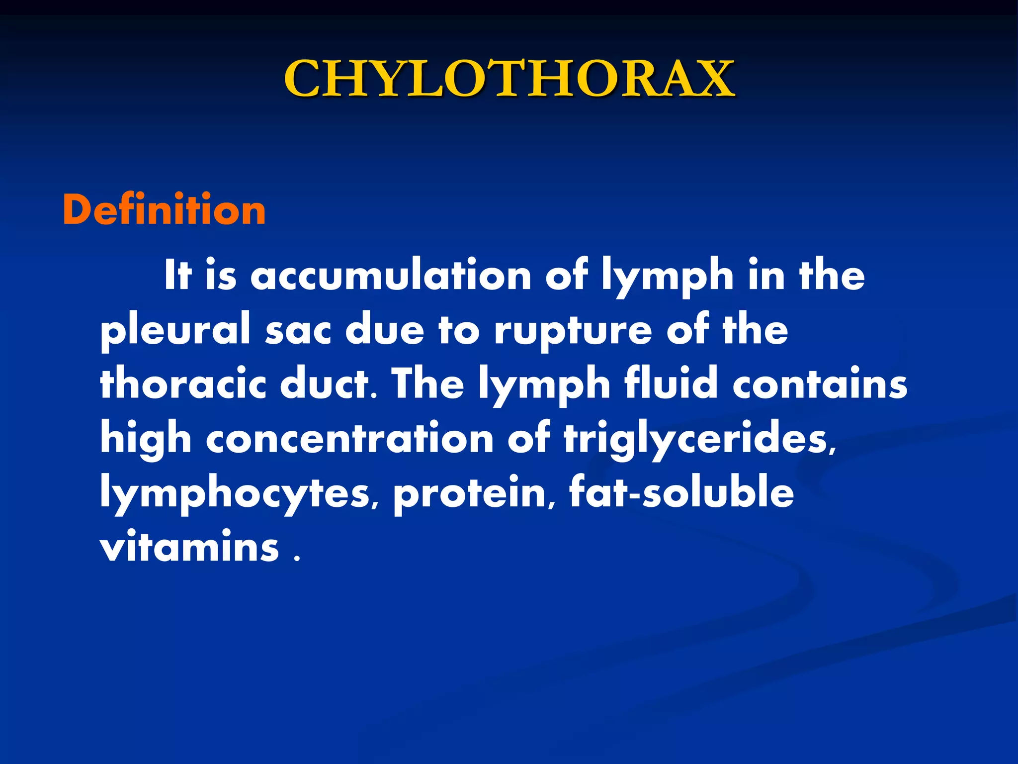
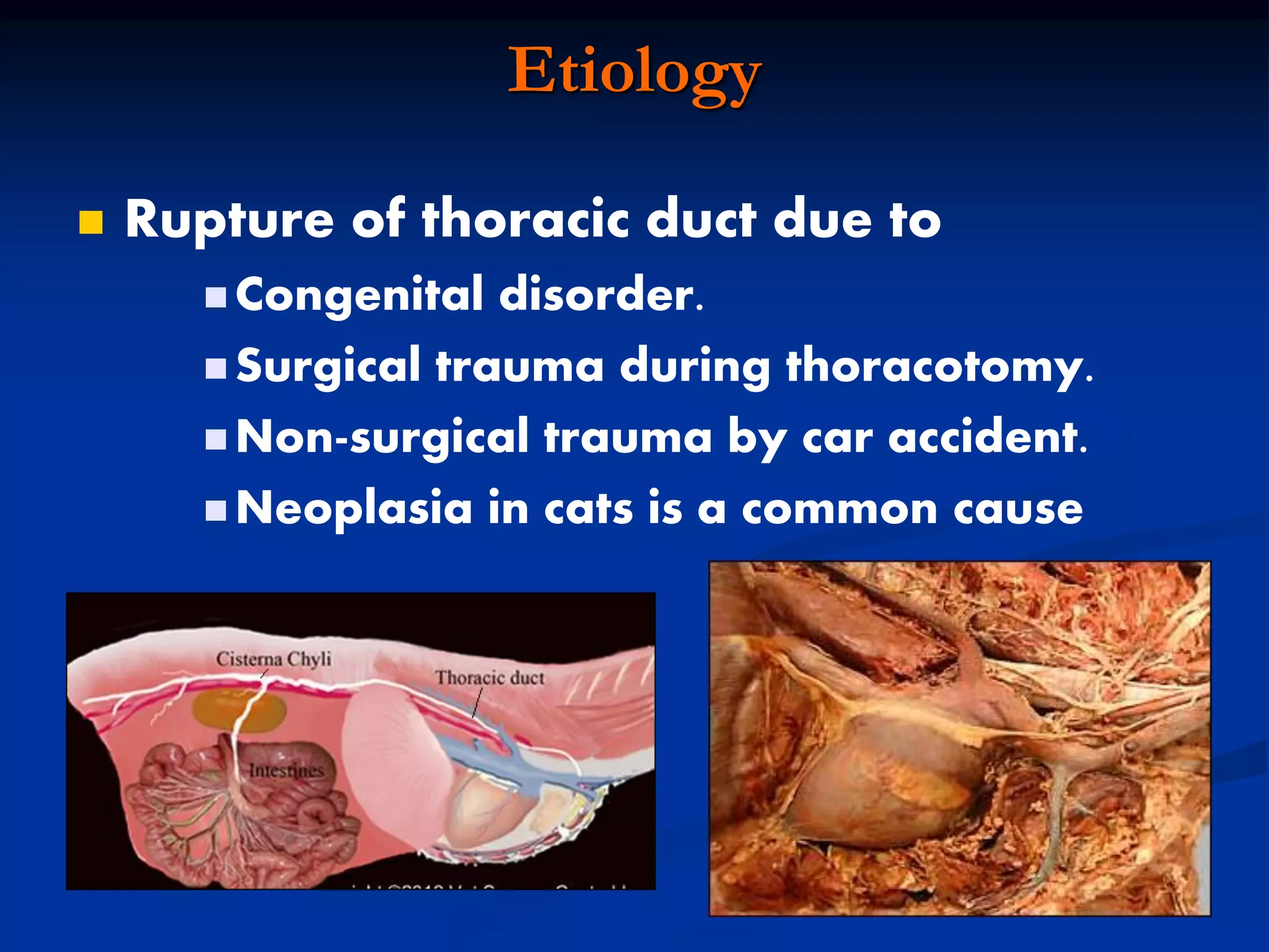


Pyothorax or empyema is the accumulation of pus and infected material within the pleural sac, causing symptoms like fever, breathing difficulties, and weight loss. It is typically caused by direct introduction of bacteria via trauma, surgery or extension from other infected sites. Diagnosis involves examining pleural fluid for bacteria, chest x-rays showing effusion, and ruling out other causes. Treatment requires tube drainage of the pleural space along with intravenous antibiotics and fluid therapy. Chylothorax is a similar condition involving accumulation of lymph fluid in the pleural space, often due to trauma or cancer causing a rupture of the thoracic duct.


![Empyema[174].pptx,and management and approch](https://cdn.slidesharecdn.com/ss_thumbnails/empyema174-240721195029-f8bee125-thumbnail.jpg?width=640&height=640&fit=bounds)


