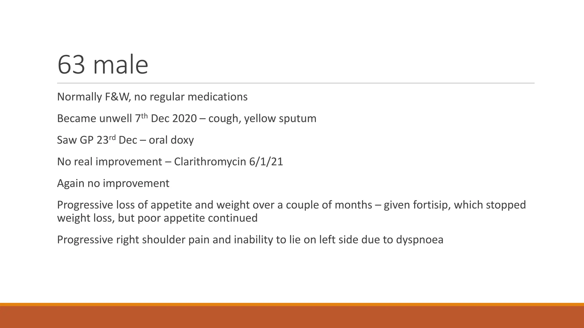
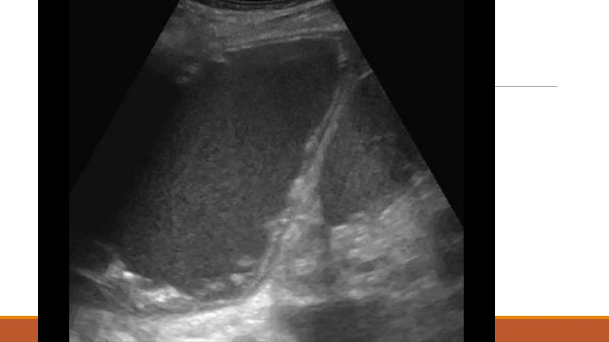
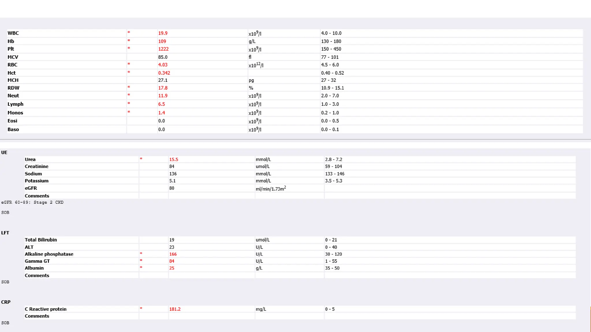
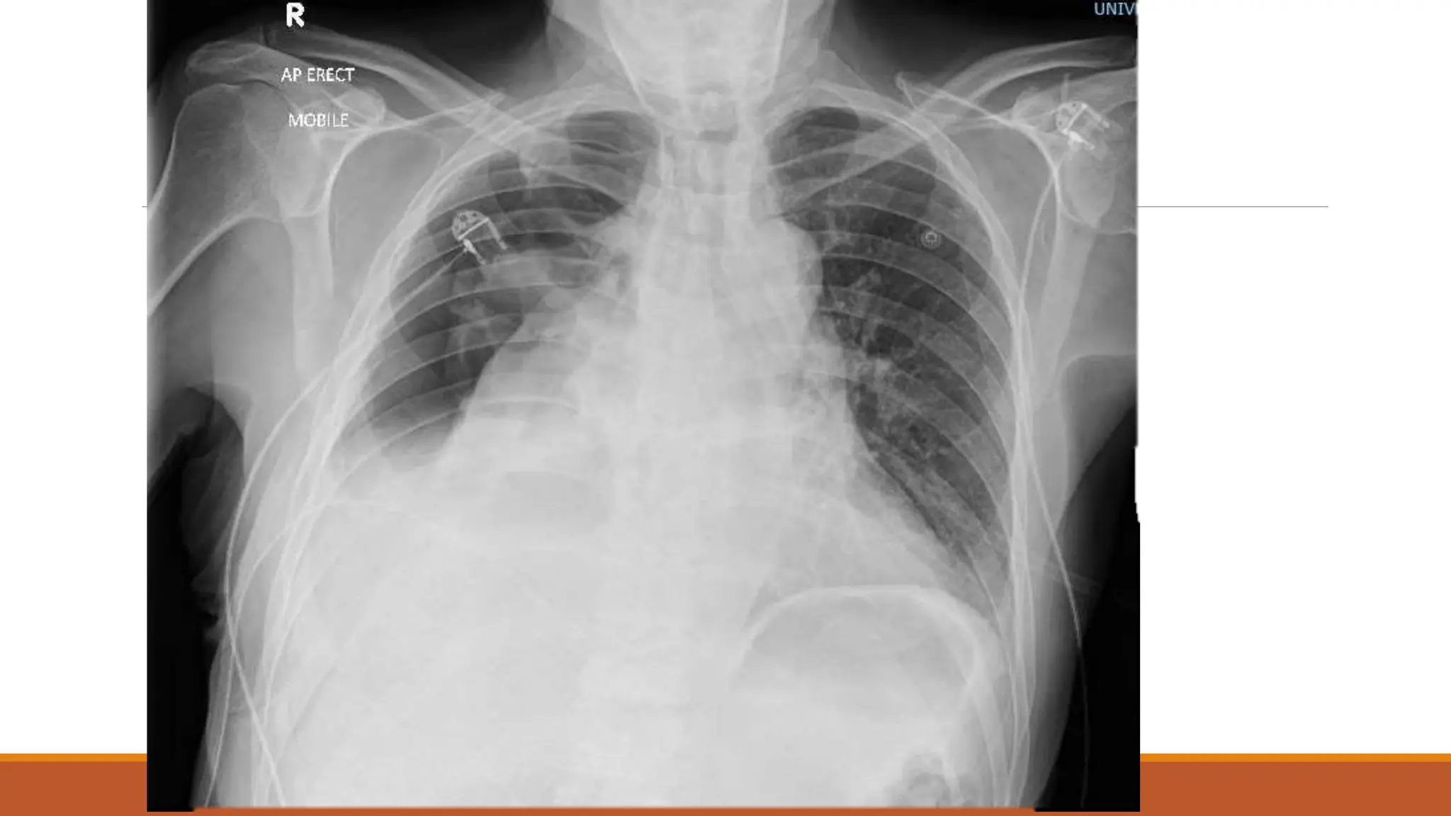
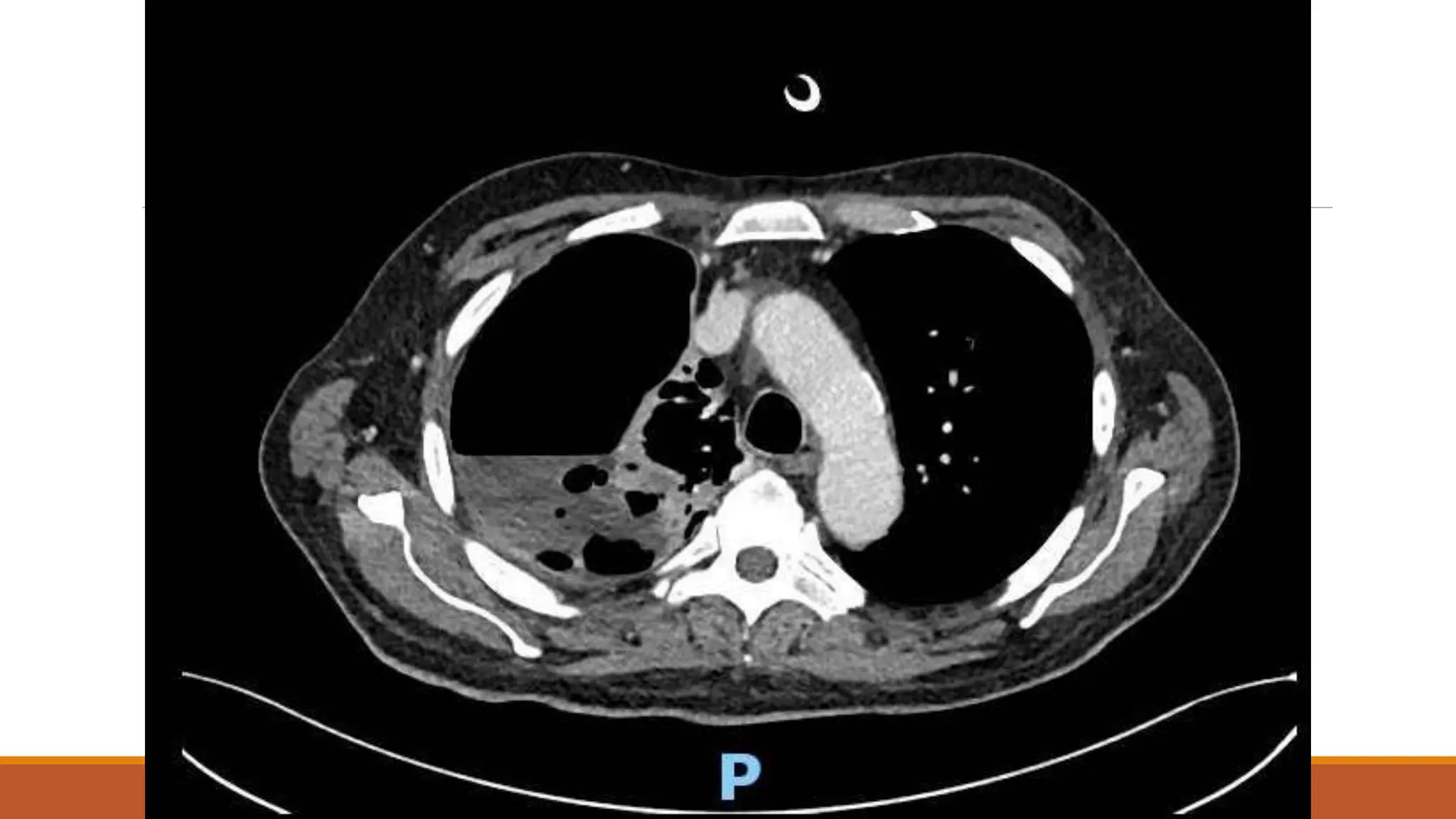
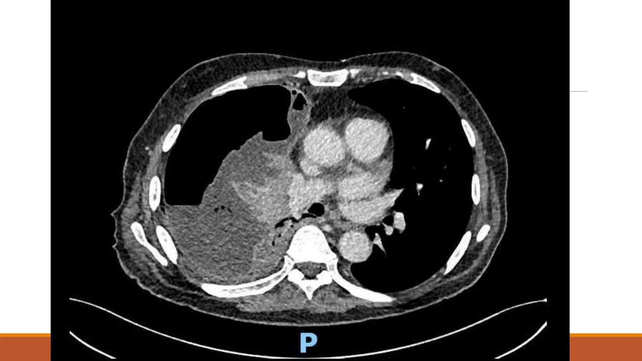
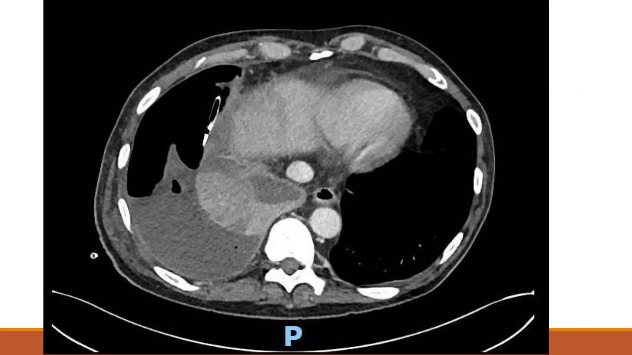
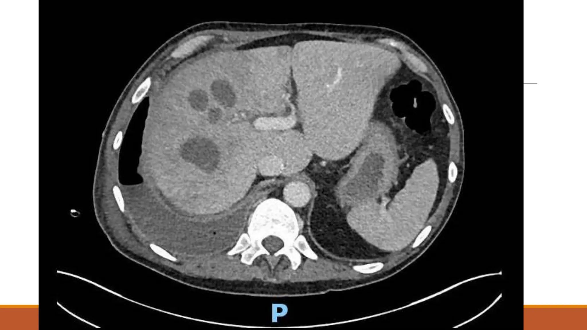
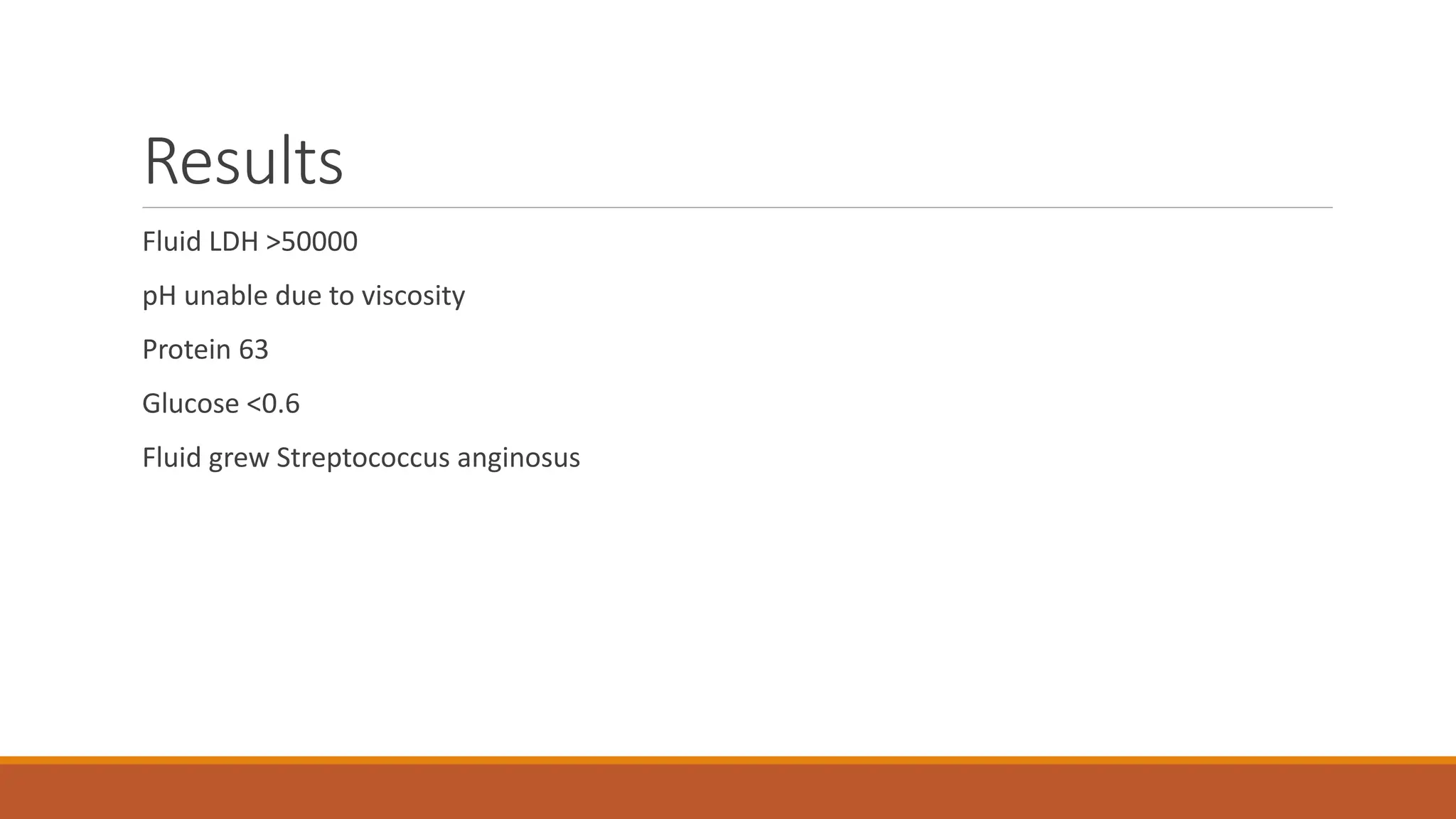
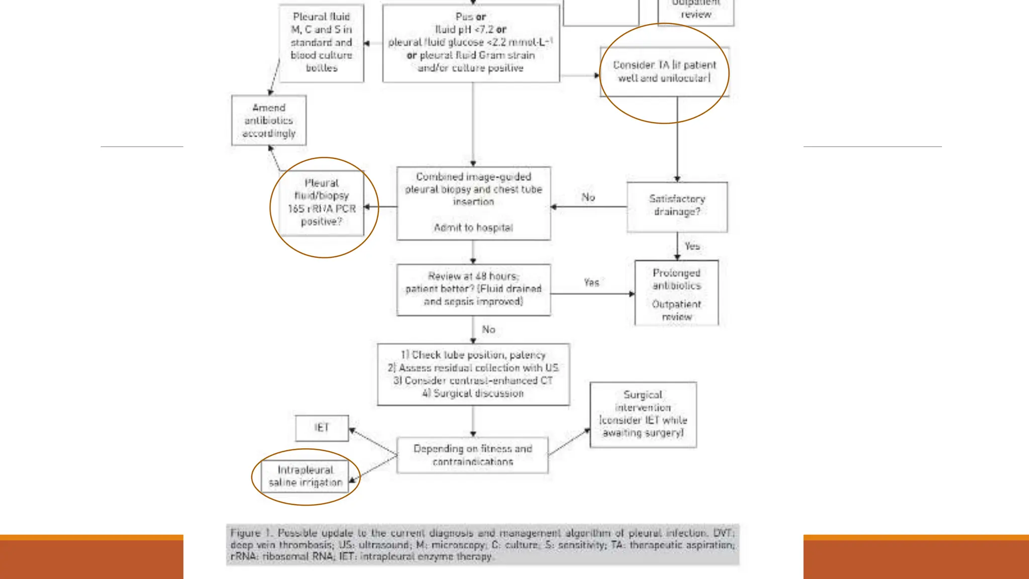
Empyema
IAN BITTINER
ST5
63 male
Normally F&W, no regular medications
Became unwell 7th Dec 2020 – cough, yellow sputum
Saw GP 23rd Dec – oral doxy
No real improvement – Clarithromycin 6/1/21
Again no improvement
Progressive loss of appetite and weight over a couple of months – given fortisip, which stopped
weight loss, but poor appetite continued
Progressive right shoulder pain and inability to lie on left side due to dyspnoea
SH
Works for government in IT
Ex-smoker, 5-10/day for 10-15 years
Lives with step daughter and her partner
Bottle wine/night up until December – nil since
On examination
OE: Acutely distressed, in significant pain and sats dropped to 80% when sat forwards, but 98%
when reclined at 60 degrees
Chest – no air entry right side
If an empyema does not rupture, death will
occur - Hippocrates
Results
Fluid LDH >50000
pH unable due to viscosity
Protein 63
Glucose <0.6
Fluid grew Streptococcus anginosus
Pleural infections - overview
From Greek, Empyein – pus producing
Incidence of pleural infection is rising
Rising most in elderly, where mortality is highest
Microbiology is inherently different from pneumonia
There has been improvement of non surgical drainage over the past decade
Surgical drainage continues to play an important role
Pleural infections - overview
Up to 57% of patients with pneumonia develop pleural effusion
The majority do not develop empyema
Simple effusions lead to increased admission, longer stays and antibiotic durations and higher
mortality
Pleural infection (complicated parapneumonic, empyema) require prompt drainage and
prolonged courses of antibiotics
Clinical presentation
Can be diverse – fevers with effusion and non-resolving pneumonia
Another pattern (more elderly) – Malaise, anorexia, weight loss (as in the case) can end up on
malignant diagnostic pathways, leading to delays
Mortality 10-20%
Risk factors
Age
Pneumonia
Alcohol/drugs
Diabetes
GORD
Poor oral hygiene
Immunosuppression
(Pleural intervention)
3 stages of Empyema
Exudative
Fibrino-purulent
Organising
These are not necessarily linear and are dependant on innumerate host-pathogen factors
Eg: patient with heavily loculated, but serous appearing effusion
Stages of Empyema
Exudative Fibrino-purulent Organising
0-14 days 7 days to 6 weeks From 2 weeks
Exudative stage (1-7 days) –
“parapneumonic effusion”
Bacterial invasion of lung parenchyma
Parenchymal inflammation leads to visceral pleural membrane permeability and leakage of
interstitial fluid
Mesothelial lining further disrupted by neutrophil infiltration, leading to mesothelial cells
releasing pro-inflammatory cytokines such as IL6, IL8 and TNF-alpha
High level of endogenous fibrinolytic enzymes
Anatomical distortion occurs, leading to increased fluid
Normal pH, glucose and culture negative
Important to recognise these early and treat
Fibrino-purulent stage (4 days – 6 weeks)
Bacteria transcend the pleural membrane
High fibrinolytic levels start to be suppressed by a rise in plasminogen activator inhibitors - PAI-1
and PAI-2
PAI-1 levels now correlate with residual pleural thickening
Progressive leukocytosis and fibrin accumulation initially at the pleural surfaces
Progressive tendency towards loculation
Bacterial and neutrophil phagocytic activity use glucose and produce lactate
LDH rises due to release from polymorphs and mononuclear cells
Organising stage (2-6+ weeks)
Fibroblasts grow onto pleural surfaces
Formation of inelastic membrane “pleural rind”
Platelet-derived growth factor and transforming growth factor beta can lead to pleural fibrosis
Pleural thickness inhibits antibiotic penetration further
Microbial biofilm occur further reducing antibiotic effectiveness
Collagen deposition in this phase reduces the effectiveness of fibrinolysis and surgery becomes
essentially inevitable
Late complications
Destruction of lung tissue, bronchopleural fistulae/pyopneumothorax
Spontaneous entry into chest wall “empyema necessitans”
Rupture into abdominal cavity
Limitations of understanding
Animal models have been largely dependant on artificial inoculation of bacteria into the pleural
space, thus bypassing the predominant route of bacterial translocation to the pleural space
Study: IV vs intranasal inoculation of S. pneumoniae in mice – intranasal formed empyema much
more quickly and reliably, suggesting direct mesothelial penetration rather than haemotogenous
spread was predominant
Microbiology
Distinct from pneumonia
Staph aureus has overtaken Strep viridans gr
Polymicrobial in estimated 23%, likely underestimate from current culture techniques
Recent metagenomics DNA studies show that primary pleural infection (without pneumonia) is more
likely polymicrobial (60%) than secondary to pneumonia (25%)
Microbiology
Gram +ve aerobes predominant in CA
Gram –ve aerobes in HA
Anaerobes hard to culture, but suited to the hypoxic pleural environment
Atypical pathogens rarely cause pleural infection
Aims of treatment
Control of infection
Expansion of lungs
Antibiotics
Penicillin and metronidazole most rapidly formed equilibrium of serum and pleural
concentrations, flowed by ceftriaxone and clindamycin (rabbit models)
It remains unclear how effective antibiotics are at preventing bacterial replication in an infected
pleural space
Drain size
Evidence suggest small drains with adequate flushing are probably non-inferior to larger drains
I’d still put in an 18Fr
2nd Multicentre Intrapleural Sepsis Trial
(MIST2)
NEJM 2011
210 patients with empyema
Randomised to 1 of 4 arms: double placebo, tpa 10mg bd, dnase 5mg bd, tpa and dnase
Primary outcome measure was reduction of opacification of hemithorax on day 7 CXR compared
to baseline
Secondary outcomes: referral for surgery, duration of hospital stay and adverse events
Eligibility
Evidence of infection (fever/high inflammatory markers)
AND one of
Purulent fluid macroscopically
Positive gram stain or culture
pH <7.2
Outcomes
Only tpa and DNase group shows significance
-29.5% vs -17.2% change in opacification of chest radiograph
77% reduction in referrals for thoracic surgery
6.7 day reduction in inpatient stay
Challenges
Rapid inactivation of enzymes by inhibitors inc plasminogen activator inhibitor 1. High levels of
this increase loculation, and dramatically effect outcome of MIST2 protocol
Future
◦ Personalised dosing of enzymes based on pleural fluid inhibitor levels?
◦ PAI-1 neutrolising antibodies?
◦ LTI-01 – a single chain urokinase plasminogen activator activator resistant to PAI-1 shows bioavailability
beyond 24 hours
Better prevention? – STOPPE trial.
Steroid therapy and outcome of parapneumonic pleural effusions
Hypothesis: Initial exudative stage could be due to exaggerated pleural inflammatory response
Rationale: Paediatric trial which showed reduced time to recovery in simple parapneumonic
effusions in children with viral pneumonia
Rationale 2: Observation of reduced numbers of empyema in patients with COPD ?steroid effect
Currently recruiting RCT looking at their role
Alternative strategies
Pleural irrigation Trial (2015) – single centre in Bristol 250ml saline infused via gravity tds vs 30ml
flush tds via 12Fr tube for 3/7
All patients put on suction (up to 20cmH20)
This showed a reduction in effusion size on CT and a reduction in referral for surgery
Multicentre RCT planned
◦ Role could be in patients who are not fit for surgery and in whom fibrinolysis is contraindicated
Surgery - VATS
RCTs show 80% can be managed medically
VATS has widened the number of patients suitable for surgery
Current guidance is for usage in treatment failure or advanced cases
In advanced cases VATS treatment failure increases with conversion to decortication up to 61%
VATS decortication mortality 2-6%
Complication rate 9-40%
Persistent air leak, pain, bleeding, infection, residual pleural space
No data for best time for surgery
Fibrinolysis v VATS
Multiple trials ongoing to investigate
MIST 3 – cannot see protocol
DICE – IR guided drain + fibrinolysis vs VATS as primary treatment for empyema
v
How long are fibrinolytics retained in the
pleural space?
1-2 hours
2-4 hours
4-6 hours
6-8 hours
8-12 hours
How long are fibrinolytics retained in the
pleural space?
1-2 hours
2-4 hours
4-6 hours
6-8 hours
8-12 hours
References
Bedawi EO, Rahman NM. Pleural infection: moving from treatment to prevention. In Maskell
Nam Laursen CB, Lee YCG et al. eds Pleural Disease (ERS Monograph). Sheffield, European
Respiratory Society, 2020; pp. 155-171

Empyema[174].pptx,and management and approch

  • 1.
  • 2.
    63 male Normally F&W,no regular medications Became unwell 7th Dec 2020 – cough, yellow sputum Saw GP 23rd Dec – oral doxy No real improvement – Clarithromycin 6/1/21 Again no improvement Progressive loss of appetite and weight over a couple of months – given fortisip, which stopped weight loss, but poor appetite continued Progressive right shoulder pain and inability to lie on left side due to dyspnoea
  • 3.
    SH Works for governmentin IT Ex-smoker, 5-10/day for 10-15 years Lives with step daughter and her partner Bottle wine/night up until December – nil since
  • 4.
    On examination OE: Acutelydistressed, in significant pain and sats dropped to 80% when sat forwards, but 98% when reclined at 60 degrees Chest – no air entry right side
  • 6.
    If an empyemadoes not rupture, death will occur - Hippocrates
  • 15.
    Results Fluid LDH >50000 pHunable due to viscosity Protein 63 Glucose <0.6 Fluid grew Streptococcus anginosus
  • 16.
    Pleural infections -overview From Greek, Empyein – pus producing Incidence of pleural infection is rising Rising most in elderly, where mortality is highest Microbiology is inherently different from pneumonia There has been improvement of non surgical drainage over the past decade Surgical drainage continues to play an important role
  • 17.
    Pleural infections -overview Up to 57% of patients with pneumonia develop pleural effusion The majority do not develop empyema Simple effusions lead to increased admission, longer stays and antibiotic durations and higher mortality Pleural infection (complicated parapneumonic, empyema) require prompt drainage and prolonged courses of antibiotics
  • 18.
    Clinical presentation Can bediverse – fevers with effusion and non-resolving pneumonia Another pattern (more elderly) – Malaise, anorexia, weight loss (as in the case) can end up on malignant diagnostic pathways, leading to delays Mortality 10-20%
  • 19.
    Risk factors Age Pneumonia Alcohol/drugs Diabetes GORD Poor oralhygiene Immunosuppression (Pleural intervention)
  • 20.
    3 stages ofEmpyema Exudative Fibrino-purulent Organising These are not necessarily linear and are dependant on innumerate host-pathogen factors Eg: patient with heavily loculated, but serous appearing effusion
  • 21.
    Stages of Empyema ExudativeFibrino-purulent Organising 0-14 days 7 days to 6 weeks From 2 weeks
  • 22.
    Exudative stage (1-7days) – “parapneumonic effusion” Bacterial invasion of lung parenchyma Parenchymal inflammation leads to visceral pleural membrane permeability and leakage of interstitial fluid Mesothelial lining further disrupted by neutrophil infiltration, leading to mesothelial cells releasing pro-inflammatory cytokines such as IL6, IL8 and TNF-alpha High level of endogenous fibrinolytic enzymes Anatomical distortion occurs, leading to increased fluid Normal pH, glucose and culture negative Important to recognise these early and treat
  • 23.
    Fibrino-purulent stage (4days – 6 weeks) Bacteria transcend the pleural membrane High fibrinolytic levels start to be suppressed by a rise in plasminogen activator inhibitors - PAI-1 and PAI-2 PAI-1 levels now correlate with residual pleural thickening Progressive leukocytosis and fibrin accumulation initially at the pleural surfaces Progressive tendency towards loculation Bacterial and neutrophil phagocytic activity use glucose and produce lactate LDH rises due to release from polymorphs and mononuclear cells
  • 24.
    Organising stage (2-6+weeks) Fibroblasts grow onto pleural surfaces Formation of inelastic membrane “pleural rind” Platelet-derived growth factor and transforming growth factor beta can lead to pleural fibrosis Pleural thickness inhibits antibiotic penetration further Microbial biofilm occur further reducing antibiotic effectiveness Collagen deposition in this phase reduces the effectiveness of fibrinolysis and surgery becomes essentially inevitable
  • 25.
    Late complications Destruction oflung tissue, bronchopleural fistulae/pyopneumothorax Spontaneous entry into chest wall “empyema necessitans” Rupture into abdominal cavity
  • 26.
    Limitations of understanding Animalmodels have been largely dependant on artificial inoculation of bacteria into the pleural space, thus bypassing the predominant route of bacterial translocation to the pleural space Study: IV vs intranasal inoculation of S. pneumoniae in mice – intranasal formed empyema much more quickly and reliably, suggesting direct mesothelial penetration rather than haemotogenous spread was predominant
  • 27.
    Microbiology Distinct from pneumonia Staphaureus has overtaken Strep viridans gr Polymicrobial in estimated 23%, likely underestimate from current culture techniques Recent metagenomics DNA studies show that primary pleural infection (without pneumonia) is more likely polymicrobial (60%) than secondary to pneumonia (25%)
  • 28.
    Microbiology Gram +ve aerobespredominant in CA Gram –ve aerobes in HA Anaerobes hard to culture, but suited to the hypoxic pleural environment Atypical pathogens rarely cause pleural infection
  • 29.
    Aims of treatment Controlof infection Expansion of lungs
  • 30.
    Antibiotics Penicillin and metronidazolemost rapidly formed equilibrium of serum and pleural concentrations, flowed by ceftriaxone and clindamycin (rabbit models) It remains unclear how effective antibiotics are at preventing bacterial replication in an infected pleural space
  • 31.
    Drain size Evidence suggestsmall drains with adequate flushing are probably non-inferior to larger drains I’d still put in an 18Fr
  • 32.
    2nd Multicentre IntrapleuralSepsis Trial (MIST2) NEJM 2011 210 patients with empyema Randomised to 1 of 4 arms: double placebo, tpa 10mg bd, dnase 5mg bd, tpa and dnase Primary outcome measure was reduction of opacification of hemithorax on day 7 CXR compared to baseline Secondary outcomes: referral for surgery, duration of hospital stay and adverse events
  • 33.
    Eligibility Evidence of infection(fever/high inflammatory markers) AND one of Purulent fluid macroscopically Positive gram stain or culture pH <7.2
  • 34.
    Outcomes Only tpa andDNase group shows significance -29.5% vs -17.2% change in opacification of chest radiograph 77% reduction in referrals for thoracic surgery 6.7 day reduction in inpatient stay
  • 35.
    Challenges Rapid inactivation ofenzymes by inhibitors inc plasminogen activator inhibitor 1. High levels of this increase loculation, and dramatically effect outcome of MIST2 protocol Future ◦ Personalised dosing of enzymes based on pleural fluid inhibitor levels? ◦ PAI-1 neutrolising antibodies? ◦ LTI-01 – a single chain urokinase plasminogen activator activator resistant to PAI-1 shows bioavailability beyond 24 hours
  • 36.
    Better prevention? –STOPPE trial. Steroid therapy and outcome of parapneumonic pleural effusions Hypothesis: Initial exudative stage could be due to exaggerated pleural inflammatory response Rationale: Paediatric trial which showed reduced time to recovery in simple parapneumonic effusions in children with viral pneumonia Rationale 2: Observation of reduced numbers of empyema in patients with COPD ?steroid effect Currently recruiting RCT looking at their role
  • 37.
    Alternative strategies Pleural irrigationTrial (2015) – single centre in Bristol 250ml saline infused via gravity tds vs 30ml flush tds via 12Fr tube for 3/7 All patients put on suction (up to 20cmH20) This showed a reduction in effusion size on CT and a reduction in referral for surgery Multicentre RCT planned ◦ Role could be in patients who are not fit for surgery and in whom fibrinolysis is contraindicated
  • 38.
    Surgery - VATS RCTsshow 80% can be managed medically VATS has widened the number of patients suitable for surgery Current guidance is for usage in treatment failure or advanced cases In advanced cases VATS treatment failure increases with conversion to decortication up to 61% VATS decortication mortality 2-6% Complication rate 9-40% Persistent air leak, pain, bleeding, infection, residual pleural space No data for best time for surgery
  • 39.
    Fibrinolysis v VATS Multipletrials ongoing to investigate MIST 3 – cannot see protocol DICE – IR guided drain + fibrinolysis vs VATS as primary treatment for empyema
  • 41.
  • 42.
    How long arefibrinolytics retained in the pleural space? 1-2 hours 2-4 hours 4-6 hours 6-8 hours 8-12 hours
  • 43.
    How long arefibrinolytics retained in the pleural space? 1-2 hours 2-4 hours 4-6 hours 6-8 hours 8-12 hours
  • 44.
    References Bedawi EO, RahmanNM. Pleural infection: moving from treatment to prevention. In Maskell Nam Laursen CB, Lee YCG et al. eds Pleural Disease (ERS Monograph). Sheffield, European Respiratory Society, 2020; pp. 155-171