Pathology of respiratory system
•
12 likes•1,777 views

This document summarizes various pathologies that can affect the respiratory system. It begins by discussing various diseases that can cause inflammation in different parts of the respiratory tract, such as epistaxis (nose bleeding), rhinitis (inflammation of the nasal mucosa), sinusitis (inflammation of the sinuses), laryngitis (inflammation of the larynx), and tracheitis (inflammation of the trachea). It then discusses pathologies that can affect the lungs, such as different types of pneumonia (suppurative bronchopneumonia, fibrinous bronchopneumonia, interstitial pneumonia, and embolic pneumonia), pulmonary edema, atelectasis, and pulmonary emphysema. It provides descriptions

Recommended
More Related Content
What's hot
What's hot (20)
Similar to Pathology of respiratory system
Similar to Pathology of respiratory system (20)
Recently uploaded
Recently uploaded (20)
Pathology of respiratory system
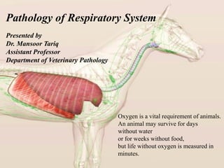
- 1. Pathology of Respiratory System Oxygen is a vital requirement of animals. An animal may survive for days without water or for weeks without food, but life without oxygen is measured in minutes. Presented by Dr. Mansoor Tariq Assistant Professor Department of Veterinary Pathology
- 3. TRANSITIONAL REGION Respiratory bronchioles Best developed in carnivores & primates First level at which gas exchange occurs Primarily air passage ways GAS EXCHANGE Alveolar ducts Alveolar sacs Alveoli
- 4. 1) Pathology of the nasal cavity 1) Epistaxis 2) Rhinitis 3) Sinusitis 2) Laryngitis 3) Tracheitis 4) Bronchi 5) Bronchioles 6) Lungs (Pneumonia) Pathology of Respiratory System
- 5. EPISTAXIS is bleeding from nasal passage due to trauma, neoplasm and ulcerative lesions as a result of infections. Hemoptysis blood comes from mouth, saliva or sputum. hemorrhage from the nose, usually due to rupture of small vessels Minor bleeding may be caused by a blow on the nose, irritation from foreign bodies. Exercise induced pulmonary hemorrhage in horses.
- 6. RHINITIS (inflammation of the nasal mucosa) may be caused by infections or allergies. & Acute infectious rhinitis is most often caused by viruses and is a typical manifestation of the common cold. Acute rhinitis is present in certain infectious diseases such as strangles, influenza and glanders of horses, and distemper in dog.
- 12. SINUSITIS is the inflammation of sinuses Frontal sinusitis is usually associated with dehorning, and maxillary sinusitis with infected teeth.
- 13. e.g. Frontal sinusitis in dehorned cattle. The larvae of botfly Oestrus ovis enters in nasal passage and migrate upto frontal sinuses and turbinate bones and cause mucopurulent inflammation. Similarly leeches (Dinobdella ferox) is known to cause nasal cavity inflammation in domestic animals and suck blood.
- 15. LARYNGITIS inflammatory condition of larynx Necrotic laryngitis ("calf diphtheria") is common in feedlots. predominantly in calves, usually >3 months old. (calf diphtheria) Diphtheria means leather, due to the type of exudate that forms. In truth. the pus that forms in affected cattle is more like curds.
- 17. TRACHEITIS is the inflammation of trachea. • In canines, it is tracheobronchitis and caused by adenovirus, influenza virus and herpes virus. • while in poultry it is manifested by laryngo-tracheitis is caused by herpes virus. OTHER DISEASE INVOLVED LARYNX AND TRACHEA • Calf Diphtheria fusiformis necrophorus • Infectious bovine rhinotracheitis (IBR) caused by a bovine herpesvirus. • Feline viral rhinotracheitis caused by feline herpesvirus
- 18. Avian infectious laryngo-tracheitis (ILT) In poultry, hemorrhage in trachea and caseous plug in trachea towards larynx causing obstruction.
- 20. Inflammation of the bronchi (bronchitis) and bronchioles (bronchiolitis) The larger bronchi, well supplied mucous-secreting and ciliated cells . The lumen is large enough to remain healthy even in the presence of abundant exudate and such exudate can be expelled by an effective cough reflex. Bronchioles walls are thin and the Small lumens are easily blocked by exudate which may be too far away for the cough reflex to be effective.
- 21. BRONCHITIS is the inflammation of bronchi, characterized by catarrhal, suppurative, fibrinous or haemorrhagic exudate. Normal Bronchial wall Inflamed Bronchial wall Tracheobronchitis is an acute or chronic inflammation of the trachea and bronchial airways; it may also extend into the lungs.
- 22. Chronic bronchitis in cattle and sheep is parasitic infection. e.g Dictyocaulus filaria in sheep/goat Dictyocaulus vivparus in ox.
- 24. smooth muscle in the wall degenerates, mucosa becomes thinner and the walls are destroyed and replaced by fibrous connective tissue
- 25. The surface area of the lungs is roughly the same size as a tennis court. The capillaries in the lungs would extend 1,600 km if you placed end to end Everyday we lose half a liter of water through breathing.
- 27. Lungs of horse, sheep, ox, and pig. L, left; R, right; A, cranial (apical); C, middle (cardiac); D, caudal (diaphragmatic); I, accessory (intermediate).
- 29. ATELECTASIS (Collapse) Atelectasis is the failure of alveoli to open or the alveoli are collapsed and thus do not have air. may affect a part of lobe or two or all of one lung. ➢ Common cause of atelectasis is a blockage of one of the tubes (bronchi) that branch off from the trachea (windpipe) and lead to the lung tissue. ➢ The blockage may be caused by something inside the bronchus, such as a plug of mucus, a tumor, or an inhaled foreign object. ➢ When a bronchus or a smaller airway (bronchiole) becomes blocked, the air in the alveoli beyond the blockage is absorbed into the bloodstream, causing the alveoli to shrink and collapse.
- 32. PULMONARY EMPHYSEMA Emphysema is the increase in amount of air in lungs characterized by over dilation of the alveoli. It may be acute or chronic and focal or generalized. Pathogenesis is not fully understood but there is 2 possibilities 1. May be degradation and weakening of the interstitium by proteolytic enzymes, e.g. elastase, released by inflammatory cells. 2. More common, the condition develops secondary to either chronic bronchitis and bronchiolitis, which cause obstruction of airways on expiration but still allow air to enter alveoli.
- 36. PULMONARY EMPHYSEMA Two major forms of pulmonary emphysema occur in the lungs 1. Alveolar (vascular) emphysema 2. Interstitial (interlobular) emphysema There are several conditions in which enlargement of airspaces is not accompanied by destruction; is called overinflation.
- 37. 1. Alveolar (Vascular) emphysema In alveolar emphysema the alveoli are distended by excessive amounts of air pressure and often times rupture. Alveolar membrane disturb
- 38. 2. Interstitial (interlobular) emphysema In interstitial emphysema the excessive air accumulates in the sub- pleural, interstitial, and interlobular regions of the lungs. when air enter in the supporting connective tissue of the lungs, (Mediastial, Subcutaneous tissues, sub pleural, interstitial spaces). May occur with a sudden increase in intra alveolar pressure cause a tear in alveolar spaces, break the alveolar sac and air come out in the tissue space of the lung (i.e. interlobular or pleural space).
- 39. Atelectasis
- 41. PULMONARY EDEMA is accumulation of serous fluid in alveoli of lungs. ➢ Characterized by accumulation of fluid in interstitium and alveoli. ➢ Failure of the left heart especially results in pulmonary edema - the pulmonary veins have seriously increased hydrostatic pressure. Normally lung produces fluid (transudate) that is rapidly removed by the lymphatic system. when fluid production exceeds lymphatic system unable to remove cause the pulmonary edema.
- 43. In heart-related pulmonary edema, when the heart’s left ventricle is not able to pump out enough of the blood it receives from the lungs, pressure increases in the left atrium, and then in the blood vessels of the lungs, causing fluid to be pushed into the lung’s air sacs.
- 44. Pulmonary Edema The lungs appear wet, heavy and fail to collapse when the thorax is opened.
- 46. PLEURITIS is the inflammation of pleura characterized by serous, fibrinous or purulent exudate. It is also known as pleurisy.
- 47. Pneumothorax Is the presence of gas or air in the thorax cavity. • Air can leak in from outside (loss of continuity of body wall) or from inside (break in visceral pleura). • Air can really compress the lungs, causing atelectasis.
- 48. Increased vascular permeability transudation of fluid body tissues (e.g. brain and lungs) and body cavities (e.g. pericardial and thoracic cavities), but mechanisms responsible for the transudation are poorly understood. Hydrothorax Hydrothorax is the accumulation of abnormal quantities of transudate (serous fluid) in thoracic cavity. Causes include congestive heart failure, Hypoproteinemia (liver, renal, intestinal disease, starvation) and lymphatic obstruction.
- 49. Hemothorax is the presence of free blood in thoracic cavity. • It is caused by severe trauma or penetrating wound into the lungs, rupture of major blood vessels (aneurism), coagulopathies. • Lesions: blood in thorax cavity.
- 50. Pyothorax is the presence of purulent material filled in thorax cavity. • Pyothorax usually seen in the dog and cats, result of bacterial infection due to injury. • Unilateral pyothorax, where one lung is affected. • Suppurative exudate present in thorax
- 51. Chylothorax ▪ is the presence of free lymph (chyle) in the thorax and it is caused by rupture of a major lymphatic duct of thorax cavity. ▪ Most common causes are thoracic trauma and neoplasia. There is also an idiopathic form of chylotorax in dogs. ▪ Lesions: Milky fluid in thorax cavity. For diagnosis, submit fluid to clinical pathology – it should be high in triglycerides and lymphocytes.
- 52. Salamander Lizard PigeonPrimate Nostrils, mouth, and throat Trachea Lung Air sac Vertebrate Lungs
- 54. PNEUMONIA "inflammation of the lung caused by bacteria, in which the air sacs (alveoli) become filled with inflammatory cells and the lungs become solid" Pneumonia is "a severe form of acute lower respiratory infection that specifically affects the lungs". During a Pneumonia infection, the alveoli of one or both lungs fill up with pus or fluid. This increases the labor of breathing, and thus gaseous exchange cannot occur as it normally would. Pneumonia is due to infections caused primarily by 1. bacteria 2. viruses 3. Fungi 4. Parasites Causes of Pneumonia primarily by bacteria or viruses and less commonly by fungi and parasites. Young animals are more susceptible to the pneumonia rather than adult.
- 55. Stages of Pneumonia Pneumonia has four stages of inflammatory response, 1. Congestion 2. Red hepatization 3. Grey hepatization 4. Resolution
- 56. Congestion • This phase represent the acute inflammatory response to bacterial infection. • Occurs in the first 24 to 48 hours • Capillaries in the surrounding alveolar walls become dilated and congested • Cellular exudates containing neutrophils, lymphocytes and fibrin replaces the alveolar air • The infections spreads to the hilum and pleura fairly rapidly • Pleurisy occurs • Grossly, the lung is heavy, enlarge and hyperemic r
- 57. Red Hepatization • Occurs in the 2-4 days after congestion • At this point the consistency of the lungs resembles that of the liver • The lungs become hyperemic • Alveolar capillaries are engorged with blood which shows the red appearance of lungs. • Fibrinous exudates fill the alveoli • This stage is "characterized by the presence of many erythrocytes, neutrophils, desquamated epithelial cells, and fibrin within the alveoli"
- 58. Grey Hepatization • Occurs in the 4-8 days after Red Hepatization • This is an avascular stage • The lung appears "gray-brown to yellow because of fibrinopurulent exudates, disintegration of red cells, and hemosiderin" • The pressure of the exudates in the alveoli causes compression of the capillaries • "Leukocytes migrate into the congested alveoli"
- 59. Resolution • It start with 8th day and completed in next 3 weeks. • Characterized by the "resorption and restoration of the pulmonary architecture" • A large number of macrophages enter the alveolar spaces • Phagocytosis of the bacteria-laden leucocytes occurs • "Fibrinous inflammation may extend to and across the pleural space and it may lead to resolution or to organization and pleural adhesions“ • Alveolar capillaries are engorged, progressive removal of cellular exudate, resulting in restoration of normal lung parenchyma with ventilation.
- 60. Pathogens Stressors Viruses Mycoplasma Bacteria Upper respiratory disease Damage Pneumonia Physical Environmental Immune System
- 61. PNEUMONIA is an infection that inflames the air sacs in one or both lungs. The gross appearance of the pneumonia will help you to classify it into one of the four categories below, which in turn will help you to think about the etiology, and consequently the treatment and/or prognosis. 1. Suppurative Bronchopnemonia 2. Fibrinous Bronchopneumonia 3. Interstitial pneumonia 4. Embolic pneumonia 5. Granulomatous pneumonia
- 63. Distribution of lesions in Pneumonias
- 64. Suppurative Bronchopneumonia Bronchopneumonia starts in the airways. It may be an extension from a bronchitis or it may be a primary inflammation at the bronchiole-alveolar junction. • Aerogenous route • Cranioventral distribution • (Acute) Red in color to grey (Chronic) • Purulent exudate in bronchi, and abscesses
- 66. The cranio-ventral areas are dark, consolidated, and on the left there is even some roughening on the surface that might be fibrin. A bacteria has come in through the airways and started this disturbance. Suppurative Bronchopneumonia a classic picture of severe bronchopneumonia in a dog.
- 67. Pulmonary abscesses and bronchiectasis are two important results of the suppurative bronchopneumonia. Note: several large abscesses in the consolidated cranial and intermediate lobes are seen whereas the Caudal lobes are essential normal Suppurative Bronchopneumonia
- 69. Note cranio- ventral consolidation affected. Affected lung is covered with fibrin. Several injury to the lung with leakage of fibrin into the airspaces. Only small portion of the lung appears grossly normal (asterisk) Fibrinous Bronchopneumonia
- 73. These lungs came from a foal suffering from septicemia Interstitial Pneumonia Most of the alveolar lining cells are responding to damage, and that is a huge amount of surface area to be affected and so they are DIFFUSELY swollen. Inflammation of alveolar interstitium is commonly caused by injury and destruction of pneumocytes. It may be the results of viral infections, deposition of antigen-antibody complexes, inhalation of toxic gases (nitrogen dioxide, sulfur dioxide).
- 76. The port of entry in embolic Pneumonia is hematogenous. Commonly, due to • jugular thrombosis, • vegetative endocarditis (right side of heart), • rupture of hepatic abscesses into the vena cava (in bovine) • Embolic foreign body (hairs, septic emboli)
- 79. Granulomatous pneumonia Chronic, sever, granulomatous Pneumonia There are some organisms that tend to incite a predominantly macrophage response and the lungs have a difficult time overcoming the organism and so lots of macrophages accumulate in GRANULOMAS. These pneumonias are characterized by having focal granulomas scattered throughout.