The document discusses pulmonary ventilation and the steps of respiration. It describes how pulmonary ventilation occurs through the alternating contraction and relaxation of respiratory muscles that create pressure differences, causing air to flow into and out of the lungs. The key muscles involved in inhalation are the diaphragm and external intercostals, while exhalation occurs passively through elastic recoil. Factors like surface tension, lung compliance, and airway resistance also affect pulmonary ventilation.
6) transport of oxygen and carbon dioxdideAyub Abdi
lecture 6: transportaion of both gases need a hemoglobin and part of them are transported by plasma. if Hb is low the saturation of oxygen also low and leads a hypoxia, fatigue, dyspnea, etc. in other hand acidosis can occur.
6) transport of oxygen and carbon dioxdideAyub Abdi
lecture 6: transportaion of both gases need a hemoglobin and part of them are transported by plasma. if Hb is low the saturation of oxygen also low and leads a hypoxia, fatigue, dyspnea, etc. in other hand acidosis can occur.
lecture 5: it's good for as to take a breif about how does atmospheric air will pass to our lungs then to blood, for transportation and utilization of oxygen and excretion of carbon dioxide. Many issue are related when gas exchange is performed.
Like heartbeat, breathing must occur in a continuous, cyclic pattern to sustain life processes.
Inspiratory muscles must rhythmically contract and relax to alternately fill the lungs with air and empty them.
The rhythmic pattern of breathing is established by cyclic neural activity to the respiratory muscles
Regulation of respiration (the guyton and hall physiology)Maryam Fida
Normal respiration is spontaneous and unconscious.
There are 4 groups of neurons on each side in the Pons and medulla oblongata which are involved in regulation of respiration. These include
1. Medullary centers
Dorsal respiratory group of neurons
Ventral respiratory group of neurons
2. Pontine centers
Pneumotaxic centre
Apneustic centre.
It contains “I”neurons which are inspiratory neurons.
It’s located in dorsal portion of medulla oblongata.
It also includes the nucleus of tractus solitarius which is the sensory termination of afferent fibers in 9th ( GLOSSOPHARYNGEAL NERVE) and 10th (VAGUS NERVE) cranial nerves.
They receive impulses from peripheral chemoreceptors, carotid and aortic baroreceptors and also other receptors in the lungs.
In this group inspiratory ramp signals are produced spontaneously.
If we cut the medulla oblongata from other parts of brain and also the afferent nerves which enter the medulla, still inspiratory ramp signals are produced which indicate it’s the inherent property of medulla.
Initially the signal is weak and then it progressively increases and then fades away.
Each ramp signal’s duration is 2 sec and then for 3 seconds there is no ramp signal.
So each cycle lasts for 5 seconds and there are 12 cycles /minute which is the respiratory rate.
Significance of the signal in the form of ramp is that it causes progressive expansion of the lungs. After production, these ramp signals are transmitted to the contra lateral motor neurons supplying the inspiratory muscles.
Rate and duration of inspiratory ramp signals is controlled by impulses from the Pneumotaxic centre and impulses from the lungs via vagi.
Introduction to respiration and mechanics of ventilation (the guyton and hall...Maryam Fida
Respiration is the process by which oxygen is taken in and carbon dioxide is given out.
Respiration is classified into two types:
1. External respiration
It involves exchange of respiratory gases, i.e. oxygen and carbon dioxide between lungs and blood.
2. Internal respiration
It involves exchange of gases between blood and tissues.
Respiration occurs in two phases:
Inspiration during which air enters the lungs from atmosphere.
2. Expiration during which air leaves the lungs.
During normal breathing, inspiration is an active
process and expiration is a passive process.
Respiratory tract is divided into two parts:
1. Upper respiratory tract that includes all the
structures from nose up to vocal cords; vocal cords are the folds of mucous membrane within larynx that vibrates to produce the voice
2. Lower respiratory tract, which includes Larynx, trachea, bronchi and lungs.
RESPIRATORY UNIT
Respiratory unit is defined as:
“The structural and functional unit of lung”. Exchange of gases occurs only in this part of the respiratory tract.
STRUCTURE OF RESPIRATORY UNIT
1. Respiratory bronchioles
2. Alveolar ducts
3. Alveolar sacs
4. Antrum
5. Alveoli
Between the trachea and alveoli airways divide 23 times
Out of 23 divisions first 16 are just to conduct air and these divisions of airways are up to terminal bronchioles.
The last 7 divisions are for the exchange of gases and these divisions which are for exchange of gases includes respiratory bronchioles, alveolar ducts and alveoli.
There are 300 million alveoli in the lungs and the alveolar surface form s an area of 70-100 square meters
FUNCTIONS OF THE BRONCHIOLES And it's uses PDF.pdfMaryphiri7
This talks about the the function of the bronchioles and the disorders of the function of the bronchioles so in this presentation I will talk about the importance and why it is important
drugs acting on respiratory system.&pathophysiology of respiratory sys.Vicky Anthony
this ppt contains a general overview of the respiratory system,its pathophysiology and common drugs that act on respiratory system .....all these topics are covered in a short overview.
micro teaching on communication m.sc nursing.pdfAnurag Sharma
Microteaching is a unique model of practice teaching. It is a viable instrument for the. desired change in the teaching behavior or the behavior potential which, in specified types of real. classroom situations, tends to facilitate the achievement of specified types of objectives.
Flu Vaccine Alert in Bangalore Karnatakaaddon Scans
As flu season approaches, health officials in Bangalore, Karnataka, are urging residents to get their flu vaccinations. The seasonal flu, while common, can lead to severe health complications, particularly for vulnerable populations such as young children, the elderly, and those with underlying health conditions.
Dr. Vidisha Kumari, a leading epidemiologist in Bangalore, emphasizes the importance of getting vaccinated. "The flu vaccine is our best defense against the influenza virus. It not only protects individuals but also helps prevent the spread of the virus in our communities," he says.
This year, the flu season is expected to coincide with a potential increase in other respiratory illnesses. The Karnataka Health Department has launched an awareness campaign highlighting the significance of flu vaccinations. They have set up multiple vaccination centers across Bangalore, making it convenient for residents to receive their shots.
To encourage widespread vaccination, the government is also collaborating with local schools, workplaces, and community centers to facilitate vaccination drives. Special attention is being given to ensuring that the vaccine is accessible to all, including marginalized communities who may have limited access to healthcare.
Residents are reminded that the flu vaccine is safe and effective. Common side effects are mild and may include soreness at the injection site, mild fever, or muscle aches. These side effects are generally short-lived and far less severe than the flu itself.
Healthcare providers are also stressing the importance of continuing COVID-19 precautions. Wearing masks, practicing good hand hygiene, and maintaining social distancing are still crucial, especially in crowded places.
Protect yourself and your loved ones by getting vaccinated. Together, we can help keep Bangalore healthy and safe this flu season. For more information on vaccination centers and schedules, residents can visit the Karnataka Health Department’s official website or follow their social media pages.
Stay informed, stay safe, and get your flu shot today!
These simplified slides by Dr. Sidra Arshad present an overview of the non-respiratory functions of the respiratory tract.
Learning objectives:
1. Enlist the non-respiratory functions of the respiratory tract
2. Briefly explain how these functions are carried out
3. Discuss the significance of dead space
4. Differentiate between minute ventilation and alveolar ventilation
5. Describe the cough and sneeze reflexes
Study Resources:
1. Chapter 39, Guyton and Hall Textbook of Medical Physiology, 14th edition
2. Chapter 34, Ganong’s Review of Medical Physiology, 26th edition
3. Chapter 17, Human Physiology by Lauralee Sherwood, 9th edition
4. Non-respiratory functions of the lungs https://academic.oup.com/bjaed/article/13/3/98/278874
Knee anatomy and clinical tests 2024.pdfvimalpl1234
This includes all relevant anatomy and clinical tests compiled from standard textbooks, Campbell,netter etc..It is comprehensive and best suited for orthopaedicians and orthopaedic residents.
TEST BANK for Operations Management, 14th Edition by William J. Stevenson, Ve...kevinkariuki227
TEST BANK for Operations Management, 14th Edition by William J. Stevenson, Verified Chapters 1 - 19, Complete Newest Version.pdf
Pulmonary Thromboembolism - etilogy, types, medical- Surgical and nursing man...VarunMahajani
Disruption of blood supply to lung alveoli due to blockage of one or more pulmonary blood vessels is called as Pulmonary thromboembolism. In this presentation we will discuss its causes, types and its management in depth.
Lung Cancer: Artificial Intelligence, Synergetics, Complex System Analysis, S...Oleg Kshivets
RESULTS: Overall life span (LS) was 2252.1±1742.5 days and cumulative 5-year survival (5YS) reached 73.2%, 10 years – 64.8%, 20 years – 42.5%. 513 LCP lived more than 5 years (LS=3124.6±1525.6 days), 148 LCP – more than 10 years (LS=5054.4±1504.1 days).199 LCP died because of LC (LS=562.7±374.5 days). 5YS of LCP after bi/lobectomies was significantly superior in comparison with LCP after pneumonectomies (78.1% vs.63.7%, P=0.00001 by log-rank test). AT significantly improved 5YS (66.3% vs. 34.8%) (P=0.00000 by log-rank test) only for LCP with N1-2. Cox modeling displayed that 5YS of LCP significantly depended on: phase transition (PT) early-invasive LC in terms of synergetics, PT N0—N12, cell ratio factors (ratio between cancer cells- CC and blood cells subpopulations), G1-3, histology, glucose, AT, blood cell circuit, prothrombin index, heparin tolerance, recalcification time (P=0.000-0.038). Neural networks, genetic algorithm selection and bootstrap simulation revealed relationships between 5YS and PT early-invasive LC (rank=1), PT N0—N12 (rank=2), thrombocytes/CC (3), erythrocytes/CC (4), eosinophils/CC (5), healthy cells/CC (6), lymphocytes/CC (7), segmented neutrophils/CC (8), stick neutrophils/CC (9), monocytes/CC (10); leucocytes/CC (11). Correct prediction of 5YS was 100% by neural networks computing (area under ROC curve=1.0; error=0.0).
CONCLUSIONS: 5YS of LCP after radical procedures significantly depended on: 1) PT early-invasive cancer; 2) PT N0--N12; 3) cell ratio factors; 4) blood cell circuit; 5) biochemical factors; 6) hemostasis system; 7) AT; 8) LC characteristics; 9) LC cell dynamics; 10) surgery type: lobectomy/pneumonectomy; 11) anthropometric data. Optimal diagnosis and treatment strategies for LC are: 1) screening and early detection of LC; 2) availability of experienced thoracic surgeons because of complexity of radical procedures; 3) aggressive en block surgery and adequate lymph node dissection for completeness; 4) precise prediction; 5) adjuvant chemoimmunoradiotherapy for LCP with unfavorable prognosis.
ARTIFICIAL INTELLIGENCE IN HEALTHCARE.pdfAnujkumaranit
Artificial intelligence (AI) refers to the simulation of human intelligence processes by machines, especially computer systems. It encompasses tasks such as learning, reasoning, problem-solving, perception, and language understanding. AI technologies are revolutionizing various fields, from healthcare to finance, by enabling machines to perform tasks that typically require human intelligence.
Recomendações da OMS sobre cuidados maternos e neonatais para uma experiência pós-natal positiva.
Em consonância com os ODS – Objetivos do Desenvolvimento Sustentável e a Estratégia Global para a Saúde das Mulheres, Crianças e Adolescentes, e aplicando uma abordagem baseada nos direitos humanos, os esforços de cuidados pós-natais devem expandir-se para além da cobertura e da simples sobrevivência, de modo a incluir cuidados de qualidade.
Estas diretrizes visam melhorar a qualidade dos cuidados pós-natais essenciais e de rotina prestados às mulheres e aos recém-nascidos, com o objetivo final de melhorar a saúde e o bem-estar materno e neonatal.
Uma “experiência pós-natal positiva” é um resultado importante para todas as mulheres que dão à luz e para os seus recém-nascidos, estabelecendo as bases para a melhoria da saúde e do bem-estar a curto e longo prazo. Uma experiência pós-natal positiva é definida como aquela em que as mulheres, pessoas que gestam, os recém-nascidos, os casais, os pais, os cuidadores e as famílias recebem informação consistente, garantia e apoio de profissionais de saúde motivados; e onde um sistema de saúde flexível e com recursos reconheça as necessidades das mulheres e dos bebês e respeite o seu contexto cultural.
Estas diretrizes consolidadas apresentam algumas recomendações novas e já bem fundamentadas sobre cuidados pós-natais de rotina para mulheres e neonatos que recebem cuidados no pós-parto em unidades de saúde ou na comunidade, independentemente dos recursos disponíveis.
É fornecido um conjunto abrangente de recomendações para cuidados durante o período puerperal, com ênfase nos cuidados essenciais que todas as mulheres e recém-nascidos devem receber, e com a devida atenção à qualidade dos cuidados; isto é, a entrega e a experiência do cuidado recebido. Estas diretrizes atualizam e ampliam as recomendações da OMS de 2014 sobre cuidados pós-natais da mãe e do recém-nascido e complementam as atuais diretrizes da OMS sobre a gestão de complicações pós-natais.
O estabelecimento da amamentação e o manejo das principais intercorrências é contemplada.
Recomendamos muito.
Vamos discutir essas recomendações no nosso curso de pós-graduação em Aleitamento no Instituto Ciclos.
Esta publicação só está disponível em inglês até o momento.
Prof. Marcus Renato de Carvalho
www.agostodourado.com
These lecture slides, by Dr Sidra Arshad, offer a quick overview of physiological basis of a normal electrocardiogram.
Learning objectives:
1. Define an electrocardiogram (ECG) and electrocardiography
2. Describe how dipoles generated by the heart produce the waveforms of the ECG
3. Describe the components of a normal electrocardiogram of a typical bipolar leads (limb II)
4. Differentiate between intervals and segments
5. Enlist some common indications for obtaining an ECG
Study Resources:
1. Chapter 11, Guyton and Hall Textbook of Medical Physiology, 14th edition
2. Chapter 9, Human Physiology - From Cells to Systems, Lauralee Sherwood, 9th edition
3. Chapter 29, Ganong’s Review of Medical Physiology, 26th edition
4. Electrocardiogram, StatPearls - https://www.ncbi.nlm.nih.gov/books/NBK549803/
5. ECG in Medical Practice by ABM Abdullah, 4th edition
6. ECG Basics, http://www.nataliescasebook.com/tag/e-c-g-basics
New Drug Discovery and Development .....NEHA GUPTA
The "New Drug Discovery and Development" process involves the identification, design, testing, and manufacturing of novel pharmaceutical compounds with the aim of introducing new and improved treatments for various medical conditions. This comprehensive endeavor encompasses various stages, including target identification, preclinical studies, clinical trials, regulatory approval, and post-market surveillance. It involves multidisciplinary collaboration among scientists, researchers, clinicians, regulatory experts, and pharmaceutical companies to bring innovative therapies to market and address unmet medical needs.
Prix Galien International 2024 Forum ProgramLevi Shapiro
June 20, 2024, Prix Galien International and Jerusalem Ethics Forum in ROME. Detailed agenda including panels:
- ADVANCES IN CARDIOLOGY: A NEW PARADIGM IS COMING
- WOMEN’S HEALTH: FERTILITY PRESERVATION
- WHAT’S NEW IN THE TREATMENT OF INFECTIOUS,
ONCOLOGICAL AND INFLAMMATORY SKIN DISEASES?
- ARTIFICIAL INTELLIGENCE AND ETHICS
- GENE THERAPY
- BEYOND BORDERS: GLOBAL INITIATIVES FOR DEMOCRATIZING LIFE SCIENCE TECHNOLOGIES AND PROMOTING ACCESS TO HEALTHCARE
- ETHICAL CHALLENGES IN LIFE SCIENCES
- Prix Galien International Awards Ceremony
2. z
TOPICS OF
DISCUSSION
PULMONARY VENTILATION
INTERNAL AND EXTERNAL RESPIRATION
EXCHANGE AND TRANSPORT OF OXYGEN
AND CARBONDIOXIDE
REPIRATORY CONTROL CENTERS IN BRAIN
LUNG CAPACITIES AND LUNG VOLUMES
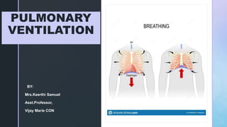
3. z
INTRODUCTION
RESPIRATION:
The process of exchange of gases
in the body . The following are the main
steps:
PULMONARY VENTILATION: or
BREATHING is the inhalation and
exhalation of the air and involves the
exchange of air between the
atmosphere and the alveoli of the
lungs.
4. z
RESPIRATION STEPS CONTD…..
1. EXTERNAL (PULMONARY ) RESPIRATION : exchange of gases between the
alveoli and blood in the pulmonary capillaries across the respiratory membrane. In
this process the capillary blood gains oxygen and loss carbon dioxide.
2. INTERNAL (TISSUE ) RESPIRATION: the exchange of gases between blood in
systemic capillaries and tissue cells.in this step the blood looses oxygen and gains
carbon dioxide.
3. CELLULAR RESPIRATION: within the cells the metabolic reaction that consume
o2 and gives off co2 during the production of ATP are termed as CELLULAR
RESPIRATION.
5.
6. z
PULMONARY VENTILATION
In the pulmonary ventilation air flows between the atmosphere and the
alveoli of the lungs because of the alternating pressure differences
created by the contraction and relaxation of the respiratory muscles.
Air moves into the lungs when the air pressure inside the lungs is less
than the air pressure in the atmosphere.
Air moves out of the lungs when the air pressure inside the lungs is
greater than the air pressure in the atmosphere.
9. z
INHALATION
Just before respiration air pressure
in the lungs and atmosphere is the
same ( 760mmhg)
For the air to flow into the lungs the
pressure inside the lungs should be
less than the atmospheric pressure.
This happens when the size of the
size of the lungs increases
following the Boyles law.
BREATHING IN
IS CALLED
INHALATION
11. z
INHALATION-DIAPHRAGM
The most important muscle of inhalation
is the DIAPHRAGM- a dome shaped
skeletal muscle that forms the floor of
the thoracic cavity.
It is innervated by fibers of the PHRENIC
NERVES which emerge from the spinal
cord at cervical levels 3,4 and 5 .
Contraction of diaphragm causes it to
flatten lowering its dome
This increases the vertical diameter of
the thoracic cavity.
12. z
INHALATION
Flattening of diaphragm
increases the vertical diameter of
the thoracic cavity.
During quiet inhalation the
diaphragm descends about 1cm
reducing 1-3mmhg pressure
thereby inhaling 500ml of air.
In strenuous breathing the
diaphragm descends 10cms
which produces a pressure
difference of 100mmhg and
inhale 2-3lit of air.
13. z
INHALATION
Flattening of diaphragm
increases the vertical diameter of
the thoracic cavity.
During quiet inhalation the
diaphragm descends about 1cm
reducing 1-3mmhg pressure
thereby inhaling 500ml of air.
In strenuous breathing the
diaphragm descends 10cms
which produces a pressure
difference of 100mmhg and
inhale 2-3lit of air.
14. z
INHALATION
Contraction of diaphragm is
responsible for 75% of the air
that enters the lungs during quiet
breathing.
Advanced pregnancy and
obesity or confining abdominal
clothing can prevent complete
decent of the diaphragm.
15. z
INHALATION-
EXTERNALINTERCOSALS
The next important muscles of
inspiration.
When these muscles contract they
elevate the ribs.
Elevation increases the anteroposterior
diameter and lateral diameters of the
chest cavity.
Contraction of external intercostals is
responsible for 25% of air that enters
into the lungs during normal quiet
breathing
16. z
During quiet inhalations the pressure between the two membranes that is the
INTRAPLEURAL PRESSURE is always sub atmospheric.
Just before inhalation it is about 4mmhg less than the atmospheric pressure
about 756mmhg at an atmospheric pressure of 760mm hg.
As the diaphragm and external intercostal contract the overall size of the
thoracic cavity increases along with the volume of the pleural cavity.
This causes the intrapleural pressure to decrease to 756mmhg
During the expansion of the thorax both the parietal and visceral pleura are adhered
tightly due to the surface tension between them. The parietal pleura is pulled in all
directions and along with it the visceral pleura and lungs are also pulled
17. z
As the volume of the lung increases this way the
alveolar pressure inside the lungs also drops
from 760 to 758mmhg establishing a pressure
difference.
Since the air flows from an area of higher
pressure to the lower pressure area INHALATION
occurs.
During forceful breathing the ACCESSORY
MUSCLES of respiration (very little contribution)
will help in inhalation
The accessory muscles are –
STERNOCLEIDOMASTOID- elevates the
sternum
SCALENE MUSCLES – elevates the first two
ribs.
PECTORALIS MINOR- elevates third to fifth
ribs
18. z
EXHALATION
Breathing out is called EXHALATION, and is also due to pressure gradient. In this
case the gradient is in opposite direction.
The pressure in the lungs is greater than the pressure in the atmosphere.
During quiet breathing exhalation is a passive process as no muscular
contractions are involved.
Exhalation results from ELASTIC RECOIL of the chest wall and lungs, which has
natural tendency to spring back after they have been stretched. The two inwardly
directed forces that contributes to elastic recoil are :
The recoil of elastic fibers that were stretched during inhalation.
The inward pull of the surface tension due to the film of the alveolar fluid.
19. z
EXHALATION
Inspiratory muscles relax; diaphragm relaxes, its
dome shape superiorly due to its elasticity.
Relaxation of external intercoastal causes
depressed ribs.
These movements decrease vertical, lateral and
anteroposterior diameters of the thoracic cavity,
which decreases the lung volume.
The alveolar pressure increases to 762mmhg.
And the air flows from the area of higher pressure
in the alveoli to the area of lower pressure into the
atmosphere.
20. z
EXHALATION
Exhalation becomes active only during forceful
breathing Ex: playing wind instrument or exercise.
During the these times muscles of exhalation the
abdominals and internal intercostals contract which
increases abdominal and thoracic pressure.
This contraction moves the inferior ribs downwards and
compresses the viscera forcing the diaphragm
superiorly.
21.
22. z
FACTORS AFFEECTING PULMONARY
VENTILATION
1. SURFACE TENSION:
alveolar fluid quotes the luminal surface of the alveoli
called surface tension.
During breathing the surface tension must be overcome to
expand the lungs during each inhalation. Surface tension
is also responsible for lung elastic recoil.
The SURFACTANT present in the alveolar fluid reduces the
surface tension .deficiency of surfactant in babies is called
Respiratory distress syndrome.
23. z
FACTORS AFFEECTING PULMONARY
VENTILATION
2. COMPLIANCE OF LUNGS:
The effort needed to stretch the lungs is called compliance.
High compliance- easy expansion of chest wall and lungs and the vice versa( analogy of an
expansion of balloon)
Compliance of lungs is related to two main factor:
Surface tension
Elasticity
The lungs generally has high compliance but the conditions which decrease the
compliance are:
Scar lung tissues( TB)
Edema of the lung tissue (pul.edema)
Deficiency of surfactant
Paralysis of muscles ( ICM)
24. z
FACTORS AFFEECTING PULMONARY
VENTILATION
3. AIRWAY RESISTANCE:
The rate of the airflow depends on the pressure difference and resistance.
The walls of the bronchioles offer some resistance to the normal airflow.
Larger diameter airways has decreased resistance and vice versa.
The airway diameter is regulated by degree of contraction and relaxation of the
smooth muscle in the walls of airways which in turn is regulated by the signals
from ANS.