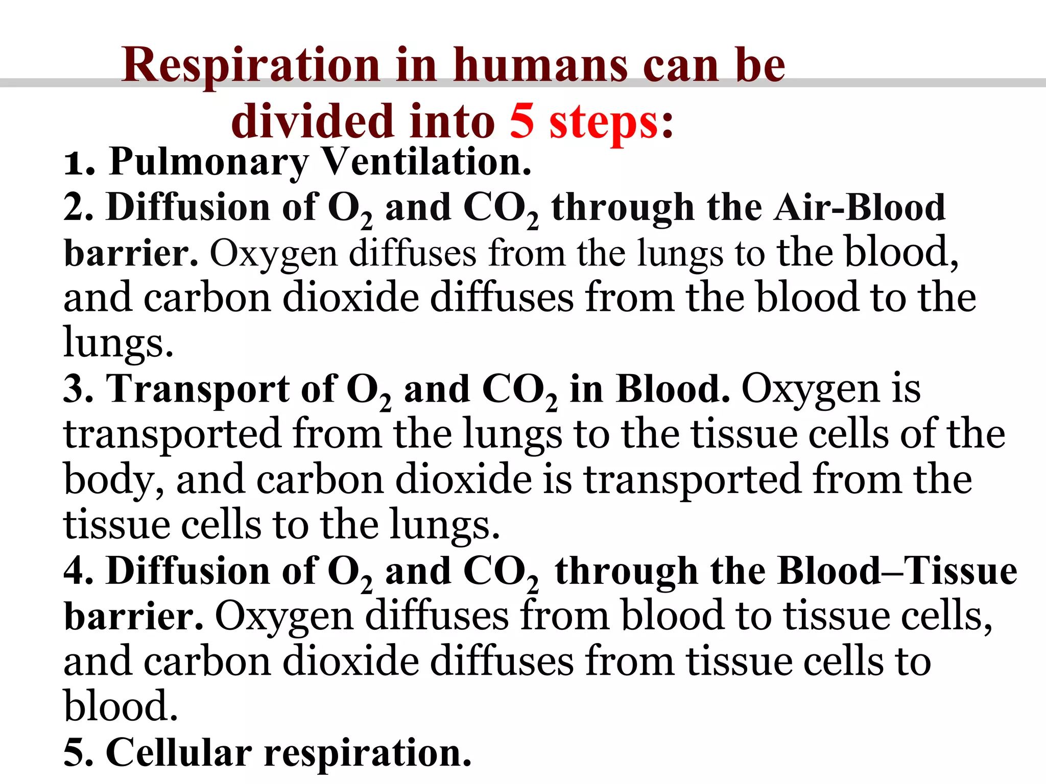
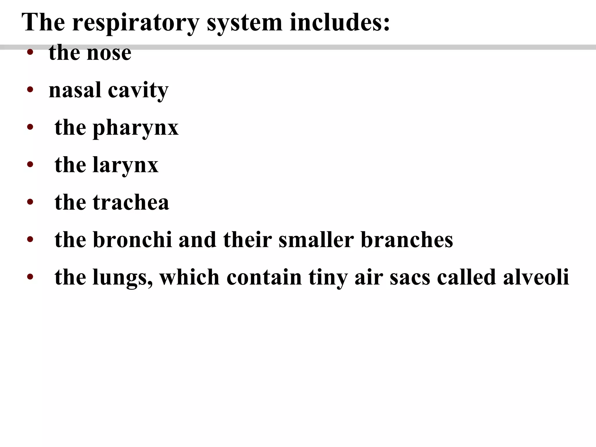
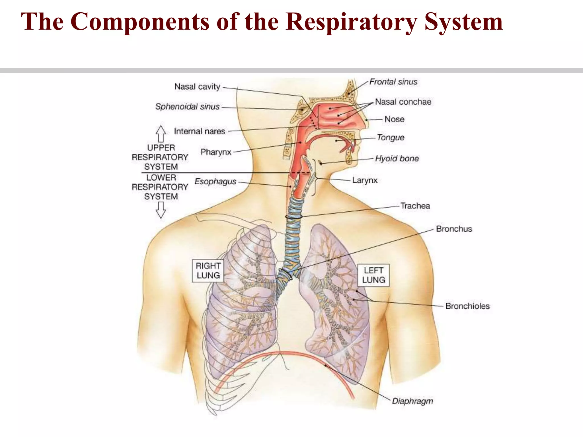
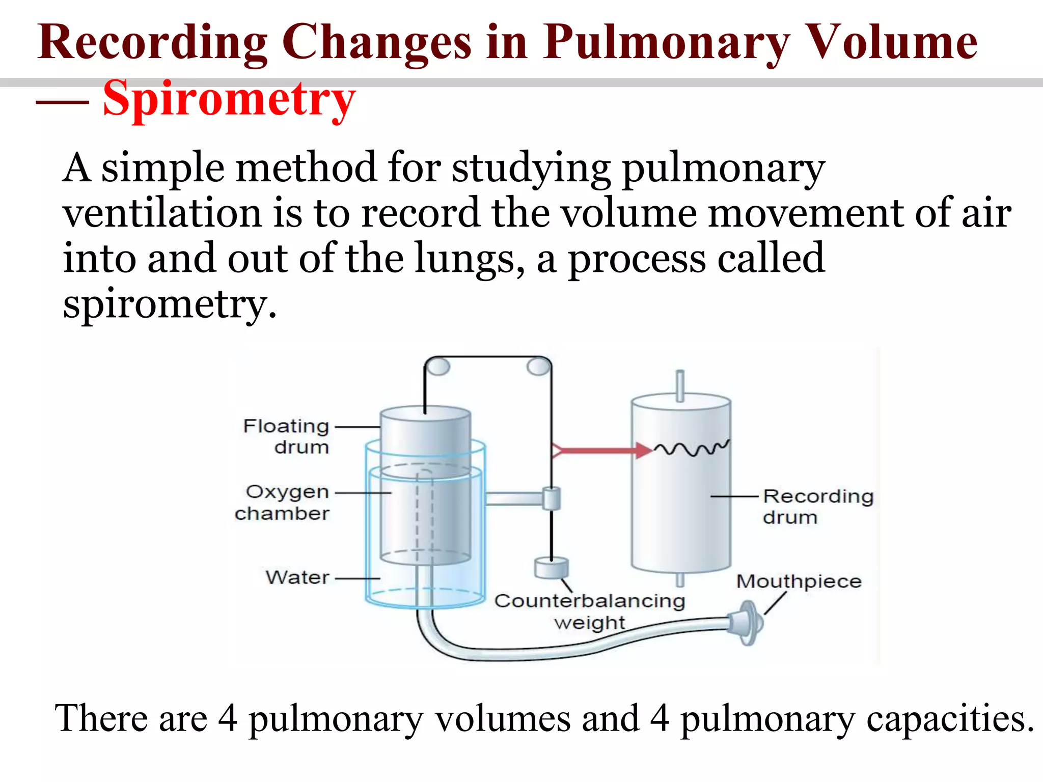
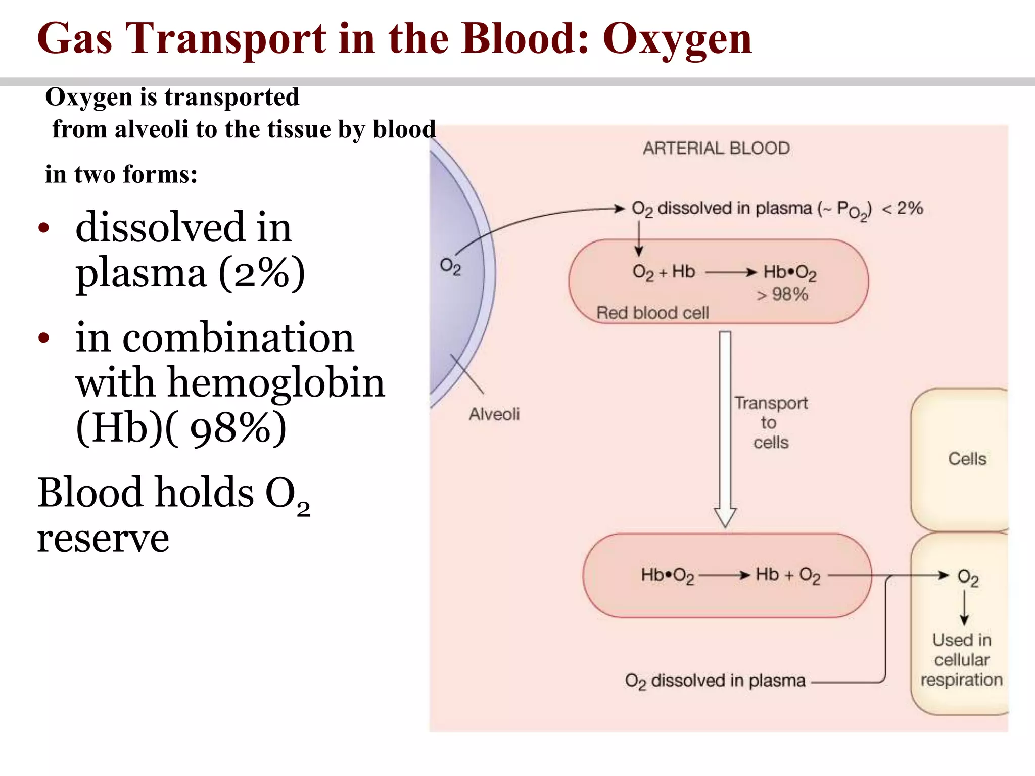
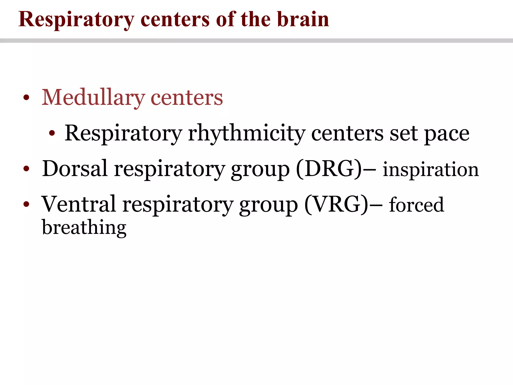
Respiratory Physiology
LECTURE
LECTURE OUTLINE
1. Respiration: Definition, Function. The Steps of respiration.
2. Conducting zone and Respiratory zone.
3. Lung Volumes and Capacities.
4. Mechanics of Breathing.
5. Gas exchange. Partial pressure. Fick’s law.
6. Gas transport in the lungs.
7. Exchange of O2 and CO2 between air –blood and blood-tissue barriers
8. Transport of oxygen in the blood.
9. Control of breathing.
RESPIRATION
• Respiration is the exchange of oxygen and
carbon dioxide between the environment
and the body cells.
The goals of respiration are to provide oxygen to the
tissues and to remove carbon dioxide.
The Stages (Steps) of
Respiration
Respiration in humans can be
divided into 5 steps:
1. Pulmonary Ventilation.
2. Diffusion of O2 and CO2 through the Air-Blood
barrier. Oxygen diffuses from the lungs to the blood,
and carbon dioxide diffuses from the blood to the
lungs.
3. Transport of O2 and CO2 in Blood. Oxygen is
transported from the lungs to the tissue cells of the
body, and carbon dioxide is transported from the
tissue cells to the lungs.
4. Diffusion of O2 and CO2 through the Blood–Tissue
barrier. Oxygen diffuses from blood to tissue cells,
and carbon dioxide diffuses from tissue cells to
blood.
5. Cellular respiration.
What is External Respiration and
Internal Respiration?
• External respiration is the first and second
stages of respiration.
• Internal respiration is the third, fourth and
fifth stages of respiration.
Physical Processes of
Gas Transport System
Organization of the Respiratory
System
The respiratory system includes:
• the nose
• nasal cavity
• the pharynx
• the larynx
• the trachea
• the bronchi and their smaller branches
• the lungs, which contain tiny air sacs called alveoli
The Components of the Respiratory System
1. Conducting Zone
2. Respiratory Zone
Functionally, the respiratory
system consists of two zones:
The Components of the Respiratory System
• The conducting zone includes respiratory
passage-ways (trachea, bronchus, bronchioles)
which provide fairly rigid conduits for air to reach
the gas exchange sites. The conducting zone
organs also cleanse, humidify, and warm
incoming air. As a result, air reaching the lungs
has fewer irritants (dust, bacteria, etc.) than when
it entered the body, and it is warm and damp.
• The respiratory zone, the actual site of gas
exchange, is composed of the respiratory
bronchioles, alveolar ducts, and alveoli.
• Region of gas
exchange
between air
and blood.
• Includes
respiratory
bronchioles
and alveolar
sacs.
Conducting zone &Respiratory Zone
The Bronchi and Lobules of the Lung
The Air-Blood barrier
(Respiratory Membrane)
This air-blood barrier is composed of:
• Alveolar and capillary walls
• Their fused basal laminas
Together, the capillary and alveolar walls and their fused basement
membranes form the respiratory membrane, a 0.5-μm-thick blood air
barrier that has blood flowing past on one side and gas on the other.
•Alveolar walls are a single layer that consists of type I
epithelial cells (permit gas exchange by simple
diffusion) and type II cells that secrete surfactant
Respiratory Membrane
Pulmonary Volumes
and Capacities
Recording Changes in Pulmonary Volume
— Spirometry
A simple method for studying pulmonary
ventilation is to record the volume movement of air
into and out of the lungs, a process called
spirometry.
There are 4 pulmonary volumes and 4 pulmonary capacities.
Respiratory Volumes
1. Tidal volume (Vt) is the volume inspired
or expired with each normal breath.
2. Inspiratory reserve volume (IRV) is the
volume that can be inspired over and
above the tidal volume. It is used during
exercise.
3. Expiratory reserve volume (ERV) is the
volume that can be expired after the
expiration of a tidal volume.
4. Residual volume (RV) is the volume that
remains in the lungs after a maximal
expiration. It cannot be measured by
spirometry.
Respiratory Capacities
1. Inspiratory capacity (IC) – total amount of air that can be inspired
after a tidal expiration (IRV + TV)
2. Functional residual capacity (FRC) – amount of air remaining in
the lungs after a tidal expiration
(RV + ERV)
3. Vital capacity (VC) – the total amount of exchangeable air (TV + IRV
+ ERV)
4. Total lung capacity (TLC) – sum of all lung volumes (approximately
6000 ml in males)
Dead Space
The volume of the airways that does not participate
in gas exchange
Anatomical dead space – volume of the
conducting respiratory passages (150 ml)
Functional dead space – alveoli that cease to act
in gas exchange due to collapse or obstruction
Physiological dead space – sum of alveolar and
anatomical dead spaces
Normal Respiration Cycle
(Normal Breathing Pattern)
Normal Respiration Cycle
(Normal Breathing Pattern)
1. Inhalation
2. Exhalation
3. Automatic Pause
Mechanics of Breathing
Inspiration
• Inspiration is active process:
• Diaphragm contracts -> increased thoracic
volume vertically.
• Intercostals muscles contract, expanding rib
cage -> increased thoracic volume laterally.
• More volume -> lowered pressure -> air in.
Expiration
• Expiration is passive process:
• Due to recoil of elastic lungs.
• Less volume -> pressure within alveoli is
above atmospheric pressure -> air leaves
lungs.
• Note: Residual volume of air is always left
behind, so alveoli do not collapse.
Mechanisms of Pulmonary Ventilation
Gas Exchange
• Daltons Law and partial pressure
Individual gases in a mixture exert
pressure proportional to their abundance
Partial pressure = Total pressure ×
Fractional
Ex: Gas concentration PO2 =760mm
Hg×0.21 =160mm Hg
Gas Exchange
Gas Exchange
The diffusion rates of O2 and CO2 depend on the partial
pressure differences across the membrane and the area
available for diffusion.
For example, the diffusion of O2 from alveolar air into the
pulmonary capillary depends on the partial pressure difference
for O2 between alveolar air and pulmonary capillary blood.
Normally, capillary blood equilibrates with alveolar gas; when
the partial pressures of O2 become equal, there is no more net
diffusion of O2.
Gas diffusion across the alveolar–pulmonary capillary barrier
occurs according to Fick’s law: Vx =DL×ΔP
where:
Vx = volume of gas transferred per minute (mL/min)
DL = lung diffusing capacity (mL/min/mm Hg)
P = partial pressure difference of gas (mm Hg)
• Gas exchange across respiratory
membrane is efficient due to:
• Differences in partial pressure
• Small diffusion distance
• Lipid-soluble gases
• Large surface area of all alveoli
• Coordination of blood flow and airflow
Diffusion and respiratory function
Gas Pickup and Delivery
An Overview of Respiratory Processes and
Partial Pressures in Respiration
Gas Exchange in the Lungs and Tissues: Oxygen
Gas Transport in the Blood: Oxygen
• dissolved in
plasma (2%)
• in combination
with hemoglobin
(Hb)( 98%)
Blood holds O2
reserve
Oxygen is transported
from alveoli to the tissue by blood
in two forms:
Hemoglobin Transport of Oxygen
• 4 binding sites per Hb molecule
• 98% saturated in alveolar arteries
• More unloaded with more need
• 75% in reserve at normal activity
Hemoglobin Saturation Curve
• Temperature, pH, PCO2, and DPG
• Increase of temperature, PCO2, and DPG and
decrease of pH :
• Decrease hemoglobin’s affinity for oxygen
• Enhance oxygen unloading from the blood
• Decreases of temperature, PCO2, and DPG and
the increase of pH act in the opposite manner
• These parameters are all high in systemic
capillaries where oxygen unloading is the goal
Factors Influencing Hemoglobin Saturation
• 7% dissolved in plasma
• 70% carried as becarbonate
• 23% bound to hemoglobin
(carbaminohemoglobin)
Carbon dioxide transport
Co2 is transported
from the tissue to alveoli by blood
in three forms:
A Summary of the Gas Transport Mechanisms
Air-Blood barrier
A Summary of the Gas Transport Mechanisms
Tissue-Blood Barrier
Control of Respiration
• Medullary centers
• Respiratory rhythmicity centers set pace
• Dorsal respiratory group (DRG)– inspiration
• Ventral respiratory group (VRG)– forced
breathing
Respiratory centers of the brain
• Pons
• Apneustic and pneumotaxic centers:
● regulate the respiratory rate and the
depth of respiration in response to
sensory stimuli or input from other centers
in the brain
Respiratory centers of the brain
Respiratory Centers and Reflex Controls
Chemoreceptors
• Chemoreceptors are located throughout the
body (in brain and arteries).
• chemoreceptors are more sensitive
to changes in PCO2
(as sensed through
changes in pH).
• Ventilation is adjusted to maintain
arterial PC02 of 40 mm Hg.
Medullary Respiratory Centers
QUESTIONS
1. Respiration: Definition, Function. The Steps of respiration.
2. Structure of the Conducting zone. Respiratory zone.
3. Site of Gas Exchange: The Alveoli.
4. Lung Volumes and Capacities.
5. Dead space. Anatomic dead space. Functional dead space..
6. Mechanics of Breathing.
7. Sequence of events during inspiration and expiration.
8. Gas exchange. Partial pressure.
9. Diffusion of gases. Form gases in solution.
10. The diagram of the changes in Po2 and Pco2 in the lungs and tissues.
11. O2- hemoglobin dissociation curve.
12. Transport of oxygen in the blood. Form of O2 in the blood.
13. Carbon dioxide transport in blood. Forms CO2 in the blood.
14. Exchange of O2 and CO2 between alveolar air and blood in lung capillaries.
15. Exchange of O2 and CO2 between blood in tissue capillaries and cells in
tissues.
16. Control of breathing.

respiratory system Physiology

  • 1.
  • 2.
    LECTURE OUTLINE 1. Respiration:Definition, Function. The Steps of respiration. 2. Conducting zone and Respiratory zone. 3. Lung Volumes and Capacities. 4. Mechanics of Breathing. 5. Gas exchange. Partial pressure. Fick’s law. 6. Gas transport in the lungs. 7. Exchange of O2 and CO2 between air –blood and blood-tissue barriers 8. Transport of oxygen in the blood. 9. Control of breathing.
  • 3.
    RESPIRATION • Respiration isthe exchange of oxygen and carbon dioxide between the environment and the body cells. The goals of respiration are to provide oxygen to the tissues and to remove carbon dioxide.
  • 4.
    The Stages (Steps)of Respiration
  • 5.
    Respiration in humanscan be divided into 5 steps: 1. Pulmonary Ventilation. 2. Diffusion of O2 and CO2 through the Air-Blood barrier. Oxygen diffuses from the lungs to the blood, and carbon dioxide diffuses from the blood to the lungs. 3. Transport of O2 and CO2 in Blood. Oxygen is transported from the lungs to the tissue cells of the body, and carbon dioxide is transported from the tissue cells to the lungs. 4. Diffusion of O2 and CO2 through the Blood–Tissue barrier. Oxygen diffuses from blood to tissue cells, and carbon dioxide diffuses from tissue cells to blood. 5. Cellular respiration.
  • 6.
    What is ExternalRespiration and Internal Respiration? • External respiration is the first and second stages of respiration. • Internal respiration is the third, fourth and fifth stages of respiration.
  • 7.
    Physical Processes of GasTransport System
  • 8.
    Organization of theRespiratory System
  • 9.
    The respiratory systemincludes: • the nose • nasal cavity • the pharynx • the larynx • the trachea • the bronchi and their smaller branches • the lungs, which contain tiny air sacs called alveoli
  • 10.
    The Components ofthe Respiratory System
  • 12.
    The Components ofthe Respiratory System 1. Conducting Zone 2. Respiratory Zone Functionally, the respiratory system consists of two zones:
  • 13.
    The Components ofthe Respiratory System • The conducting zone includes respiratory passage-ways (trachea, bronchus, bronchioles) which provide fairly rigid conduits for air to reach the gas exchange sites. The conducting zone organs also cleanse, humidify, and warm incoming air. As a result, air reaching the lungs has fewer irritants (dust, bacteria, etc.) than when it entered the body, and it is warm and damp. • The respiratory zone, the actual site of gas exchange, is composed of the respiratory bronchioles, alveolar ducts, and alveoli.
  • 14.
    • Region ofgas exchange between air and blood. • Includes respiratory bronchioles and alveolar sacs. Conducting zone &Respiratory Zone
  • 15.
    The Bronchi andLobules of the Lung
  • 16.
    The Air-Blood barrier (RespiratoryMembrane) This air-blood barrier is composed of: • Alveolar and capillary walls • Their fused basal laminas Together, the capillary and alveolar walls and their fused basement membranes form the respiratory membrane, a 0.5-μm-thick blood air barrier that has blood flowing past on one side and gas on the other. •Alveolar walls are a single layer that consists of type I epithelial cells (permit gas exchange by simple diffusion) and type II cells that secrete surfactant
  • 17.
  • 18.
  • 19.
    Recording Changes inPulmonary Volume — Spirometry A simple method for studying pulmonary ventilation is to record the volume movement of air into and out of the lungs, a process called spirometry. There are 4 pulmonary volumes and 4 pulmonary capacities.
  • 20.
    Respiratory Volumes 1. Tidalvolume (Vt) is the volume inspired or expired with each normal breath. 2. Inspiratory reserve volume (IRV) is the volume that can be inspired over and above the tidal volume. It is used during exercise. 3. Expiratory reserve volume (ERV) is the volume that can be expired after the expiration of a tidal volume. 4. Residual volume (RV) is the volume that remains in the lungs after a maximal expiration. It cannot be measured by spirometry.
  • 21.
    Respiratory Capacities 1. Inspiratorycapacity (IC) – total amount of air that can be inspired after a tidal expiration (IRV + TV) 2. Functional residual capacity (FRC) – amount of air remaining in the lungs after a tidal expiration (RV + ERV) 3. Vital capacity (VC) – the total amount of exchangeable air (TV + IRV + ERV) 4. Total lung capacity (TLC) – sum of all lung volumes (approximately 6000 ml in males)
  • 22.
    Dead Space The volumeof the airways that does not participate in gas exchange Anatomical dead space – volume of the conducting respiratory passages (150 ml) Functional dead space – alveoli that cease to act in gas exchange due to collapse or obstruction Physiological dead space – sum of alveolar and anatomical dead spaces
  • 23.
  • 24.
    Normal Respiration Cycle (NormalBreathing Pattern) 1. Inhalation 2. Exhalation 3. Automatic Pause
  • 25.
  • 26.
    Inspiration • Inspiration isactive process: • Diaphragm contracts -> increased thoracic volume vertically. • Intercostals muscles contract, expanding rib cage -> increased thoracic volume laterally. • More volume -> lowered pressure -> air in.
  • 27.
    Expiration • Expiration ispassive process: • Due to recoil of elastic lungs. • Less volume -> pressure within alveoli is above atmospheric pressure -> air leaves lungs. • Note: Residual volume of air is always left behind, so alveoli do not collapse.
  • 28.
  • 29.
  • 30.
    • Daltons Lawand partial pressure Individual gases in a mixture exert pressure proportional to their abundance Partial pressure = Total pressure × Fractional Ex: Gas concentration PO2 =760mm Hg×0.21 =160mm Hg Gas Exchange
  • 31.
    Gas Exchange The diffusionrates of O2 and CO2 depend on the partial pressure differences across the membrane and the area available for diffusion. For example, the diffusion of O2 from alveolar air into the pulmonary capillary depends on the partial pressure difference for O2 between alveolar air and pulmonary capillary blood. Normally, capillary blood equilibrates with alveolar gas; when the partial pressures of O2 become equal, there is no more net diffusion of O2. Gas diffusion across the alveolar–pulmonary capillary barrier occurs according to Fick’s law: Vx =DL×ΔP where: Vx = volume of gas transferred per minute (mL/min) DL = lung diffusing capacity (mL/min/mm Hg) P = partial pressure difference of gas (mm Hg)
  • 32.
    • Gas exchangeacross respiratory membrane is efficient due to: • Differences in partial pressure • Small diffusion distance • Lipid-soluble gases • Large surface area of all alveoli • Coordination of blood flow and airflow Diffusion and respiratory function
  • 33.
  • 34.
    An Overview ofRespiratory Processes and Partial Pressures in Respiration
  • 35.
    Gas Exchange inthe Lungs and Tissues: Oxygen
  • 36.
    Gas Transport inthe Blood: Oxygen • dissolved in plasma (2%) • in combination with hemoglobin (Hb)( 98%) Blood holds O2 reserve Oxygen is transported from alveoli to the tissue by blood in two forms:
  • 37.
    Hemoglobin Transport ofOxygen • 4 binding sites per Hb molecule • 98% saturated in alveolar arteries • More unloaded with more need • 75% in reserve at normal activity
  • 38.
  • 39.
    • Temperature, pH,PCO2, and DPG • Increase of temperature, PCO2, and DPG and decrease of pH : • Decrease hemoglobin’s affinity for oxygen • Enhance oxygen unloading from the blood • Decreases of temperature, PCO2, and DPG and the increase of pH act in the opposite manner • These parameters are all high in systemic capillaries where oxygen unloading is the goal Factors Influencing Hemoglobin Saturation
  • 41.
    • 7% dissolvedin plasma • 70% carried as becarbonate • 23% bound to hemoglobin (carbaminohemoglobin) Carbon dioxide transport Co2 is transported from the tissue to alveoli by blood in three forms:
  • 42.
    A Summary ofthe Gas Transport Mechanisms Air-Blood barrier
  • 43.
    A Summary ofthe Gas Transport Mechanisms Tissue-Blood Barrier
  • 44.
  • 45.
    • Medullary centers •Respiratory rhythmicity centers set pace • Dorsal respiratory group (DRG)– inspiration • Ventral respiratory group (VRG)– forced breathing Respiratory centers of the brain
  • 46.
    • Pons • Apneusticand pneumotaxic centers: ● regulate the respiratory rate and the depth of respiration in response to sensory stimuli or input from other centers in the brain Respiratory centers of the brain
  • 47.
    Respiratory Centers andReflex Controls
  • 48.
    Chemoreceptors • Chemoreceptors arelocated throughout the body (in brain and arteries). • chemoreceptors are more sensitive to changes in PCO2 (as sensed through changes in pH). • Ventilation is adjusted to maintain arterial PC02 of 40 mm Hg.
  • 49.
  • 50.
    QUESTIONS 1. Respiration: Definition,Function. The Steps of respiration. 2. Structure of the Conducting zone. Respiratory zone. 3. Site of Gas Exchange: The Alveoli. 4. Lung Volumes and Capacities. 5. Dead space. Anatomic dead space. Functional dead space.. 6. Mechanics of Breathing. 7. Sequence of events during inspiration and expiration. 8. Gas exchange. Partial pressure. 9. Diffusion of gases. Form gases in solution. 10. The diagram of the changes in Po2 and Pco2 in the lungs and tissues. 11. O2- hemoglobin dissociation curve. 12. Transport of oxygen in the blood. Form of O2 in the blood. 13. Carbon dioxide transport in blood. Forms CO2 in the blood. 14. Exchange of O2 and CO2 between alveolar air and blood in lung capillaries. 15. Exchange of O2 and CO2 between blood in tissue capillaries and cells in tissues. 16. Control of breathing.