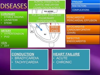
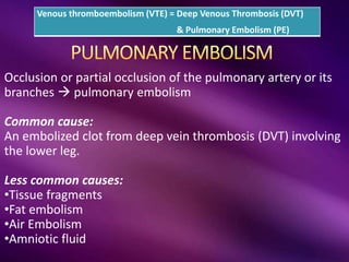
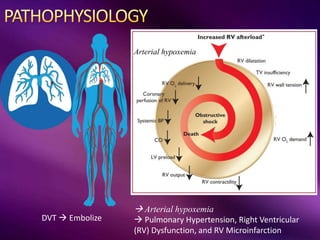
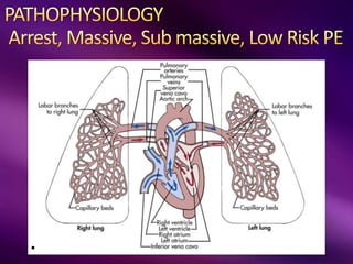
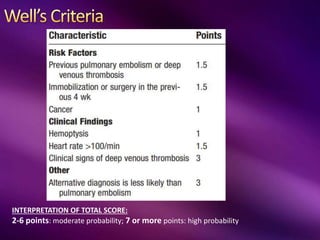
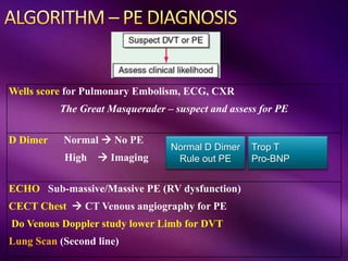
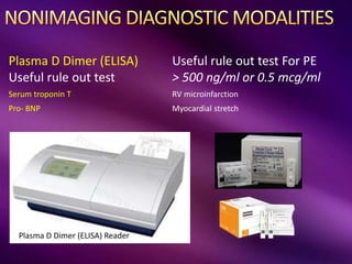
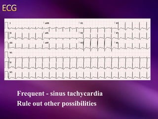
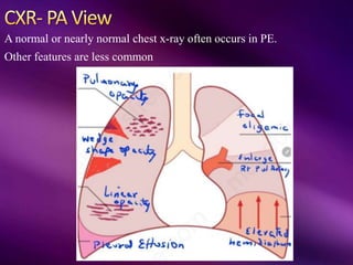
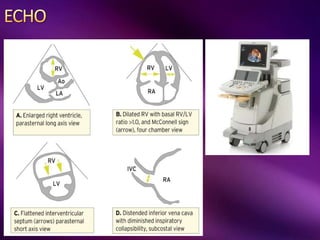
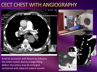
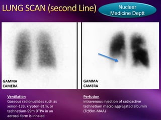
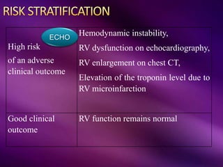
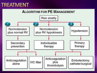
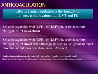
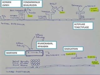
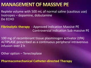
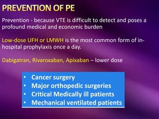
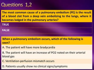
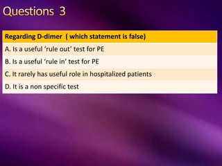
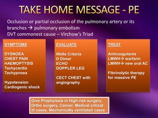
Col Bharat Malhotra
HOD & Senior Advisor (Medicine)
PERICARDITIS
PERICARDIAL EFFUSION
MYOCARDITIS
CARDIOMYOPATHY
(D/H/R)
INFECTIVE
ENDOCARDITIS
HEART FAILURE
• ACUTE
• CHRONIC
CORONARY
• STABLE ANGINA
• UA/NSTEMI
• STEMI
RHD
CHD
PUL ARTERY EMBOLISM
PULMONARY
HYPERTENSION
ETIOLOGY
RISK FACTORS
COMPLICATIONS
ARTERY
• HYPERTENSION
• PVD
VEIN
• DVT
AORTIC DISSECTION
AORTIC ANEURYSM
COARCTATION AORTA
CONDUCTION
• BRADYCARDIA
• TACHYCARDIA
PLEURAL EFFUSION
EMPYEMA
PNEUMOTHORAX
PNEUMONIA
LUNG ABCESS
ACUTE RESPIRATORY
FAILURE
ACUTE RESPIRATORY
DISTRESS SYNDROME
MECHANICAL VENTILATION
PULMONARY EMBOLISM
PULMONARY HYPERTENSION
VENTILATION
HYPOVENTILATION
SYNDROMES
SLEEP APNEAS
MEDIASTINAL
ACUTE MEDIASTINITIS
CHRONIC MEDIASTINITIS
MEDIASTINAL MASS
PNEUMOMEDIASTINUM
INTERSTITIAL LUNG DISEASE
HYPERSENSITIVITY
PNEUMONITIS
OCCUPATIONAL LUNG DISEASE
ASTHMA
COPD
Occlusion or partial occlusion of the pulmonary artery or its
branches  pulmonary embolism
Common cause:
An embolized clot from deep vein thrombosis (DVT) involving
the lower leg.
Less common causes:
•Tissue fragments
•Fat embolism
•Air Embolism
•Amniotic fluid
Venous thromboembolism (VTE) = Deep Venous Thrombosis (DVT)
& Pulmonary Embolism (PE)
PE is the most common preventable cause
of death among hospitalized patients
“the Great
Masquerader”
Diagnosis is difficult because symptoms and
signs are nonspecific
 Arterial hypoxemia
 Pulmonary Hypertension, Right Ventricular
(RV) Dysfunction, and RV Microinfarction
DVT  Embolize
Arterial hypoxemia
Virchow’s Triad
Massive PE
5-10%
Extensive thrombosis affecting at
least half of the pulmonary
vasculature
Sub-massive PE
20-25%
Characterized by RV dysfunction
despite normal systemic arterial
pressure
Low-risk PE
65-75%
No RV dysfunction
No hypotension
“the Great
Masquerader”
Diagnosis is difficult because symptoms and signs are
nonspecific
Asymptomatic
or discovered incidentally
Worsening dyspnea
Cough, Sputum
Hemoptysis
Syncope
Chest pain – Anginal,
Pleuritic
Fever, Diaphoresis
Cardiogenic shock
multiorgan dysfunction
Evidence of DVT
Normal Exam with sinus
tachycardia
Sinus Tachycardia
Tachypnoea
Loud S2, RV dysfunction
Crackles, Pleural rub
Hypotension,
Cardiogenic shock in massive
PE
INTERPRETATION OF TOTAL SCORE:
2-6 points: moderate probability; 7 or more points: high probability
Wells score for Pulmonary Embolism, ECG, CXR
The Great Masquerader – suspect and assess for PE
D Dimer Normal  No PE
High  Imaging
ECHO Sub-massive/Massive PE (RV dysfunction)
CECT Chest  CT Venous angiography for PE
Do Venous Doppler study lower Limb for DVT
Lung Scan (Second line)
Normal D Dimer
Rule out PE
Trop T
Pro-BNP
Plasma D Dimer (ELISA)
Useful rule out test
Useful rule out test For PE
> 500 ng/ml or 0.5 mcg/ml
Serum troponin T RV microinfarction
Pro- BNP Myocardial stretch
Plasma D Dimer (ELISA) Reader
Frequent - sinus tachycardia
Rule out other possibilities
Frequent - sinus tachycardia
RV strain – RBBB, T-wave inversion in leads V1 to V4
S1Q3T3 sign
A normal or nearly normal chest x-ray often occurs in PE.
Other features are less common Rule out other possibilities
A normal or nearly normal chest x-ray often occurs in PE.
Other features are less common
Hampton’s Sign
Wedge shaped
density
A normal or nearly normal chest x-ray often occurs in PE.
Other features are less common
Westermark Sign
Focal oligemia
A normal or nearly normal chest x-ray often occurs in PE.
Other features are less common
Pallas Sign
Enlarge
Rt Pul Artery
A normal or nearly normal chest x-ray often occurs in PE.
Other features are less common
Arterial occlusion with failure to enhance
the entire lumen due to a large filling
defect; the artery may be enlarged
compared with adjacent patent vessels
Ventilation
Gaseous radionuclides such as
xenon-133, krypton-81m, or
technetium-99m DTPA in an
aerosol form is inhaled
Nuclear
Medicine Deptt
Perfusion
intravenous injection of radioactive
technetium macro aggregated albumin
(Tc99m-MAA)
GAMMA
CAMERA
GAMMA
CAMERA
High risk
of an adverse
clinical outcome
Hemodynamic instability,
RV dysfunction on echocardiography,
RV enlargement on chest CT,
Elevation of the troponin level due to
RV microinfarction
Good clinical
outcome
RV function remains normal
ECHO
1 2 3
S/C anticoagulation with (UFH), or (LMWH), or fondaparinux
“bridged” 5d  to warfarin
S/C anticoagulation with (UFH), or (LMWH), or fondaparinux
“bridged” 5d  novel oral anticoagulant such as dabigatran (a direct
thrombin inhibitor) or apixaban (an anti-Xa agent)
Oral anticoagulation monotherapy with rivaroxaban (3week) or apixaban (1 week)
(both are anti-Xa agents) Loading dose  maintenance dose (without S/C anticoagulation)
Effective anticoagulation is the foundation
for successful treatment of DVT and PE
HEPARIN
LMWH
FONDAPARINUX
BIVALIRUDIN
WARFARIN
RIVAROXABAN,
APIXABAN
DABIGATRAN
ALTEPLASE
TENECTIPLASE
Complication
Hemorrhage
How long to give
Initial anticoagulants for 3 months
(monitor INR monthly) (Keep INR 2-3)
Indefinite – in Unprovoked PE, Recurrent PE, APLA
( keep INR 2-3)
Low risk cases if requiring Rx after 3 months
Anticoagulants ( INR 1.5-2)
Low dose Aspirin in low risk
Few
Cases
Replete volume with 500 mL of normal saline (cautious use)
Inotropes – dopamine, dobutamine
Do ECHO
Fibrinolytic therapy - Approved indication Massive PE
Controversial indication Sub massive PE
100 mg of recombinant tissue plasminogen activator (tPA)
ALTIPLASE prescribed as a continuous peripheral intravenous
infusion over 2 h
Other option – Tenecteplase
Pharmacomechanical Catheter-directed Therapy
Prevention - because VTE is difficult to detect and poses a
profound medical and economic burden
Low-dose UFH or LMWH is the most common form of in-
hospital prophylaxis once a day.
Dabigatran, Rivaroxaban, Apixaban – lower dose
• Cancer surgery
• Major orthopedic surgeries
• Critical Medically ill patients
• Mechanical ventilated patients
The most common cause of a pulmonary embolism (PE) is the result
of a blood clot from a deep vein embolizing to the lungs, where it
becomes lodged in the pulmonary arteries?
TRUE
FALSE
When a pulmonary embolism occurs, which of the following is
seen?
A. The patient will have more bradycardia
B. The patient will have an increase of PO2 noted on their arterial
blood gas
C. Ventilation-perfusion mismatch occurs
D. Patients usually show no clinical signs/symptoms
Regarding D-dimer ( which statement is false)
A. Is a useful ‘rule out’ test for PE
B. Is a useful ‘rule in’ test for PE
C. It rarely has useful role in hospitalized patients
D. It is a non specific test
When a pulmonary embolism occurs, we prefer doing following test
immediately. Pick the test which is a second line option.
A. D dimer
B. ABG
C. ECHO
D. Lung scan
Treatment of PE is done by all except
A. Antibiotics (IV Augmentin) and IV Normal Saline
B. Enoxaparin followed by warfarin
C. Enoxaparin followed by Apixaban
D. Fibrinolytic therapy
SYMPTOMS
DYSNOEA
CHEST PAIN
HAEMOPTYSIS
Tachycardia
Tachypnoea
Hypotension
Cardiogenic shock
EVALUATE
Wells Criteria
D Dimer
ECHO
DOPPLER LEG
CECT CHEST with
angiography
TREAT
Anticoagulants
LMWH warfarin
LMWH new oral AC
Fibrinolytic therapy
for massive PE
Give Prophylaxis in High risk surgery,
Ortho surgery, Cancer, Medical critical
ill cases, Mechanically ventilated cases
Occlusion or partial occlusion of the pulmonary artery or its
branches  pulmonary embolism
DVT commonest cause – Virchow’s Triad

Pulmonary embolism 21jan21

  • 1.
    Col Bharat Malhotra HOD& Senior Advisor (Medicine)
  • 2.
    PERICARDITIS PERICARDIAL EFFUSION MYOCARDITIS CARDIOMYOPATHY (D/H/R) INFECTIVE ENDOCARDITIS HEART FAILURE •ACUTE • CHRONIC CORONARY • STABLE ANGINA • UA/NSTEMI • STEMI RHD CHD PUL ARTERY EMBOLISM PULMONARY HYPERTENSION ETIOLOGY RISK FACTORS COMPLICATIONS ARTERY • HYPERTENSION • PVD VEIN • DVT AORTIC DISSECTION AORTIC ANEURYSM COARCTATION AORTA CONDUCTION • BRADYCARDIA • TACHYCARDIA
  • 3.
    PLEURAL EFFUSION EMPYEMA PNEUMOTHORAX PNEUMONIA LUNG ABCESS ACUTERESPIRATORY FAILURE ACUTE RESPIRATORY DISTRESS SYNDROME MECHANICAL VENTILATION PULMONARY EMBOLISM PULMONARY HYPERTENSION VENTILATION HYPOVENTILATION SYNDROMES SLEEP APNEAS MEDIASTINAL ACUTE MEDIASTINITIS CHRONIC MEDIASTINITIS MEDIASTINAL MASS PNEUMOMEDIASTINUM INTERSTITIAL LUNG DISEASE HYPERSENSITIVITY PNEUMONITIS OCCUPATIONAL LUNG DISEASE ASTHMA COPD
  • 4.
    Occlusion or partialocclusion of the pulmonary artery or its branches  pulmonary embolism Common cause: An embolized clot from deep vein thrombosis (DVT) involving the lower leg. Less common causes: •Tissue fragments •Fat embolism •Air Embolism •Amniotic fluid Venous thromboembolism (VTE) = Deep Venous Thrombosis (DVT) & Pulmonary Embolism (PE)
  • 5.
    PE is themost common preventable cause of death among hospitalized patients “the Great Masquerader” Diagnosis is difficult because symptoms and signs are nonspecific
  • 6.
     Arterial hypoxemia Pulmonary Hypertension, Right Ventricular (RV) Dysfunction, and RV Microinfarction DVT  Embolize Arterial hypoxemia
  • 8.
  • 9.
    Massive PE 5-10% Extensive thrombosisaffecting at least half of the pulmonary vasculature Sub-massive PE 20-25% Characterized by RV dysfunction despite normal systemic arterial pressure Low-risk PE 65-75% No RV dysfunction No hypotension
  • 10.
    “the Great Masquerader” Diagnosis isdifficult because symptoms and signs are nonspecific Asymptomatic or discovered incidentally Worsening dyspnea Cough, Sputum Hemoptysis Syncope Chest pain – Anginal, Pleuritic Fever, Diaphoresis Cardiogenic shock multiorgan dysfunction Evidence of DVT Normal Exam with sinus tachycardia Sinus Tachycardia Tachypnoea Loud S2, RV dysfunction Crackles, Pleural rub Hypotension, Cardiogenic shock in massive PE
  • 11.
    INTERPRETATION OF TOTALSCORE: 2-6 points: moderate probability; 7 or more points: high probability
  • 12.
    Wells score forPulmonary Embolism, ECG, CXR The Great Masquerader – suspect and assess for PE D Dimer Normal  No PE High  Imaging ECHO Sub-massive/Massive PE (RV dysfunction) CECT Chest  CT Venous angiography for PE Do Venous Doppler study lower Limb for DVT Lung Scan (Second line) Normal D Dimer Rule out PE Trop T Pro-BNP
  • 13.
    Plasma D Dimer(ELISA) Useful rule out test Useful rule out test For PE > 500 ng/ml or 0.5 mcg/ml Serum troponin T RV microinfarction Pro- BNP Myocardial stretch Plasma D Dimer (ELISA) Reader
  • 14.
    Frequent - sinustachycardia Rule out other possibilities
  • 15.
    Frequent - sinustachycardia RV strain – RBBB, T-wave inversion in leads V1 to V4 S1Q3T3 sign
  • 16.
    A normal ornearly normal chest x-ray often occurs in PE. Other features are less common Rule out other possibilities
  • 17.
    A normal ornearly normal chest x-ray often occurs in PE. Other features are less common Hampton’s Sign Wedge shaped density
  • 18.
    A normal ornearly normal chest x-ray often occurs in PE. Other features are less common Westermark Sign Focal oligemia
  • 19.
    A normal ornearly normal chest x-ray often occurs in PE. Other features are less common Pallas Sign Enlarge Rt Pul Artery
  • 20.
    A normal ornearly normal chest x-ray often occurs in PE. Other features are less common
  • 22.
    Arterial occlusion withfailure to enhance the entire lumen due to a large filling defect; the artery may be enlarged compared with adjacent patent vessels
  • 23.
    Ventilation Gaseous radionuclides suchas xenon-133, krypton-81m, or technetium-99m DTPA in an aerosol form is inhaled Nuclear Medicine Deptt Perfusion intravenous injection of radioactive technetium macro aggregated albumin (Tc99m-MAA) GAMMA CAMERA GAMMA CAMERA
  • 24.
    High risk of anadverse clinical outcome Hemodynamic instability, RV dysfunction on echocardiography, RV enlargement on chest CT, Elevation of the troponin level due to RV microinfarction Good clinical outcome RV function remains normal ECHO
  • 25.
  • 26.
    S/C anticoagulation with(UFH), or (LMWH), or fondaparinux “bridged” 5d  to warfarin S/C anticoagulation with (UFH), or (LMWH), or fondaparinux “bridged” 5d  novel oral anticoagulant such as dabigatran (a direct thrombin inhibitor) or apixaban (an anti-Xa agent) Oral anticoagulation monotherapy with rivaroxaban (3week) or apixaban (1 week) (both are anti-Xa agents) Loading dose  maintenance dose (without S/C anticoagulation) Effective anticoagulation is the foundation for successful treatment of DVT and PE
  • 27.
  • 28.
    Complication Hemorrhage How long togive Initial anticoagulants for 3 months (monitor INR monthly) (Keep INR 2-3) Indefinite – in Unprovoked PE, Recurrent PE, APLA ( keep INR 2-3) Low risk cases if requiring Rx after 3 months Anticoagulants ( INR 1.5-2) Low dose Aspirin in low risk Few Cases
  • 29.
    Replete volume with500 mL of normal saline (cautious use) Inotropes – dopamine, dobutamine Do ECHO Fibrinolytic therapy - Approved indication Massive PE Controversial indication Sub massive PE 100 mg of recombinant tissue plasminogen activator (tPA) ALTIPLASE prescribed as a continuous peripheral intravenous infusion over 2 h Other option – Tenecteplase Pharmacomechanical Catheter-directed Therapy
  • 30.
    Prevention - becauseVTE is difficult to detect and poses a profound medical and economic burden Low-dose UFH or LMWH is the most common form of in- hospital prophylaxis once a day. Dabigatran, Rivaroxaban, Apixaban – lower dose • Cancer surgery • Major orthopedic surgeries • Critical Medically ill patients • Mechanical ventilated patients
  • 32.
    The most commoncause of a pulmonary embolism (PE) is the result of a blood clot from a deep vein embolizing to the lungs, where it becomes lodged in the pulmonary arteries? TRUE FALSE When a pulmonary embolism occurs, which of the following is seen? A. The patient will have more bradycardia B. The patient will have an increase of PO2 noted on their arterial blood gas C. Ventilation-perfusion mismatch occurs D. Patients usually show no clinical signs/symptoms
  • 33.
    Regarding D-dimer (which statement is false) A. Is a useful ‘rule out’ test for PE B. Is a useful ‘rule in’ test for PE C. It rarely has useful role in hospitalized patients D. It is a non specific test
  • 34.
    When a pulmonaryembolism occurs, we prefer doing following test immediately. Pick the test which is a second line option. A. D dimer B. ABG C. ECHO D. Lung scan
  • 35.
    Treatment of PEis done by all except A. Antibiotics (IV Augmentin) and IV Normal Saline B. Enoxaparin followed by warfarin C. Enoxaparin followed by Apixaban D. Fibrinolytic therapy
  • 36.
    SYMPTOMS DYSNOEA CHEST PAIN HAEMOPTYSIS Tachycardia Tachypnoea Hypotension Cardiogenic shock EVALUATE WellsCriteria D Dimer ECHO DOPPLER LEG CECT CHEST with angiography TREAT Anticoagulants LMWH warfarin LMWH new oral AC Fibrinolytic therapy for massive PE Give Prophylaxis in High risk surgery, Ortho surgery, Cancer, Medical critical ill cases, Mechanically ventilated cases Occlusion or partial occlusion of the pulmonary artery or its branches  pulmonary embolism DVT commonest cause – Virchow’s Triad