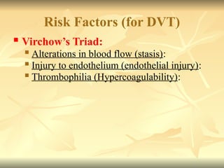
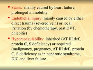
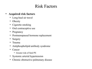
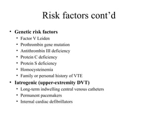
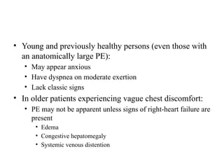
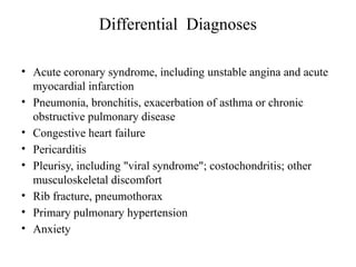
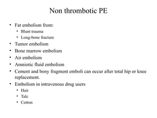
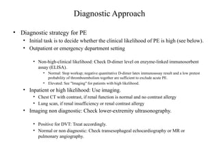
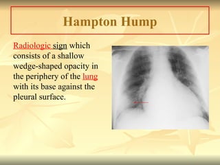
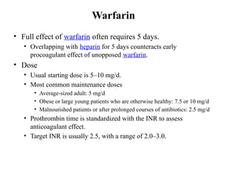
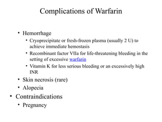
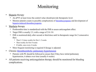
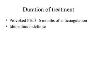
Pulmonary Embolism
Dr.Makhoba
Definition
• A condition in which venous thrombi dislodge from their
site of formation and embolize to the pulmonary arterial
circulation
• Clinical syndromes
– Massive pulmonary thromboembolism (PE)
• Systemic arterial hypotension and anatomically widespread
thromboembolism
– Moderate to large PE
• Right ventricular (RV) hypokinesis, but normal systemic arterial
pressure
– Small to moderate PE
• Normal right-heart function and normal systemic arterial pressure
Epidemiology
• PE
• Incidence
• 69 cases per 100,000 persons annually
• Age
• More common with advancing age
• Sex
• More common in women than in men; ratio, 1.3:1
• Venous thromboembolism (VTE): encompasses deep venous
thrombosis (DVT) and PE
• VTE-related deaths in the U.S. are estimated at 300,000 annually.
• 7% diagnosed with VTE and treated
• 34% sudden fatal PE
• 59% as undetected PE
Epidemiology cont’d
• Symptomatic VTE events
• Approximately two-thirds are hospital acquired.
• One-third are community acquired.
• Residents of nursing facilities are especially vulnerable.
• Estimated hospitalized patients at risk for VTE in
the U.S.
• 13.4 million patients annually
• 5.8 million surgical patients at moderate–high risk
• 7.6 million medical patients with comorbidities such as heart
failure, cancer, and stroke
Risk Factors (for DVT)
 Virchow’s Triad:
 Alterations in blood flow (stasis):
 Injury to endothelium (endothelial injury):
 Thrombophilia (Hypercoagulability):
 Stasis: mainly caused by heart failure,
prolonged immobility
 Endothelial injury: mainly caused by either
direct trauma (severed vein) or local
irritation (by chemotherapy, past DVT,
phlebitis)
 Hypercoagulability: inherited (AT III def.,
protein C, S deficiency) or acquired
(malignancy, pregnancy, AT III def., protein
C, S deficiency as in nephrotic syndrome,
DIC and liver failure.
Risk Factors
• Acquired risk factors
• Long-haul air travel
• Obesity
• Cigarette smoking
• Oral contraceptive use
• Pregnancy
• Postmenopausal hormone replacement
• Surgery
• Trauma
• Antiphospholipid antibody syndrome
• Cancer
• Greater risk of fatal PE
• Systemic arterial hypertension
• Chronic obstructive pulmonary disease
Risk factors cont’d
• Genetic risk factors
• Factor V Leiden
• Prothrombin gene mutation
• Antithrombin III deficiency
• Protein C deficiency
• Protein S deficiency
• Homocysteinemia
• Family or personal history of VTE
• Iatrogenic (upper-extremity DVT)
• Long-term indwelling central venous catheters
• Permanent pacemakers
• Internal cardiac defibrillators
Risk factors cont’d
• Other risk factors
• Varicose veins
• Oral corticosteroids
• Inflammatory bowel disease
• PE (not DVT specific) risk factors
• Ischemic heart disease
• Heart failure
• Cerebrovascular disease
• Greater risk of fatal PE
Etiology
• Caused by dislodgement of venous thrombi traveling to
pulmonary arterial circulation
• About half of patients with pelvic vein thrombosis or proximal leg DVT
have PE.
• Isolated calf vein thrombi pose a lower risk of PE.
• Upper-extremity DVTs are becoming a more common problem with
increased use of:
• Long-term indwelling central venous catheters for hyperalimentation and
chemotherapy
• Frequent insertion of permanent pacemakers and internal cardiac defibrillators
• Thrombi occur due to:
• Blood vessel wall injury
• Blood stasis/venous stasis
• Hypercoagulability
Symptoms & Signs
• Dyspnea (most frequent symptom)
• Tachycardia (most frequent sign)
• Chest pain
• In massive PE, pain may resemble pain of myocardial infarction.
• Pleuritic pain in PE patients with pleural involvement
• Tachypnea
• Low-grade fever
• Neck vein distention
• Accentuated pulmonic component of the second heart sound
(P2)
• Massive PE is often indicated by:
• Dyspnea
• Syncope
• Hypotension
• Cyanosis
• Small PE located distally near the pleura is often indicated
by:
• Pleuritic pain
• Cough
• Hemoptysis
• Pulmonary infarction
• Young and previously healthy persons (even those with
an anatomically large PE):
• May appear anxious
• Have dyspnea on moderate exertion
• Lack classic signs
• In older patients experiencing vague chest discomfort:
• PE may not be apparent unless signs of right-heart failure are
present
• Edema
• Congestive hepatomegaly
• Systemic venous distention
Differential Diagnoses
• Acute coronary syndrome, including unstable angina and acute
myocardial infarction
• Pneumonia, bronchitis, exacerbation of asthma or chronic
obstructive pulmonary disease
• Congestive heart failure
• Pericarditis
• Pleurisy, including "viral syndrome"; costochondritis; other
musculoskeletal discomfort
• Rib fracture, pneumothorax
• Primary pulmonary hypertension
• Anxiety
Non thrombotic PE
• Fat embolism from:
• Blunt trauma
• Long-bone fracture
• Tumor embolism
• Bone marrow embolism
• Air embolism
• Amniotic fluid embolism
• Cement and bony fragment emboli can occur after total hip or knee
replacement.
• Embolism in intravenous drug users
• Hair
• Talc
• Cotton
Diagnostic Approach
• Diagnostic strategy for PE
• Initial task is to decide whether the clinical likelihood of PE is high (see below).
• Outpatient or emergency department setting
• Non-high-clinical likelihood: Check D-dimer level on enzyme-linked immunosorbent
assay (ELISA).
• Normal: Stop workup; negative quantitative D-dimer latex immunoassay result and a low pretest
probability of thromboembolism together are sufficient to exclude acute PE.
• Elevated: See "Imaging" for patients with high likelihood.
• Inpatient or high likelihood: Use imaging.
• Chest CT with contrast, if renal function is normal and no contrast allergy
• Lung scan, if renal insufficiency or renal contrast allergy
• Imaging non diagnostic: Check lower-extremity ultrasonography.
• Positive for DVT: Treat accordingly.
• Normal or non diagnostic: Check transesophageal echocardiography or MR or
pulmonary angiography.
Assessment of clinical likelihood (modified
Wells score)
• High clinical likelihood of PE if the point score
exceeds 4.
• Signs and symptoms of DVT: 3
• Alternative diagnosis less likely than PE: 3
• Heart rate >100/min: 1.5
• Immobilization >3 days; surgery within 4 weeks: 1.5
• Prior PE or DVT: 1.5
• Hemoptysis: 1
• Cancer: 1
• Non-high likelihood if score ≤ 4
Laboratory Tests
• Plasma D-dimer ELISA
• Elevated level (>500 ng/mL) in > 95% of patients with PE sensitive, but
nonspecific for PE
• Levels increase in patients with myocardial infarction, sepsis, or almost any
systemic illness.
• Therefore, test has no useful role for people who are already hospitalized.
• Can be used to help exclude PE: negative predictive value of up to 99.6%
• Cardiac biomarkers
• Serum troponin levels increase in RV microinfarction.
• Myocardial stretch often results in elevation of brain natriuretic peptide
(BNP) or NT-pro-brain natriuretic peptide.
• Elevated cardiac biomarkers predict an increase in major complications
and mortality from PE.
Imaging
• Chest radiography (CXR)
• Normal or near-normal result in a dyspneic
patient occurs in PE.
• Well-established abnormalities
• Focal oligemia (Westermark’s sign)
• Peripheral wedge-shaped density above the
diaphragm (Hampton’s hump)
• Enlarged right descending pulmonary artery
(Palla’s sign)
Westermark Sign
 represents a focus of oligemia (
vasoconstriction) seen distal to a
pulmonary embolism.While the
chest x-ray is normal in the
majority of PE cases, the
Westermark sign is seen in 2% of
patients.
 The sign results from a
combination of:
 The dilation of the pulmonary arteries
proximal to the embolus and
 the collapse of the distal vasculature
creating the appearance of a sharp cut
off on chest radiography.
Hampton Hump
Radiologic sign which
consists of a shallow
wedge-shaped opacity in
the periphery of the lung
with its base against the
pleural surface.
Venous ultrasonography
• Confirmed DVT is usually an adequate surrogate for
PE.
• Loss of vein compressibility is the primary criterion
for DVT.
• Half of patients with PE have no imaging evidence of
DVT, probably because the clot has already embolized
to the lung or is in the pelvic veins.
• Workup for PE should continue if there is high clinical
suspicion, despite a normal result on ultrasonography.
Chest CT
• CT with intravenous contrast
• Can effectively diagnose large, central PE
• Can detect peripherally located thrombi in
sixth-order branches
• In patients without PE, lung parenchymal
images may establish alternative diagnoses not
apparent on chest radiography.
Bilateral PE
Pulmonary angiography
• Selective pulmonary angiography is the most specific
examination available for definitively diagnosing PE.
• Detects emboli as small as 1–2 mm
• Definitive diagnosis of PE depends on visualization
of intraluminal filling defect in > 1 projection.
• Secondary signs of PE
• Abrupt occlusion ("cut-off") of vessels
• Segmental oligemia or avascularity
• Prolonged arterial phase with slow filling
• Tortuous, tapering peripheral vessels
PE on pulmonary angiogram
Lung scanning: V/Q
• Second-line diagnostic test for PE
• Mostly used for patients who cannot tolerate intravenous contrast
• Perfusion scan defect indicates absent or decreased blood flow,
possibly due to PE.
• Ventilation scans, obtained with radiolabeled-inhaled gases such as
xenon or krypton, improve the specificity of the perfusion scan.
• Abnormal ventilation scans indicate an abnormal nonventilated lung, providing possible
explanations for perfusion defects other than acute PE.
• High probability of PE is defined as ≥ 2 segmental perfusion defects in the presence of
normal ventilation.
• Diagnosis of PE is very unlikely in patients with normal and near-
normal scans but is ~90% certain in patients with high-probability
scans.
• Most patients have nondiagnostic scans, and fewer than half of patients with
angiographically confirmed PE have a high-probability scan.
• As many as 40% of patients with high clinical suspicion for PE and low-probability
scans have PE confirmed by angiography.
V/Q Scan
 Identifies mismatches between areas that are ventilated but not
perfused
 Best initial test in patients with clear CXR
 Scan can be interpreted as High, Intermediate, or Low
probability of PE, or normal
 Normal rules out PE
 High-probability scan is diagnostic of PE if the clinical suspicion is
also high
 Low-probability scan rules out PE only in a pt with low pretest clinical
probability (because PE is found in roughly 15% of pts with low-
probability scans)
 Intermediate-probability scan requires further evaluation (16-66%
chance of PE depending on pretest probability)
V/Q Scan
CARDIAC Echocardiography
• >50% of patients with PE have normal echocardiograms.
• Helps with rapid triage of extremely ill patients (can usually reliably
differentiate among illnesses that have radically different treatment)
– Acute myocardial infarction
– Pericardial tamponade
– Dissection of the aorta
– PE complicated by right-heart failure
• Transthoracic echocardiography
– Rarely images thrombus directly
– McConnell’s sign (best known indirect sign of PE on transthoracic echo)
• RV free-wall hypokinesis with normal RV apical motion, appears to be specific for PE.
• Transesophageal echocardiography
– Should be considered when CT scanning facilities are not available or when a
patient has renal failure or severe contrast allergy that precludes administration
of contrast despite premedication with high-dose corticosteroids
– Can directly visualize large proximal PE
• Detection of RV dysfunction due to PE helps to stratify risk, delineate
prognosis, and plan optimal management.
EKG Findings
Classic findings may include:
•Sinus tachycardia
•New onset Atrial fibrillation or flutter
•Rt axis deviation
•S1Q3T3 sign: S wave in lead I, a Q wave in lead III, and an inverted T wave in lead
III
•T-wave inversion in leads V1 to V4: reflects RV strain
Treatment
General guidelines
• Primary therapy
• Treatment options
• Clot dissolution with thrombolysis or
• Removal of PE by embolectomy
• Primary therapy is reserved for patients at high risk of an adverse clinical
outcome.
• Hemodynamic instability
• RV dysfunction
• Detection of RV hypokinesis on echocardiography is the most widely used approach to risk
stratification.
• Elevation of troponin level due to RV microinfarction
• Secondary prevention of recurrent PE
• Anticoagulation with heparin and warfarin
• Placement of an inferior vena caval (IVC) filter
Heparin
• Heparin prevents additional thrombus formation and permits
endogenous fibrinolytic mechanisms to lyse clot that has already
formed.
• After 5–7 days of heparin, residual thrombus begins to stabilize in the
endothelium of the vein or pulmonary artery.
• Does not directly dissolve thrombus that already exists.
• UFH (Unfractionated heparin)
• IV bolus, 5000–10,000 U, then continuous infusion of 1000–1500 U/h
• Activated partial thromboplastin time (aPTT) at least twice the control
value should provide a therapeutic level.
• Nomograms based on a patient’s weight may assist in adjusting the dose.
• Weight based: initial bolus of 80 U/kg, then initial infusion rate of 18 U/kg
hourly
LMWH(Low molecular weight heparin)
• Enoxaparin
• Preferred dose: 1 mg/kg SC twice daily
• Alternate dose: 1.5 mg/kg daily
• Tinzaparin 175 units/kg once daily with normal renal function
• Greater bioavailability, more predictable dose response, few
complications, and longer half-life than UFH
• No laboratory monitoring or dose adjustment is needed unless
the patient is markedly obese or has renal dysfunction.
• Overall 29% reduction in mortality and major bleeding rates
compared with UFH
• Weight-adjusted doses must be adjusted downward in renal
insufficiency because the kidneys excrete LMWH.
Complications of heparins
• Hemorrhage
• If life-threatening or intracranial hemorrhage, administer
protamine sulfate 1–1.5 mg IV per 100 U heparin
• Maximum, 50 mg/dose; rate, 5 mg/min
• Monitor aPTT
• Osteopenia
• Less frequent with LMWH
• Heparin-induced thrombocytopenia (HIT)
• Thrombosis due to heparin-induced thrombocytopenia
• Elevations in aminotransferase levels
Alternatives to Heparin
• Fondaparinux (Anti-Xa
pentasaccharide) .Given Subcutaneously
• Rivaroxaban, Apixaban; Oral direct Factor Xa
inhibitors.
• No laboratory monitoring is required
• Have no antidotes
Warfarin
• Full effect of warfarin often requires 5 days.
• Overlapping with heparin for 5 days counteracts early
procoagulant effect of unopposed warfarin.
• Dose
• Usual starting dose is 5–10 mg/d.
• Most common maintenance doses
• Average-sized adult: 5 mg/d
• Obese or large young patients who are otherwise healthy: 7.5 or 10 mg/d
• Malnourished patients or after prolonged courses of antibiotics: 2.5 mg/d
• Prothrombin time is standardized with the INR to assess
anticoagulant effect.
• Target INR is usually 2.5, with a range of 2.0–3.0.
Complications of Warfarin
• Hemorrhage
• Cryoprecipitate or fresh-frozen plasma (usually 2 U) to
achieve immediate hemostasis
• Recombinant factor VIIa for life-threatening bleeding in the
setting of excessive warfarin
• Vitamin K for less serious bleeding or an excessively high
INR
• Skin necrosis (rare)
• Alopecia
• Contraindications
• Pregnancy
Monitoring
• Heparin therapy
• An aPTT at least twice the control value should provide therapeutic level.
• Monitor platelet counts to possible complications of thrombocytopenia and development of
heparin-induced thrombocytopenia.
• Warfarin therapy
• Prothrombin time is standardized with the INR to assess anticoagulant effect.
• Target INR is usually 2.5, with a range of 2.0–3.0.
• INR is monitored daily after second or third dose until in therapeutic range for 2 consecutive
days.
• Then 2–3 times weekly for first 1–2 weeks
• Then weekly for first 4 weeks
• If stable, once every 4 weeks
• More frequent monitoring is required if dosage is adjusted.
• Chronic thromboembolic pulmonary hypertension
• Patients with PE should be followed to ensure that if they have initial pulmonary
hypertension, it abates over time (usually 6 weeks).
• All patients receiving anticoagulation therapy should be monitored for bleeding
complications.
Duration of treatment
• Provoked PE: 3–6 months of anticoagulation
• Idiopathic: indefinite
Complications
• Hypoxemia
• Right-heart failure
• Hypotension
• Lung hemorrhage
• Chronic thromboembolic
pulmonary hypertension
Prognosis
• Progressive RV failure is the usual cause of death from
PE.
• RV dysfunction on baseline echocardiography of patients with
PE who presented with a systolic blood pressure >90 mmHg
was associated with a doubling of the 3-month mortality rate.
• Combination of RV dysfunction plus elevated biomarkers
such as troponin portends an especially ominous
prognosis.
• Elevated cardiac biomarkers predict an increase in major
complications and mortality from PE.
• Successful thrombolytic therapy rapidly reverses RV
failure and leads to a lower rate of death and recurrent PE.
Prevention
• Mechanical and pharmacologic measures often
succeed in preventing PE.
• Minidose UFH: 5000 U SC tid
• LMWH
• Enoxaparin: 40 mg SC qd
• Dalteparin: 2500 or 5000 U SC qd
• Graduated compression stockings (GCS): 10–18
mmHg
• Intermittent pneumatic compression devices (IPC)

Pulmonary Embolism PowerPoint presentation

  • 1.
  • 2.
    Definition • A conditionin which venous thrombi dislodge from their site of formation and embolize to the pulmonary arterial circulation • Clinical syndromes – Massive pulmonary thromboembolism (PE) • Systemic arterial hypotension and anatomically widespread thromboembolism – Moderate to large PE • Right ventricular (RV) hypokinesis, but normal systemic arterial pressure – Small to moderate PE • Normal right-heart function and normal systemic arterial pressure
  • 3.
    Epidemiology • PE • Incidence •69 cases per 100,000 persons annually • Age • More common with advancing age • Sex • More common in women than in men; ratio, 1.3:1 • Venous thromboembolism (VTE): encompasses deep venous thrombosis (DVT) and PE • VTE-related deaths in the U.S. are estimated at 300,000 annually. • 7% diagnosed with VTE and treated • 34% sudden fatal PE • 59% as undetected PE
  • 4.
    Epidemiology cont’d • SymptomaticVTE events • Approximately two-thirds are hospital acquired. • One-third are community acquired. • Residents of nursing facilities are especially vulnerable. • Estimated hospitalized patients at risk for VTE in the U.S. • 13.4 million patients annually • 5.8 million surgical patients at moderate–high risk • 7.6 million medical patients with comorbidities such as heart failure, cancer, and stroke
  • 5.
    Risk Factors (forDVT)  Virchow’s Triad:  Alterations in blood flow (stasis):  Injury to endothelium (endothelial injury):  Thrombophilia (Hypercoagulability):
  • 6.
     Stasis: mainlycaused by heart failure, prolonged immobility  Endothelial injury: mainly caused by either direct trauma (severed vein) or local irritation (by chemotherapy, past DVT, phlebitis)  Hypercoagulability: inherited (AT III def., protein C, S deficiency) or acquired (malignancy, pregnancy, AT III def., protein C, S deficiency as in nephrotic syndrome, DIC and liver failure.
  • 7.
    Risk Factors • Acquiredrisk factors • Long-haul air travel • Obesity • Cigarette smoking • Oral contraceptive use • Pregnancy • Postmenopausal hormone replacement • Surgery • Trauma • Antiphospholipid antibody syndrome • Cancer • Greater risk of fatal PE • Systemic arterial hypertension • Chronic obstructive pulmonary disease
  • 8.
    Risk factors cont’d •Genetic risk factors • Factor V Leiden • Prothrombin gene mutation • Antithrombin III deficiency • Protein C deficiency • Protein S deficiency • Homocysteinemia • Family or personal history of VTE • Iatrogenic (upper-extremity DVT) • Long-term indwelling central venous catheters • Permanent pacemakers • Internal cardiac defibrillators
  • 9.
    Risk factors cont’d •Other risk factors • Varicose veins • Oral corticosteroids • Inflammatory bowel disease • PE (not DVT specific) risk factors • Ischemic heart disease • Heart failure • Cerebrovascular disease • Greater risk of fatal PE
  • 10.
    Etiology • Caused bydislodgement of venous thrombi traveling to pulmonary arterial circulation • About half of patients with pelvic vein thrombosis or proximal leg DVT have PE. • Isolated calf vein thrombi pose a lower risk of PE. • Upper-extremity DVTs are becoming a more common problem with increased use of: • Long-term indwelling central venous catheters for hyperalimentation and chemotherapy • Frequent insertion of permanent pacemakers and internal cardiac defibrillators • Thrombi occur due to: • Blood vessel wall injury • Blood stasis/venous stasis • Hypercoagulability
  • 11.
    Symptoms & Signs •Dyspnea (most frequent symptom) • Tachycardia (most frequent sign) • Chest pain • In massive PE, pain may resemble pain of myocardial infarction. • Pleuritic pain in PE patients with pleural involvement • Tachypnea • Low-grade fever • Neck vein distention • Accentuated pulmonic component of the second heart sound (P2)
  • 12.
    • Massive PEis often indicated by: • Dyspnea • Syncope • Hypotension • Cyanosis • Small PE located distally near the pleura is often indicated by: • Pleuritic pain • Cough • Hemoptysis • Pulmonary infarction
  • 13.
    • Young andpreviously healthy persons (even those with an anatomically large PE): • May appear anxious • Have dyspnea on moderate exertion • Lack classic signs • In older patients experiencing vague chest discomfort: • PE may not be apparent unless signs of right-heart failure are present • Edema • Congestive hepatomegaly • Systemic venous distention
  • 14.
    Differential Diagnoses • Acutecoronary syndrome, including unstable angina and acute myocardial infarction • Pneumonia, bronchitis, exacerbation of asthma or chronic obstructive pulmonary disease • Congestive heart failure • Pericarditis • Pleurisy, including "viral syndrome"; costochondritis; other musculoskeletal discomfort • Rib fracture, pneumothorax • Primary pulmonary hypertension • Anxiety
  • 15.
    Non thrombotic PE •Fat embolism from: • Blunt trauma • Long-bone fracture • Tumor embolism • Bone marrow embolism • Air embolism • Amniotic fluid embolism • Cement and bony fragment emboli can occur after total hip or knee replacement. • Embolism in intravenous drug users • Hair • Talc • Cotton
  • 16.
    Diagnostic Approach • Diagnosticstrategy for PE • Initial task is to decide whether the clinical likelihood of PE is high (see below). • Outpatient or emergency department setting • Non-high-clinical likelihood: Check D-dimer level on enzyme-linked immunosorbent assay (ELISA). • Normal: Stop workup; negative quantitative D-dimer latex immunoassay result and a low pretest probability of thromboembolism together are sufficient to exclude acute PE. • Elevated: See "Imaging" for patients with high likelihood. • Inpatient or high likelihood: Use imaging. • Chest CT with contrast, if renal function is normal and no contrast allergy • Lung scan, if renal insufficiency or renal contrast allergy • Imaging non diagnostic: Check lower-extremity ultrasonography. • Positive for DVT: Treat accordingly. • Normal or non diagnostic: Check transesophageal echocardiography or MR or pulmonary angiography.
  • 17.
    Assessment of clinicallikelihood (modified Wells score) • High clinical likelihood of PE if the point score exceeds 4. • Signs and symptoms of DVT: 3 • Alternative diagnosis less likely than PE: 3 • Heart rate >100/min: 1.5 • Immobilization >3 days; surgery within 4 weeks: 1.5 • Prior PE or DVT: 1.5 • Hemoptysis: 1 • Cancer: 1 • Non-high likelihood if score ≤ 4
  • 18.
    Laboratory Tests • PlasmaD-dimer ELISA • Elevated level (>500 ng/mL) in > 95% of patients with PE sensitive, but nonspecific for PE • Levels increase in patients with myocardial infarction, sepsis, or almost any systemic illness. • Therefore, test has no useful role for people who are already hospitalized. • Can be used to help exclude PE: negative predictive value of up to 99.6% • Cardiac biomarkers • Serum troponin levels increase in RV microinfarction. • Myocardial stretch often results in elevation of brain natriuretic peptide (BNP) or NT-pro-brain natriuretic peptide. • Elevated cardiac biomarkers predict an increase in major complications and mortality from PE.
  • 19.
    Imaging • Chest radiography(CXR) • Normal or near-normal result in a dyspneic patient occurs in PE. • Well-established abnormalities • Focal oligemia (Westermark’s sign) • Peripheral wedge-shaped density above the diaphragm (Hampton’s hump) • Enlarged right descending pulmonary artery (Palla’s sign)
  • 20.
    Westermark Sign  representsa focus of oligemia ( vasoconstriction) seen distal to a pulmonary embolism.While the chest x-ray is normal in the majority of PE cases, the Westermark sign is seen in 2% of patients.  The sign results from a combination of:  The dilation of the pulmonary arteries proximal to the embolus and  the collapse of the distal vasculature creating the appearance of a sharp cut off on chest radiography.
  • 21.
    Hampton Hump Radiologic signwhich consists of a shallow wedge-shaped opacity in the periphery of the lung with its base against the pleural surface.
  • 22.
    Venous ultrasonography • ConfirmedDVT is usually an adequate surrogate for PE. • Loss of vein compressibility is the primary criterion for DVT. • Half of patients with PE have no imaging evidence of DVT, probably because the clot has already embolized to the lung or is in the pelvic veins. • Workup for PE should continue if there is high clinical suspicion, despite a normal result on ultrasonography.
  • 23.
    Chest CT • CTwith intravenous contrast • Can effectively diagnose large, central PE • Can detect peripherally located thrombi in sixth-order branches • In patients without PE, lung parenchymal images may establish alternative diagnoses not apparent on chest radiography.
  • 24.
  • 25.
    Pulmonary angiography • Selectivepulmonary angiography is the most specific examination available for definitively diagnosing PE. • Detects emboli as small as 1–2 mm • Definitive diagnosis of PE depends on visualization of intraluminal filling defect in > 1 projection. • Secondary signs of PE • Abrupt occlusion ("cut-off") of vessels • Segmental oligemia or avascularity • Prolonged arterial phase with slow filling • Tortuous, tapering peripheral vessels
  • 26.
    PE on pulmonaryangiogram
  • 27.
    Lung scanning: V/Q •Second-line diagnostic test for PE • Mostly used for patients who cannot tolerate intravenous contrast • Perfusion scan defect indicates absent or decreased blood flow, possibly due to PE. • Ventilation scans, obtained with radiolabeled-inhaled gases such as xenon or krypton, improve the specificity of the perfusion scan. • Abnormal ventilation scans indicate an abnormal nonventilated lung, providing possible explanations for perfusion defects other than acute PE. • High probability of PE is defined as ≥ 2 segmental perfusion defects in the presence of normal ventilation. • Diagnosis of PE is very unlikely in patients with normal and near- normal scans but is ~90% certain in patients with high-probability scans. • Most patients have nondiagnostic scans, and fewer than half of patients with angiographically confirmed PE have a high-probability scan. • As many as 40% of patients with high clinical suspicion for PE and low-probability scans have PE confirmed by angiography.
  • 28.
    V/Q Scan  Identifiesmismatches between areas that are ventilated but not perfused  Best initial test in patients with clear CXR  Scan can be interpreted as High, Intermediate, or Low probability of PE, or normal  Normal rules out PE  High-probability scan is diagnostic of PE if the clinical suspicion is also high  Low-probability scan rules out PE only in a pt with low pretest clinical probability (because PE is found in roughly 15% of pts with low- probability scans)  Intermediate-probability scan requires further evaluation (16-66% chance of PE depending on pretest probability)
  • 29.
  • 30.
    CARDIAC Echocardiography • >50%of patients with PE have normal echocardiograms. • Helps with rapid triage of extremely ill patients (can usually reliably differentiate among illnesses that have radically different treatment) – Acute myocardial infarction – Pericardial tamponade – Dissection of the aorta – PE complicated by right-heart failure • Transthoracic echocardiography – Rarely images thrombus directly – McConnell’s sign (best known indirect sign of PE on transthoracic echo) • RV free-wall hypokinesis with normal RV apical motion, appears to be specific for PE. • Transesophageal echocardiography – Should be considered when CT scanning facilities are not available or when a patient has renal failure or severe contrast allergy that precludes administration of contrast despite premedication with high-dose corticosteroids – Can directly visualize large proximal PE • Detection of RV dysfunction due to PE helps to stratify risk, delineate prognosis, and plan optimal management.
  • 31.
    EKG Findings Classic findingsmay include: •Sinus tachycardia •New onset Atrial fibrillation or flutter •Rt axis deviation •S1Q3T3 sign: S wave in lead I, a Q wave in lead III, and an inverted T wave in lead III •T-wave inversion in leads V1 to V4: reflects RV strain
  • 32.
    Treatment General guidelines • Primarytherapy • Treatment options • Clot dissolution with thrombolysis or • Removal of PE by embolectomy • Primary therapy is reserved for patients at high risk of an adverse clinical outcome. • Hemodynamic instability • RV dysfunction • Detection of RV hypokinesis on echocardiography is the most widely used approach to risk stratification. • Elevation of troponin level due to RV microinfarction • Secondary prevention of recurrent PE • Anticoagulation with heparin and warfarin • Placement of an inferior vena caval (IVC) filter
  • 33.
    Heparin • Heparin preventsadditional thrombus formation and permits endogenous fibrinolytic mechanisms to lyse clot that has already formed. • After 5–7 days of heparin, residual thrombus begins to stabilize in the endothelium of the vein or pulmonary artery. • Does not directly dissolve thrombus that already exists. • UFH (Unfractionated heparin) • IV bolus, 5000–10,000 U, then continuous infusion of 1000–1500 U/h • Activated partial thromboplastin time (aPTT) at least twice the control value should provide a therapeutic level. • Nomograms based on a patient’s weight may assist in adjusting the dose. • Weight based: initial bolus of 80 U/kg, then initial infusion rate of 18 U/kg hourly
  • 34.
    LMWH(Low molecular weightheparin) • Enoxaparin • Preferred dose: 1 mg/kg SC twice daily • Alternate dose: 1.5 mg/kg daily • Tinzaparin 175 units/kg once daily with normal renal function • Greater bioavailability, more predictable dose response, few complications, and longer half-life than UFH • No laboratory monitoring or dose adjustment is needed unless the patient is markedly obese or has renal dysfunction. • Overall 29% reduction in mortality and major bleeding rates compared with UFH • Weight-adjusted doses must be adjusted downward in renal insufficiency because the kidneys excrete LMWH.
  • 35.
    Complications of heparins •Hemorrhage • If life-threatening or intracranial hemorrhage, administer protamine sulfate 1–1.5 mg IV per 100 U heparin • Maximum, 50 mg/dose; rate, 5 mg/min • Monitor aPTT • Osteopenia • Less frequent with LMWH • Heparin-induced thrombocytopenia (HIT) • Thrombosis due to heparin-induced thrombocytopenia • Elevations in aminotransferase levels
  • 36.
    Alternatives to Heparin •Fondaparinux (Anti-Xa pentasaccharide) .Given Subcutaneously • Rivaroxaban, Apixaban; Oral direct Factor Xa inhibitors. • No laboratory monitoring is required • Have no antidotes
  • 37.
    Warfarin • Full effectof warfarin often requires 5 days. • Overlapping with heparin for 5 days counteracts early procoagulant effect of unopposed warfarin. • Dose • Usual starting dose is 5–10 mg/d. • Most common maintenance doses • Average-sized adult: 5 mg/d • Obese or large young patients who are otherwise healthy: 7.5 or 10 mg/d • Malnourished patients or after prolonged courses of antibiotics: 2.5 mg/d • Prothrombin time is standardized with the INR to assess anticoagulant effect. • Target INR is usually 2.5, with a range of 2.0–3.0.
  • 38.
    Complications of Warfarin •Hemorrhage • Cryoprecipitate or fresh-frozen plasma (usually 2 U) to achieve immediate hemostasis • Recombinant factor VIIa for life-threatening bleeding in the setting of excessive warfarin • Vitamin K for less serious bleeding or an excessively high INR • Skin necrosis (rare) • Alopecia • Contraindications • Pregnancy
  • 39.
    Monitoring • Heparin therapy •An aPTT at least twice the control value should provide therapeutic level. • Monitor platelet counts to possible complications of thrombocytopenia and development of heparin-induced thrombocytopenia. • Warfarin therapy • Prothrombin time is standardized with the INR to assess anticoagulant effect. • Target INR is usually 2.5, with a range of 2.0–3.0. • INR is monitored daily after second or third dose until in therapeutic range for 2 consecutive days. • Then 2–3 times weekly for first 1–2 weeks • Then weekly for first 4 weeks • If stable, once every 4 weeks • More frequent monitoring is required if dosage is adjusted. • Chronic thromboembolic pulmonary hypertension • Patients with PE should be followed to ensure that if they have initial pulmonary hypertension, it abates over time (usually 6 weeks). • All patients receiving anticoagulation therapy should be monitored for bleeding complications.
  • 40.
    Duration of treatment •Provoked PE: 3–6 months of anticoagulation • Idiopathic: indefinite
  • 41.
    Complications • Hypoxemia • Right-heartfailure • Hypotension • Lung hemorrhage • Chronic thromboembolic pulmonary hypertension
  • 42.
    Prognosis • Progressive RVfailure is the usual cause of death from PE. • RV dysfunction on baseline echocardiography of patients with PE who presented with a systolic blood pressure >90 mmHg was associated with a doubling of the 3-month mortality rate. • Combination of RV dysfunction plus elevated biomarkers such as troponin portends an especially ominous prognosis. • Elevated cardiac biomarkers predict an increase in major complications and mortality from PE. • Successful thrombolytic therapy rapidly reverses RV failure and leads to a lower rate of death and recurrent PE.
  • 43.
    Prevention • Mechanical andpharmacologic measures often succeed in preventing PE. • Minidose UFH: 5000 U SC tid • LMWH • Enoxaparin: 40 mg SC qd • Dalteparin: 2500 or 5000 U SC qd • Graduated compression stockings (GCS): 10–18 mmHg • Intermittent pneumatic compression devices (IPC)