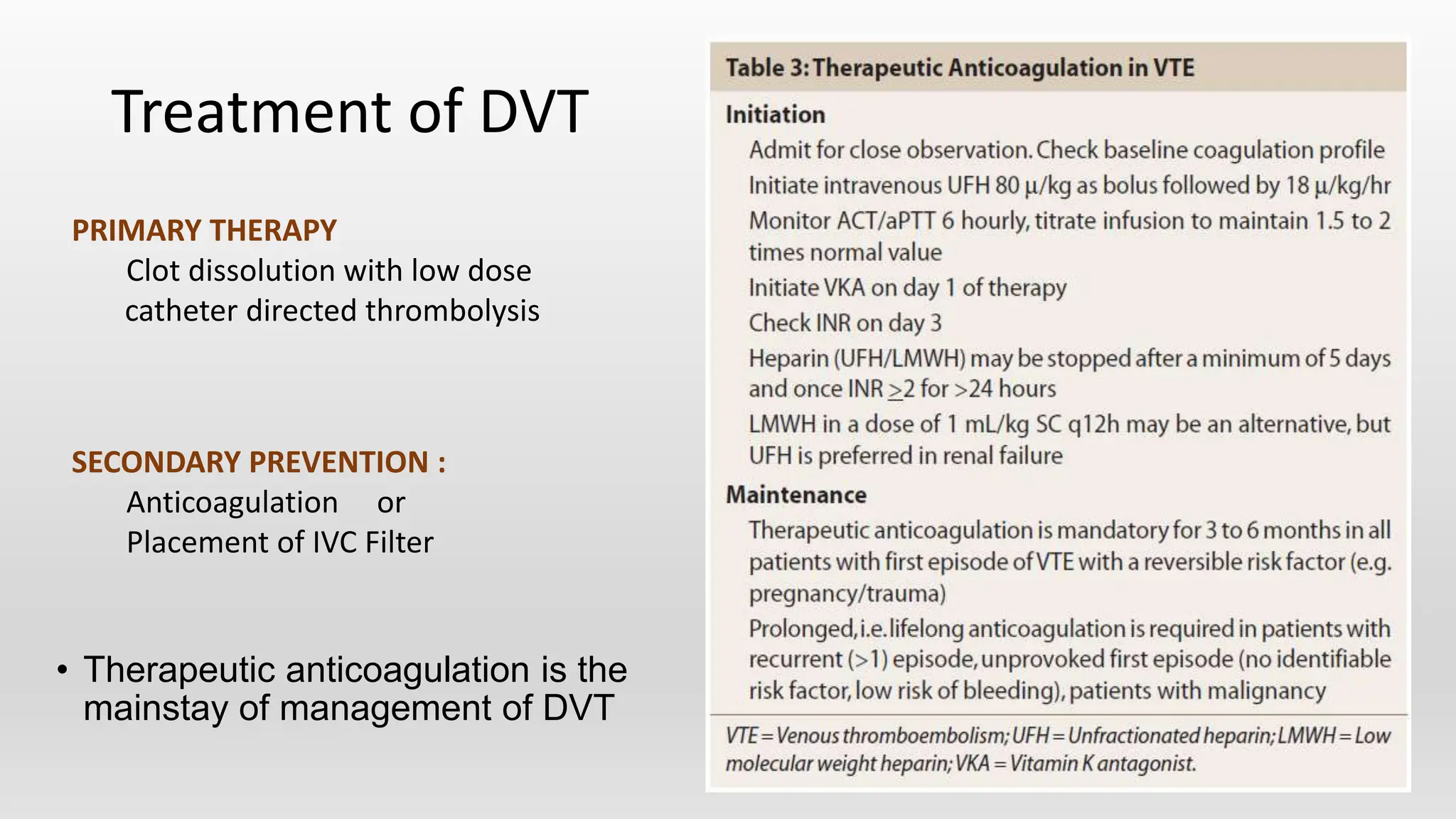
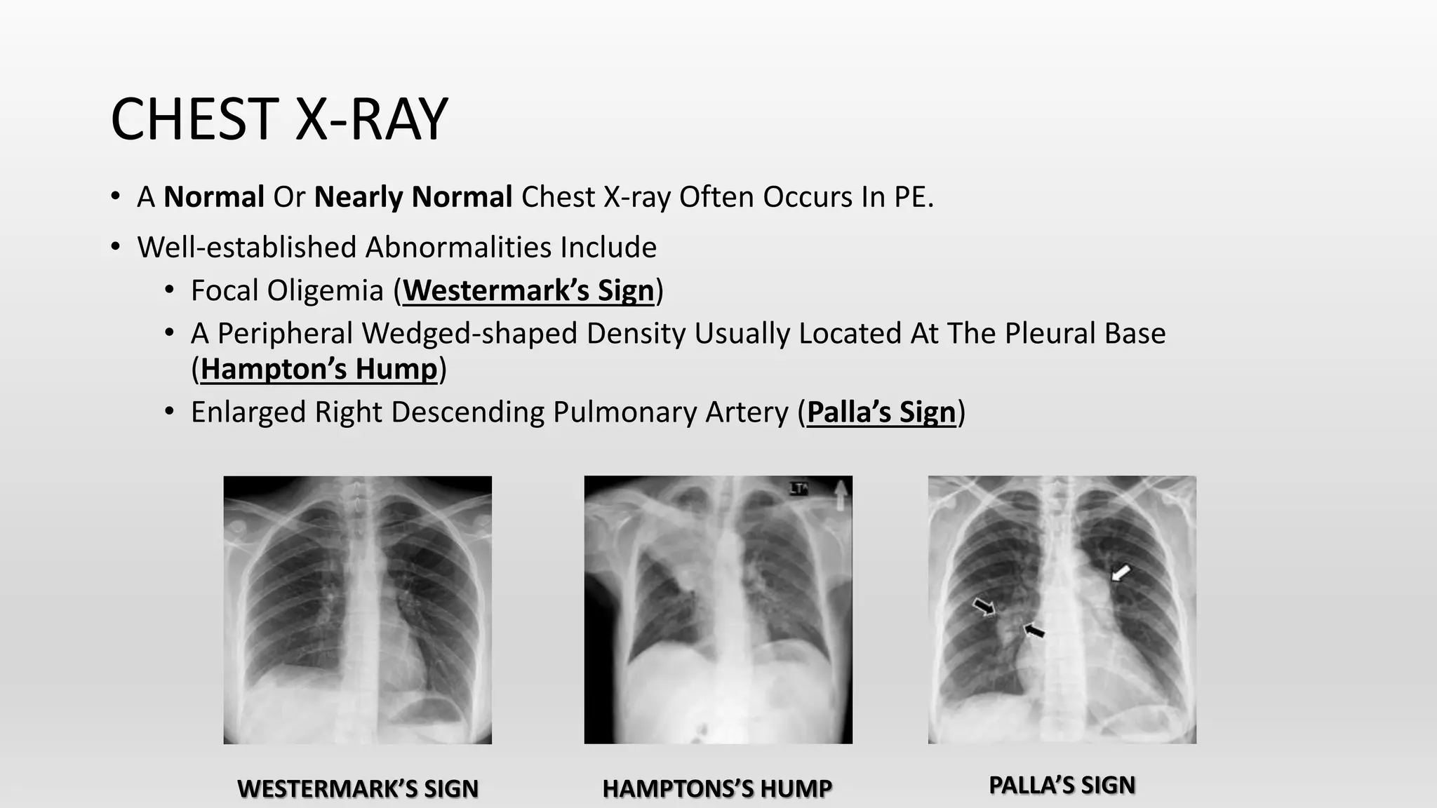
Venous Thromboembolism
Dr. Aditya Ashok Mahankar
Junior Resident
Dept. of Medicine
Venous Thromboembolism (VTE)
•VTE emcompasses
1) Deep Venous Thrombosis (DVT)
2) Pulmonary embolism (PE)
Introduction
• Pulmonary Thromboembolism is 3rd most common cause of
cardiovascular death after acute myocardial infarction and stroke.
• Leading to PE and DVT frequently results in Post-Thrombotic
Syndrome, Which is a major cause of Long-term disability.
Defination (PTE)
• Pulmonary thromboembolism (PTE) is defined as a condition where
there is significant obstruction of a part or whole of the pulmonary
arterial tree by a thrombus, migrating from a site outside the lung.
Risk Factors
• Prior DVT or PE
• Prolonged Immobility
• Surgery
• Major Abdominal / Pelvic
• Orthopaedics
• Obstetrics
• Pregnancy
• Estrogen Containing Contraceptives
• Postmenopausal Hormone Replacement
Therapy
• Cardiorespiratory
• COPD
• Congestive Cardiac Failure
• Systemic Arterial Hypertension
• Lower Limb Fractures
• Long Air Travel
• Factor V Leiden Deficiency
• Protein C Deficiency
• Protein S Deficiency
• Anti Thrombin Deficiency
• Hyperhomocystinemia
• SLE, APLA Syndrome
• Malignant Disease
• Severe Sepsis
• Obesity
• Age >40 yrs
• Cigarette Smoking
API
Kumar & clarks clinical
medicine
Risk factor differ in Degree to which they increases the likely hood of VTE occurring
• Major Surgery,
• Trauma,
• Absolute Bed rest
Risk by 10-50
fold
• Pregnancy,
• Oestrogen therapy,
• Minor Sx.,
• Heritable
thrombophilias
Risk by 3-10
fold
• Obesity,
• Long haul travel.
Risk by 3 fold
PATHOPHYSIOLOGY
COAGULATION PATHWAYS
I. Fibrinogen
II. Prothrombin
III. Tissue
Thromboplastin
IV. Ionized Calcium
V. Labile Factor /
Proaccelerin
VI. Unassigned
VII. Stable Factor /
Proconvertin
VIII. Antihemophilic
Factor A
IX. Plasma
Thromboplastin
Component/
Christmas Factor
X. Stuart-Prower
Factor
XI. Plasma
Thromboplastin
Antecedent
Inflamation
• Inflamation takes
centre stage as a
trigger of Acute PE
and DVT
VIRCHOW’S TRIAD
-Trauma
- Surgery
- Venepuncture
- Chemical
Irritation
-Atherosclerosis
- Heart Valve
Disease or
Replacement
- Indwelling
Catheters
Endothelial
Injury
-Immobility
-Bed Rest
-Anaesthesia
-CCF / Cor
pulmonale
-AF
-prior venous
thrombosis
Stasis
Hypercoagulability
2. Prothrombotic States
• The Two Most Common Autosomal Dominant Genetic Mutations Are
i. Factor V Leiden (Causes Resistance To The Endogenous Anticoagulant,
Activated Protein C)
ii. Prothrombin Gene (Increases The Plasma Prothrombin Concentration)
• Antithrombin, Protein C, And Protein S Are Naturally Occurring Coagulation
Inhibitors. Deficiencies Of These Inhibitors Are Associated With VTE But Are Rare.
• Antiphospholipid Antibody Syndrome Is The Most Common Acquired Cause Of
Thrombophilia And Is Associated With Venous Or Arterial Thrombosis.
3. Embolization
• When Deep Venous Thrombi Detach From Their Site Of Formation,
They Embolize To The Vena Cava, Right Atrium, And Right Ventricle,
And Lodge In The Pulmonary Arterial Circulation, Thereby Causing
Acute PE.
• Many Patients With PE Have No Evidence Of DVT Because The Clot
Has Already Embolized To The Lungs.
• Paradoxically, These Thrombi Occasionally Embolize To The Arterial
Circulation Through A Patent Foramen Ovale Or Atrial Septal Defect.
4. Pulmonary Hypertension
&
Right Ventricular Dysfunction
Pulmonary Artery Occlusion
Decrease Pulmonary
Blood Flow
RA/ RV Dilation Increase RV Contraction
Decrease LA & LV
Filling
Decrease
Stroke Volume
Decrease
Cardiac output
Hypotension, obstructive shock,
cardiac arrest
Decrease LA &
LV Volume
Increase Rv Demand
Supply Demand
Mismatch-----Ischemia
decrease Blood Supply
Decrease Coronary Blood Flow
CLASSIFICATION
Deep Venous Thrombosis
• Lower Extremity DVT –
• Usually Begins In The Calf And Propagates Proximally To The Popliteal Vein,
Femoral Vein, And Iliac Veins.
• Leg DVT Is About 10 Times More Common Than Upper Extremity DVT.
• Upper Extremity DVT –
• Often Precipitated By Placement Of Pacemakers, Internal Cardiac Defibrillators,
Or Indwelling Central Venous Catheters.
• The Likelihood Of Upper Extremity DVT Increases As The Catheter Diameter And
Number Of Lumens Increase.
• Superficial Venous Thrombosis –
• Usually Presents With Erythema, Tenderness, And A “Palpable Cord.”
• Patients Are At Risk For Extension Of The Thrombosis To The Deep-venous
System.
Pathological classification (Depending on what embolises)
• Thrombus (mainly from DVt of Lower limnb)
• NON thrombotic (rare)
1. Amniotic Fluid
2. Tumor (d/t Rt. Atrial Myxoma)
3. Fat Embolism (d/t Long Bone Fracture)
4. Septic Embolism (in Septicemia)
5. Air Embolism (during central line insertion, pacemaker etc)
Pulmonary Embolism
Hemodynamic Classification
• Massive PE – occlusion of pulmonary tree >50%
• Submassive PE – occlusion of pulmonary tree 30 % to 50%
• Low-risk PE – also known as non massive PE
occlusion of pulmonary tree <30 %
Clinical Evaluation / Presentation
PTE
• The Most Common Symptom Is Unexplained Dyspnoea (with Normal Chest X-ray)
• Dyspnea > Chest Pain (Pleuritic : non massive)
• Cough with hemoptysis : non massive
• Tachypnea >> Tachycardia > Rales / Decrease breath sounds
• Shock : RV Failure
• Raised JVP/ s3 gallop rhythm
• Dyspnoea within seconds
• Hypoxemia / respiratory alkalosis/ type 1 respiratory failure
DVT
1) Cramp ( Charley Horse ) : in lower calf
Most common symptom
2) Moses sign : Redness, warmth, and tenderness on calf compression
3) Homan’s Sign : Calf pain on dorsiflexion of foot with the knee bent
Differential
Diagnosis
Diagnostic Modalities
1) Non –Imaging Diagnostic Modalities.
• Wells Point Score Criteria Help Estimate The Clinical Likelihood Of
DVT And PE.
The diagnostic process start with assessment of clinical
probability using a clinical prediction score : 1st step
Measurement of D-Dimer
• Fibrin degradation product by plasmin.
• Elevation of D-dimer indicates endogenous ineffective thrombolysis.
• Sensetivity : >80% for DVT & >95% for PE
• Highly sensitive but not specific (Because increase levels seen in MI, Pneumonia,
Sepsis, Cancer, Post operative state, 2nd & 3rd trimester of pregnancy) ,
So Not Usefull in IPD patient (only Usefull in OPD Patient)
• Normal D-Dimer Value = Rule out test for PE
However, Patients With A High Clinical Likelihood Of VTE Should Skip D-dimer Testing And Undergo
Imaging As The Next Step In The Diagnostic Algorithm
2. a) Imaging Modalities in DVT
Non Invasive
(I)) Duplex Ultrasound : Venous compression Ultrasound
(CUS) + Doppler Ultrasound
Highly sensitive & specific for Thigh DVT
Less sensitive & Specific for calf DVT
(II) Impendance Plethysmography : More sensitive & specific for thigh
DVT than Calf DVT
Invasive
(I) Contrast Venography : Gold Standard (Seldom used presently
(CT & MRI alternative imaging modalities for Pt. with technically poor & nondiagnostic
Venous ultrasound)
Treatment of DVT
• Therapeutic anticoagulation is the
mainstay of management of DVT
PRIMARY THERAPY
Clot dissolution with low dose
catheter directed thrombolysis
SECONDARY PREVENTION :
Anticoagulation or
Placement of IVC Filter
• Indication of IVC Fiilters :
(a) Pt. with Absolute contraindication to anticoagulation.
(b) Pt. with recurrent VTE while receiving therapeutic dose of
anticoagulation.
For Patient with Acute DVT with Leg swelling : Below knee compression stocking
( to lessen the pt. discomfort.)
30-40mmHg or 20 to 30 mmHg
Replace every 3 month (because they loose there elasticity)
Prophylaxis for DVT
• Increased RV Wall Tension Also Compresses The Right Coronary Artery, Limits
Myocardial Oxygen Supply, And Precipitates Right Coronary Artery Ischemia And
RV Microinfarction, With Release Of Cardiac Biomarkers Such As Troponin.
• Serum Troponin And Plasma Heart-type Fatty Acid–Binding Protein (H-FABP)
Levels Increase Because Of RV Microinfarction.
• Myocardial Stretch Causes Release Of Brain Natriuretic Peptide (BNP), NT-pro-
BNP.
ELEVATED CARDIAC BIOMARKERS
ELECTROCARDIOGRAM
The Most Frequently Cited Abnormality Is The S1Q3T3 Sign
Seen In Approximately 20% Of PE Patients Suggesting High
Pressure In Right Ventricle.
• S Wave In Lead I
• Q Wave In Lead III
• Inverted T Wave In Lead III
• PE May Also Show
• Sinus Tachycardia
• Transient RBBB / RAD
• New Onset AF Or Afib.
• Pulmonary Strain Pattern
• i.e. T Wave Inversion In Leads V1 - V4.
Arterial Blood Gas Abnormalities
• Hypoxia
• Hypocapnia
Massive PE may be presented with Hypercapnia (Increase in dead
space) & respiratory acidosis with low end tidal co2
Findings are non specific and not always present
CHEST X-RAY
• A Normal Or Nearly Normal Chest X-ray Often Occurs In PE.
• Well-established Abnormalities Include
• Focal Oligemia (Westermark’s Sign)
• A Peripheral Wedged-shaped Density Usually Located At The Pleural Base
(Hampton’s Hump)
• Enlarged Right Descending Pulmonary Artery (Palla’s Sign)
WESTERMARK’S SIGN HAMPTONS’S HUMP PALLA’S SIGN
LUNG SCANNING (Ventilation – perfusion scan)
• Lung Scanning Is A Second-line Diagnostic Test For PE, Used Mostly For Patients Who
Cannot Tolerate Intravenous Contrast.
1. Perfusion Scans, Obtained With Small Particulate Aggregates Of Albumin Labelled
With A Gamma-Emitting Radionuclide Are Injected Intravenously And Are Trapped In
The Pulmonary Capillary Bed.
• The Perfusion Scan Defect Indicates Absent Or Decreased Blood Flow, Possibly Due
To PE.
2. Ventilation Scans, Obtained With A Radiolabeled Inhaled Gas Such As Xenon Or
Krypton, Improve The Specificity Of The Perfusion Scan.
• Abnormal Ventilation Scans Indicate Abnormal Nonventilated Lung, Thereby
Providing Possible Explanations For Perfusion Defects Other Than Acute PE, Such As
Asthma And COPD.
• A High-Probability Scan For PE Is Defined As Two Or More Segmental Perfusion Defects
In The Presence Of Normal Ventilation.
ECHOCARDIOGRAPHY
• 2D ECHO Is Not A Reliable Diagnostic Imaging Tool For Acute PE.
• However, Echocardiography Is A Very Useful Diagnostic Tool For Detecting Conditions That
May Mimic PE, Such As
-Acute MI -Pericardial Tamponade -Aortic Dissection
• One Should Consider Transesophageal Echocardiography When CT Is Not Available Or
When Patient Has Renal Failure Or Contrast Allergy.
• RA RV Dilatation
• The Best Indirect Sign Of PE
On Transthoracic 2D ECHO Is
*McConnell’s Sign:
RV free wall hypokinesia + RV apex hyperkineis
Since entireRV is Hypokinetic  hence LV increases contrac tion adjacent RV
apex is pull by hyperkinetic LV
Seen in Massive PE
INVASIVE DIAGNOSTIC MODALITIES
PULMONARY ANGIOGRAPHY
• A Definitive Diagnosis Of PE Requires Visualization Of An Intraluminal Filling Defect In More Than One Projection.
• Pulmonary artery dilatation
• Secondary Signs Of PE Include
• Abrupt Occlusion (“Cut-off”) Of Vessels,
• Segmental Oligemia Or Avascularity,
• Prolonged Arterial Phase With Slow Filling
• Tortuous, Tapering Peripheral Vessels.
N
O
R
M
A
L
P
E
MANAGEMENT
• Anticoagulant therapy is the standard treatment for VTE.
•
• IT is traditionally divided into three phases:
• the acute phase, lasting 5–10 days
• a maintenance phase, lasting a minimum of 3 months
• a long-term phase beyond this.
• The aim of treatment in the initial phase is to prevent thrombusextension and hence reduce
the risk of embolization; the goal thereafter is to prevent thrombus recurrence
Risk Stratification
ANTICOAGULATION
• Effective Anticoagulation Is The Foundation For Successful Treatment Of DVT And PE.
• There Are Three Major Strategies:
• (1) The Classical But Waning Strategy Of Parenteral Anticoagulation Bridged To
Warfarin.
• (2) Parenteral Therapy Switched After 5 Days To A Novel Oral Anticoagulant.
• (3) Oral Anticoagulation Monotherapy.
ANTICOAGULANTS
Parenteral
Indirect
Thrombin
Inhibitors
Heparin
(UFH)
LMWH
Fondaparinu
x
Direct
Thrombin
Inhibitors
Bivalirudin
Argatroban
Oral
Vit K
Antagonis
ts
Warfarin
Direct
Thrombin
Inhibitor
Dabigatra
n
Direct
Factor Xa
Inhibitor
Rivaroxaba
n
Apixaban
UNFRACTIONATED HEPARIN (UFH)
• Anticoagulates By Binding To And Accelerating The Activity Of Antithrombin, Thus Preventing
Additional Thrombus Formation.
• UFH Is Dosed To Achieve A Target aPTT Of 60–80 s.
• Dose: Initial Bolus 80U/Kg , f/b 18 U/Kg/hr in Pt with Normal LFT.
• The Major Advantage Of UFH Is Its Short Half-life, Useful In Patients In Hour-to-hour Control Of The
Intensity Of Anticoagulation.
LOW-MOLECULAR-WEIGHT HEPARINS
• Less Binding To Plasma Proteins And Endothelial Cells And Greater Bioavailability, A More Predictable
Dose Response, And A Longer Half-life Than Does UFH.
• No Monitoring Or Dose Adjustment Is Needed Unless The Patient Is Markedly Obese Or Has Chronic
Kidney Disease.
FONDAPARINUX
• Fondaparinux, An Anti-Xa Pentasaccharide, Is Administered As A Weight-Based Once-Daily
Subcutaneous Injection In A Prefilled Syringe.
• No Laboratory Monitoring Of aPTT Is Required.
• Fondaparinux Is Synthesized In A Laboratory And, Unlike LMWH Or UFH, Is Not Derived From
Animal Products.
• It Does Not Cause Heparin-Induced Thrombocytopenia.
• The Dose Must Be Adjusted Downward For Patients With Renal Dysfunction.
WARFARIN
• This Vitamin K Antagonist Prevents Activation Of Coagulation Factors II, VII, IX, And X.
• The Full Effect Of Warfarin Requires At Least 5 Days.
• Warfarin Dosing
• In An Average-Size Adult, Warfarin Is Often Initiated In A Dose Of 5 mg.
• The PT Is Standardized By INR, Which Assesses The Anticoagulant Effect.
• The Target INR Is Usually 2.5, With A Range Of 2.0–3.0.
• The Warfarin Dose Is Usually Titrated Empirically To Achieve The Target INR.
• Warfarin Can Cause Major Haemorrhage, Including Intracranial Haemorrhage, Even When The
INR Remains Within The Desired Therapeutic Range.
NOVEL ORAL ANTICOAGULANTS
• Novel Oral Anticoagulants (NOACs)
o Administered In A Fixed Dose
o Establish Effective Anticoagulation Within Hours
o Require No Laboratory Coagulation Monitoring
o Have Fewer Drug-Drug Or Drug-Food Interactions.
• BETRIXABAN, A Direct Factor Xa Inhibitor, Was Approved By The FDA In 2017 For VTE
Prophylaxis In Acutely Ill Medical Patients During Hospitalization.
• RIVAROXABAN And APIXABAN, Direct Factor Xa Inhibitors, Are Approved As Monotherapy.
• DABIGATRAN, A Direct Thrombin Inhibitor, And EDOXABAN, A Factor Xa Inhibitor, Are Approved
After An Initial 5-day Course Of Parenteral Anticoagulation.
FIBRINOLYSIS
• Successful Fibrinolytic Therapy Rapidly Reverses Right Heart Failure By
1. Dissolving Much Of The Thrombus
2. Preventing The Release Of Serotonin And Other Factors That Exacerbate Pulmonary
Hypertension
3. Lysing Much Of The Source Thrombus In The Pelvic Or Deep Leg Veins
• The Preferred Fibrinolytic Regimen Is 100 mg Of Recombinant Tissue Plasminogen
Activator (tPa) As A Continuous Peripheral IV Infusion Over 2 Hrs.
• The Only FDA Approved Indication For PE Fibrinolysis Is Massive PE.
• Contraindications Include Intracranial Disease, Recent Surgery, And Trauma.
• The Overall Major Bleeding Rate Is About 10%.
Special circumstances
• Pregnant Women :
• Warfarin & DOAC’s cross the placenta so avoided.
• Warfarin causes embryoathy between 6th to 12th weeks of pregnancywith
skeletal abnormality with nasal hypoplasia & later in preganacy it causes
bleeding in fetus with neurological abnormalities with risk of IUD. So drug is safe
until 6th week.
• LMWH is Treatment of choice.
Breast feeding Mother :
Both warfarin & LMWH safe
PULMONARY THROMBOENDARTERECTOMY
• PE Patients With Pulmonary Hypertension Should Be Followed Up At About 6 Weeks With A
Repeat Echocardiogram To Determine Whether Pulmonary Arterial Pressure Has Normalized.
• Dyspnea Due To Chronic Thromboembolic Pulmonary Hypertension Can Be Markedly Reduced,
And Sometimes Even Cure Pulmonary Hypertension.
• Inoperable Patients Should Be Managed With Pulmonary Vasodilator Therapy And Balloon
Angioplasty Of Pulmonary Arterial Webs.
PULMONARY EMBOLECTOMY
• The Risk Of Major Haemorrhage With Systemically Administered Fibrinolysis Has Prompted A
Renaissance Of Interest In Surgical Embolectomy, An Operation That Had Almost Become Extinct.
• Prophylactic Therapy May Include
• Anticoagulation
• Early Ambulation For Postoperative And Postparturient Patients
• Application Of Elastic Stockings
• Intermittent Pneumatic Calf Compression
• Isometric Leg Exercises For Bedridden Patients.
• Low-dose UFH Or LMWH Is The Most Common Form Of In-hospital Prophylaxis.
• Extended-Duration Prophylaxis With Betrixaban, Appears To Be Both Effective And Safe In
Medically Ill Patients, And Is Undergoing FDA Review.
PREVENTION
Management of Bleeding Caused by
Anticoagulant Treatment
Warfarin • Vit k(10mg iv slowly), FFP, rVIIa (in emergency)
Heparin
• Protamine (1mg of protamine for every 100 units of standard heparin if
the last dose of heparin was given within 30 minutes and 0.5 to 0.75
mg/100 units of heparin if it was 30 to 60 minutes since the last dose)
LMWH • 1 mg of protamine for every 1 mg of LMWH.
Dabigatran • Idarucizumab (Monoclonal antibody)
Pulmonary and venous thromboembolism.pptx

Pulmonary and venous thromboembolism.pptx

  • 1.
    Venous Thromboembolism Dr. AdityaAshok Mahankar Junior Resident Dept. of Medicine
  • 2.
    Venous Thromboembolism (VTE) •VTEemcompasses 1) Deep Venous Thrombosis (DVT) 2) Pulmonary embolism (PE)
  • 3.
    Introduction • Pulmonary Thromboembolismis 3rd most common cause of cardiovascular death after acute myocardial infarction and stroke. • Leading to PE and DVT frequently results in Post-Thrombotic Syndrome, Which is a major cause of Long-term disability.
  • 4.
    Defination (PTE) • Pulmonarythromboembolism (PTE) is defined as a condition where there is significant obstruction of a part or whole of the pulmonary arterial tree by a thrombus, migrating from a site outside the lung.
  • 5.
    Risk Factors • PriorDVT or PE • Prolonged Immobility • Surgery • Major Abdominal / Pelvic • Orthopaedics • Obstetrics • Pregnancy • Estrogen Containing Contraceptives • Postmenopausal Hormone Replacement Therapy • Cardiorespiratory • COPD • Congestive Cardiac Failure • Systemic Arterial Hypertension • Lower Limb Fractures • Long Air Travel • Factor V Leiden Deficiency • Protein C Deficiency • Protein S Deficiency • Anti Thrombin Deficiency • Hyperhomocystinemia • SLE, APLA Syndrome • Malignant Disease • Severe Sepsis • Obesity • Age >40 yrs • Cigarette Smoking
  • 6.
    API Kumar & clarksclinical medicine
  • 7.
    Risk factor differin Degree to which they increases the likely hood of VTE occurring • Major Surgery, • Trauma, • Absolute Bed rest Risk by 10-50 fold • Pregnancy, • Oestrogen therapy, • Minor Sx., • Heritable thrombophilias Risk by 3-10 fold • Obesity, • Long haul travel. Risk by 3 fold
  • 8.
  • 9.
    COAGULATION PATHWAYS I. Fibrinogen II.Prothrombin III. Tissue Thromboplastin IV. Ionized Calcium V. Labile Factor / Proaccelerin VI. Unassigned VII. Stable Factor / Proconvertin VIII. Antihemophilic Factor A IX. Plasma Thromboplastin Component/ Christmas Factor X. Stuart-Prower Factor XI. Plasma Thromboplastin Antecedent
  • 10.
    Inflamation • Inflamation takes centrestage as a trigger of Acute PE and DVT
  • 11.
    VIRCHOW’S TRIAD -Trauma - Surgery -Venepuncture - Chemical Irritation -Atherosclerosis - Heart Valve Disease or Replacement - Indwelling Catheters Endothelial Injury -Immobility -Bed Rest -Anaesthesia -CCF / Cor pulmonale -AF -prior venous thrombosis Stasis
  • 12.
  • 13.
    2. Prothrombotic States •The Two Most Common Autosomal Dominant Genetic Mutations Are i. Factor V Leiden (Causes Resistance To The Endogenous Anticoagulant, Activated Protein C) ii. Prothrombin Gene (Increases The Plasma Prothrombin Concentration) • Antithrombin, Protein C, And Protein S Are Naturally Occurring Coagulation Inhibitors. Deficiencies Of These Inhibitors Are Associated With VTE But Are Rare. • Antiphospholipid Antibody Syndrome Is The Most Common Acquired Cause Of Thrombophilia And Is Associated With Venous Or Arterial Thrombosis.
  • 14.
    3. Embolization • WhenDeep Venous Thrombi Detach From Their Site Of Formation, They Embolize To The Vena Cava, Right Atrium, And Right Ventricle, And Lodge In The Pulmonary Arterial Circulation, Thereby Causing Acute PE. • Many Patients With PE Have No Evidence Of DVT Because The Clot Has Already Embolized To The Lungs. • Paradoxically, These Thrombi Occasionally Embolize To The Arterial Circulation Through A Patent Foramen Ovale Or Atrial Septal Defect.
  • 15.
    4. Pulmonary Hypertension & RightVentricular Dysfunction
  • 16.
    Pulmonary Artery Occlusion DecreasePulmonary Blood Flow RA/ RV Dilation Increase RV Contraction Decrease LA & LV Filling Decrease Stroke Volume Decrease Cardiac output Hypotension, obstructive shock, cardiac arrest Decrease LA & LV Volume Increase Rv Demand Supply Demand Mismatch-----Ischemia decrease Blood Supply Decrease Coronary Blood Flow
  • 17.
  • 18.
    Deep Venous Thrombosis •Lower Extremity DVT – • Usually Begins In The Calf And Propagates Proximally To The Popliteal Vein, Femoral Vein, And Iliac Veins. • Leg DVT Is About 10 Times More Common Than Upper Extremity DVT. • Upper Extremity DVT – • Often Precipitated By Placement Of Pacemakers, Internal Cardiac Defibrillators, Or Indwelling Central Venous Catheters. • The Likelihood Of Upper Extremity DVT Increases As The Catheter Diameter And Number Of Lumens Increase. • Superficial Venous Thrombosis – • Usually Presents With Erythema, Tenderness, And A “Palpable Cord.” • Patients Are At Risk For Extension Of The Thrombosis To The Deep-venous System.
  • 19.
    Pathological classification (Dependingon what embolises) • Thrombus (mainly from DVt of Lower limnb) • NON thrombotic (rare) 1. Amniotic Fluid 2. Tumor (d/t Rt. Atrial Myxoma) 3. Fat Embolism (d/t Long Bone Fracture) 4. Septic Embolism (in Septicemia) 5. Air Embolism (during central line insertion, pacemaker etc) Pulmonary Embolism
  • 20.
    Hemodynamic Classification • MassivePE – occlusion of pulmonary tree >50% • Submassive PE – occlusion of pulmonary tree 30 % to 50% • Low-risk PE – also known as non massive PE occlusion of pulmonary tree <30 %
  • 21.
  • 22.
    PTE • The MostCommon Symptom Is Unexplained Dyspnoea (with Normal Chest X-ray) • Dyspnea > Chest Pain (Pleuritic : non massive) • Cough with hemoptysis : non massive • Tachypnea >> Tachycardia > Rales / Decrease breath sounds • Shock : RV Failure • Raised JVP/ s3 gallop rhythm • Dyspnoea within seconds • Hypoxemia / respiratory alkalosis/ type 1 respiratory failure
  • 23.
    DVT 1) Cramp (Charley Horse ) : in lower calf Most common symptom 2) Moses sign : Redness, warmth, and tenderness on calf compression 3) Homan’s Sign : Calf pain on dorsiflexion of foot with the knee bent
  • 24.
  • 25.
    Diagnostic Modalities 1) Non–Imaging Diagnostic Modalities.
  • 26.
    • Wells PointScore Criteria Help Estimate The Clinical Likelihood Of DVT And PE. The diagnostic process start with assessment of clinical probability using a clinical prediction score : 1st step
  • 27.
    Measurement of D-Dimer •Fibrin degradation product by plasmin. • Elevation of D-dimer indicates endogenous ineffective thrombolysis. • Sensetivity : >80% for DVT & >95% for PE • Highly sensitive but not specific (Because increase levels seen in MI, Pneumonia, Sepsis, Cancer, Post operative state, 2nd & 3rd trimester of pregnancy) , So Not Usefull in IPD patient (only Usefull in OPD Patient) • Normal D-Dimer Value = Rule out test for PE However, Patients With A High Clinical Likelihood Of VTE Should Skip D-dimer Testing And Undergo Imaging As The Next Step In The Diagnostic Algorithm
  • 28.
    2. a) ImagingModalities in DVT Non Invasive (I)) Duplex Ultrasound : Venous compression Ultrasound (CUS) + Doppler Ultrasound Highly sensitive & specific for Thigh DVT Less sensitive & Specific for calf DVT (II) Impendance Plethysmography : More sensitive & specific for thigh DVT than Calf DVT Invasive (I) Contrast Venography : Gold Standard (Seldom used presently (CT & MRI alternative imaging modalities for Pt. with technically poor & nondiagnostic Venous ultrasound)
  • 30.
    Treatment of DVT •Therapeutic anticoagulation is the mainstay of management of DVT PRIMARY THERAPY Clot dissolution with low dose catheter directed thrombolysis SECONDARY PREVENTION : Anticoagulation or Placement of IVC Filter
  • 31.
    • Indication ofIVC Fiilters : (a) Pt. with Absolute contraindication to anticoagulation. (b) Pt. with recurrent VTE while receiving therapeutic dose of anticoagulation. For Patient with Acute DVT with Leg swelling : Below knee compression stocking ( to lessen the pt. discomfort.) 30-40mmHg or 20 to 30 mmHg Replace every 3 month (because they loose there elasticity)
  • 32.
  • 33.
    • Increased RVWall Tension Also Compresses The Right Coronary Artery, Limits Myocardial Oxygen Supply, And Precipitates Right Coronary Artery Ischemia And RV Microinfarction, With Release Of Cardiac Biomarkers Such As Troponin. • Serum Troponin And Plasma Heart-type Fatty Acid–Binding Protein (H-FABP) Levels Increase Because Of RV Microinfarction. • Myocardial Stretch Causes Release Of Brain Natriuretic Peptide (BNP), NT-pro- BNP. ELEVATED CARDIAC BIOMARKERS
  • 34.
    ELECTROCARDIOGRAM The Most FrequentlyCited Abnormality Is The S1Q3T3 Sign Seen In Approximately 20% Of PE Patients Suggesting High Pressure In Right Ventricle. • S Wave In Lead I • Q Wave In Lead III • Inverted T Wave In Lead III • PE May Also Show • Sinus Tachycardia • Transient RBBB / RAD • New Onset AF Or Afib. • Pulmonary Strain Pattern • i.e. T Wave Inversion In Leads V1 - V4.
  • 35.
    Arterial Blood GasAbnormalities • Hypoxia • Hypocapnia Massive PE may be presented with Hypercapnia (Increase in dead space) & respiratory acidosis with low end tidal co2 Findings are non specific and not always present
  • 36.
    CHEST X-RAY • ANormal Or Nearly Normal Chest X-ray Often Occurs In PE. • Well-established Abnormalities Include • Focal Oligemia (Westermark’s Sign) • A Peripheral Wedged-shaped Density Usually Located At The Pleural Base (Hampton’s Hump) • Enlarged Right Descending Pulmonary Artery (Palla’s Sign) WESTERMARK’S SIGN HAMPTONS’S HUMP PALLA’S SIGN
  • 37.
    LUNG SCANNING (Ventilation– perfusion scan) • Lung Scanning Is A Second-line Diagnostic Test For PE, Used Mostly For Patients Who Cannot Tolerate Intravenous Contrast. 1. Perfusion Scans, Obtained With Small Particulate Aggregates Of Albumin Labelled With A Gamma-Emitting Radionuclide Are Injected Intravenously And Are Trapped In The Pulmonary Capillary Bed. • The Perfusion Scan Defect Indicates Absent Or Decreased Blood Flow, Possibly Due To PE. 2. Ventilation Scans, Obtained With A Radiolabeled Inhaled Gas Such As Xenon Or Krypton, Improve The Specificity Of The Perfusion Scan. • Abnormal Ventilation Scans Indicate Abnormal Nonventilated Lung, Thereby Providing Possible Explanations For Perfusion Defects Other Than Acute PE, Such As Asthma And COPD. • A High-Probability Scan For PE Is Defined As Two Or More Segmental Perfusion Defects In The Presence Of Normal Ventilation.
  • 38.
    ECHOCARDIOGRAPHY • 2D ECHOIs Not A Reliable Diagnostic Imaging Tool For Acute PE. • However, Echocardiography Is A Very Useful Diagnostic Tool For Detecting Conditions That May Mimic PE, Such As -Acute MI -Pericardial Tamponade -Aortic Dissection • One Should Consider Transesophageal Echocardiography When CT Is Not Available Or When Patient Has Renal Failure Or Contrast Allergy. • RA RV Dilatation • The Best Indirect Sign Of PE On Transthoracic 2D ECHO Is *McConnell’s Sign: RV free wall hypokinesia + RV apex hyperkineis Since entireRV is Hypokinetic  hence LV increases contrac tion adjacent RV apex is pull by hyperkinetic LV Seen in Massive PE
  • 39.
    INVASIVE DIAGNOSTIC MODALITIES PULMONARYANGIOGRAPHY • A Definitive Diagnosis Of PE Requires Visualization Of An Intraluminal Filling Defect In More Than One Projection. • Pulmonary artery dilatation • Secondary Signs Of PE Include • Abrupt Occlusion (“Cut-off”) Of Vessels, • Segmental Oligemia Or Avascularity, • Prolonged Arterial Phase With Slow Filling • Tortuous, Tapering Peripheral Vessels. N O R M A L P E
  • 42.
  • 43.
    • Anticoagulant therapyis the standard treatment for VTE. • • IT is traditionally divided into three phases: • the acute phase, lasting 5–10 days • a maintenance phase, lasting a minimum of 3 months • a long-term phase beyond this. • The aim of treatment in the initial phase is to prevent thrombusextension and hence reduce the risk of embolization; the goal thereafter is to prevent thrombus recurrence
  • 44.
  • 45.
    ANTICOAGULATION • Effective AnticoagulationIs The Foundation For Successful Treatment Of DVT And PE. • There Are Three Major Strategies: • (1) The Classical But Waning Strategy Of Parenteral Anticoagulation Bridged To Warfarin. • (2) Parenteral Therapy Switched After 5 Days To A Novel Oral Anticoagulant. • (3) Oral Anticoagulation Monotherapy.
  • 46.
  • 48.
    UNFRACTIONATED HEPARIN (UFH) •Anticoagulates By Binding To And Accelerating The Activity Of Antithrombin, Thus Preventing Additional Thrombus Formation. • UFH Is Dosed To Achieve A Target aPTT Of 60–80 s. • Dose: Initial Bolus 80U/Kg , f/b 18 U/Kg/hr in Pt with Normal LFT. • The Major Advantage Of UFH Is Its Short Half-life, Useful In Patients In Hour-to-hour Control Of The Intensity Of Anticoagulation. LOW-MOLECULAR-WEIGHT HEPARINS • Less Binding To Plasma Proteins And Endothelial Cells And Greater Bioavailability, A More Predictable Dose Response, And A Longer Half-life Than Does UFH. • No Monitoring Or Dose Adjustment Is Needed Unless The Patient Is Markedly Obese Or Has Chronic Kidney Disease.
  • 49.
    FONDAPARINUX • Fondaparinux, AnAnti-Xa Pentasaccharide, Is Administered As A Weight-Based Once-Daily Subcutaneous Injection In A Prefilled Syringe. • No Laboratory Monitoring Of aPTT Is Required. • Fondaparinux Is Synthesized In A Laboratory And, Unlike LMWH Or UFH, Is Not Derived From Animal Products. • It Does Not Cause Heparin-Induced Thrombocytopenia. • The Dose Must Be Adjusted Downward For Patients With Renal Dysfunction.
  • 51.
    WARFARIN • This VitaminK Antagonist Prevents Activation Of Coagulation Factors II, VII, IX, And X. • The Full Effect Of Warfarin Requires At Least 5 Days. • Warfarin Dosing • In An Average-Size Adult, Warfarin Is Often Initiated In A Dose Of 5 mg. • The PT Is Standardized By INR, Which Assesses The Anticoagulant Effect. • The Target INR Is Usually 2.5, With A Range Of 2.0–3.0. • The Warfarin Dose Is Usually Titrated Empirically To Achieve The Target INR. • Warfarin Can Cause Major Haemorrhage, Including Intracranial Haemorrhage, Even When The INR Remains Within The Desired Therapeutic Range.
  • 52.
    NOVEL ORAL ANTICOAGULANTS •Novel Oral Anticoagulants (NOACs) o Administered In A Fixed Dose o Establish Effective Anticoagulation Within Hours o Require No Laboratory Coagulation Monitoring o Have Fewer Drug-Drug Or Drug-Food Interactions. • BETRIXABAN, A Direct Factor Xa Inhibitor, Was Approved By The FDA In 2017 For VTE Prophylaxis In Acutely Ill Medical Patients During Hospitalization. • RIVAROXABAN And APIXABAN, Direct Factor Xa Inhibitors, Are Approved As Monotherapy. • DABIGATRAN, A Direct Thrombin Inhibitor, And EDOXABAN, A Factor Xa Inhibitor, Are Approved After An Initial 5-day Course Of Parenteral Anticoagulation.
  • 54.
    FIBRINOLYSIS • Successful FibrinolyticTherapy Rapidly Reverses Right Heart Failure By 1. Dissolving Much Of The Thrombus 2. Preventing The Release Of Serotonin And Other Factors That Exacerbate Pulmonary Hypertension 3. Lysing Much Of The Source Thrombus In The Pelvic Or Deep Leg Veins • The Preferred Fibrinolytic Regimen Is 100 mg Of Recombinant Tissue Plasminogen Activator (tPa) As A Continuous Peripheral IV Infusion Over 2 Hrs. • The Only FDA Approved Indication For PE Fibrinolysis Is Massive PE. • Contraindications Include Intracranial Disease, Recent Surgery, And Trauma. • The Overall Major Bleeding Rate Is About 10%.
  • 56.
    Special circumstances • PregnantWomen : • Warfarin & DOAC’s cross the placenta so avoided. • Warfarin causes embryoathy between 6th to 12th weeks of pregnancywith skeletal abnormality with nasal hypoplasia & later in preganacy it causes bleeding in fetus with neurological abnormalities with risk of IUD. So drug is safe until 6th week. • LMWH is Treatment of choice. Breast feeding Mother : Both warfarin & LMWH safe
  • 57.
    PULMONARY THROMBOENDARTERECTOMY • PEPatients With Pulmonary Hypertension Should Be Followed Up At About 6 Weeks With A Repeat Echocardiogram To Determine Whether Pulmonary Arterial Pressure Has Normalized. • Dyspnea Due To Chronic Thromboembolic Pulmonary Hypertension Can Be Markedly Reduced, And Sometimes Even Cure Pulmonary Hypertension. • Inoperable Patients Should Be Managed With Pulmonary Vasodilator Therapy And Balloon Angioplasty Of Pulmonary Arterial Webs. PULMONARY EMBOLECTOMY • The Risk Of Major Haemorrhage With Systemically Administered Fibrinolysis Has Prompted A Renaissance Of Interest In Surgical Embolectomy, An Operation That Had Almost Become Extinct.
  • 58.
    • Prophylactic TherapyMay Include • Anticoagulation • Early Ambulation For Postoperative And Postparturient Patients • Application Of Elastic Stockings • Intermittent Pneumatic Calf Compression • Isometric Leg Exercises For Bedridden Patients. • Low-dose UFH Or LMWH Is The Most Common Form Of In-hospital Prophylaxis. • Extended-Duration Prophylaxis With Betrixaban, Appears To Be Both Effective And Safe In Medically Ill Patients, And Is Undergoing FDA Review. PREVENTION
  • 59.
    Management of BleedingCaused by Anticoagulant Treatment Warfarin • Vit k(10mg iv slowly), FFP, rVIIa (in emergency) Heparin • Protamine (1mg of protamine for every 100 units of standard heparin if the last dose of heparin was given within 30 minutes and 0.5 to 0.75 mg/100 units of heparin if it was 30 to 60 minutes since the last dose) LMWH • 1 mg of protamine for every 1 mg of LMWH. Dabigatran • Idarucizumab (Monoclonal antibody)