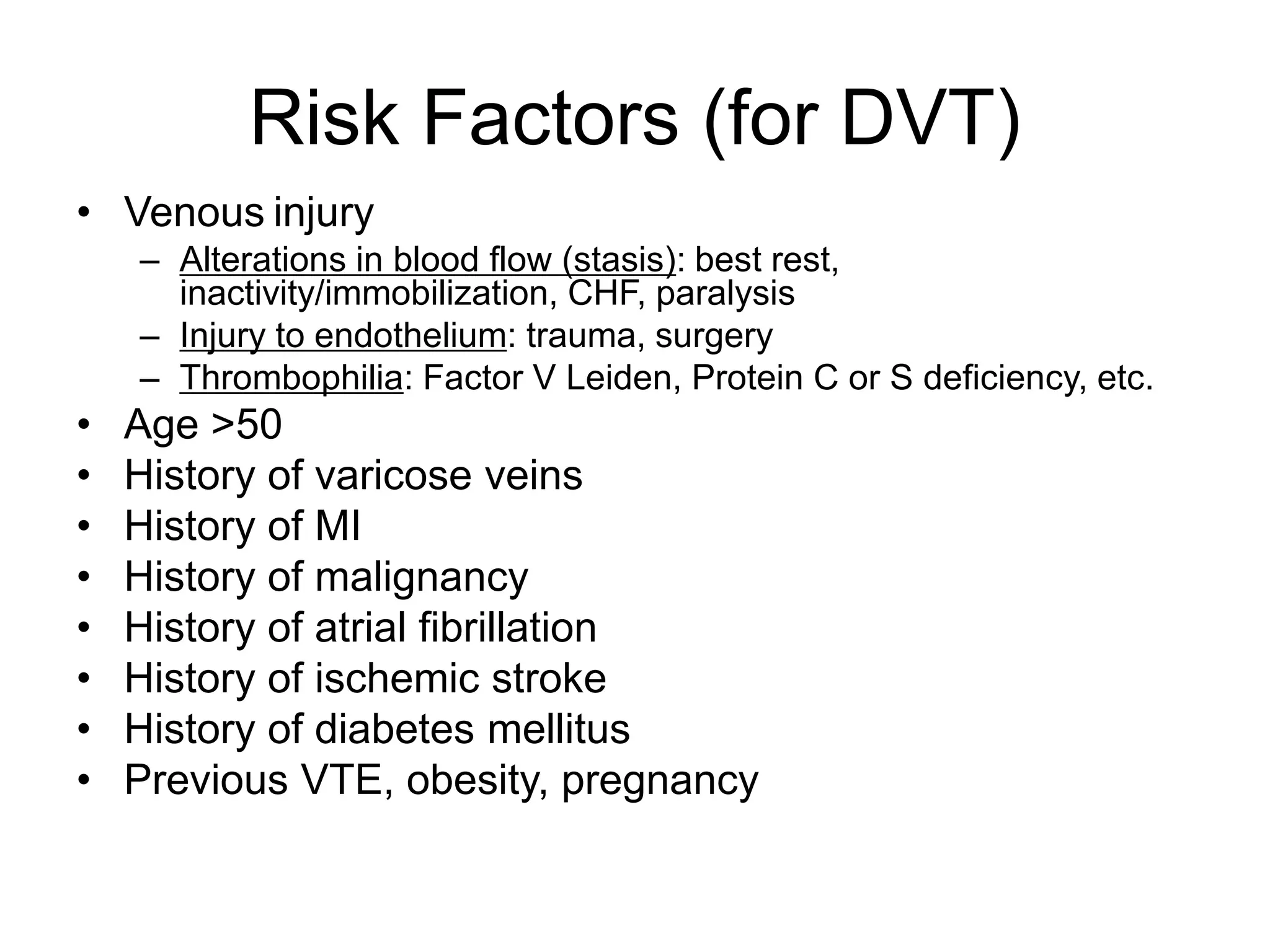
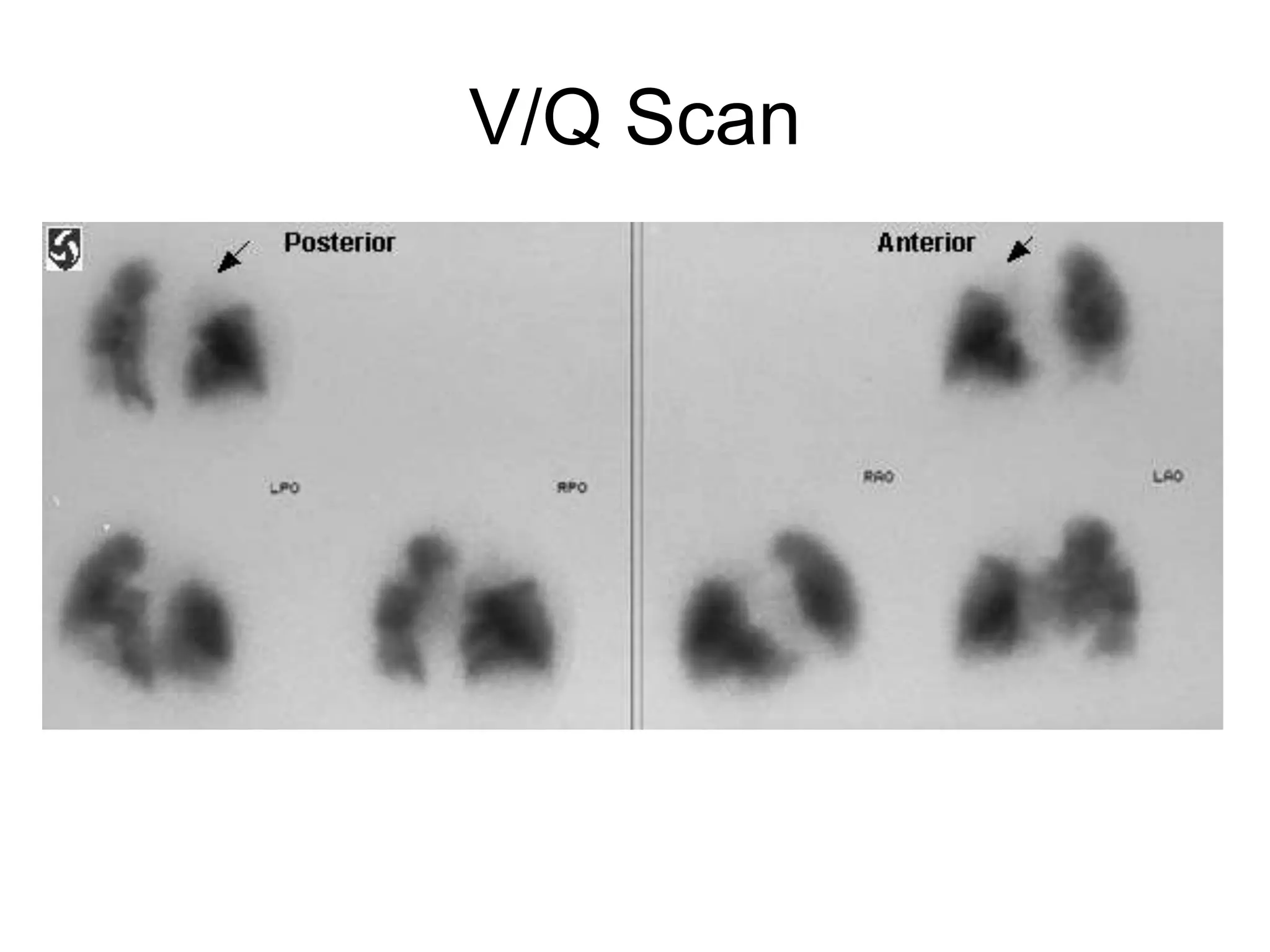
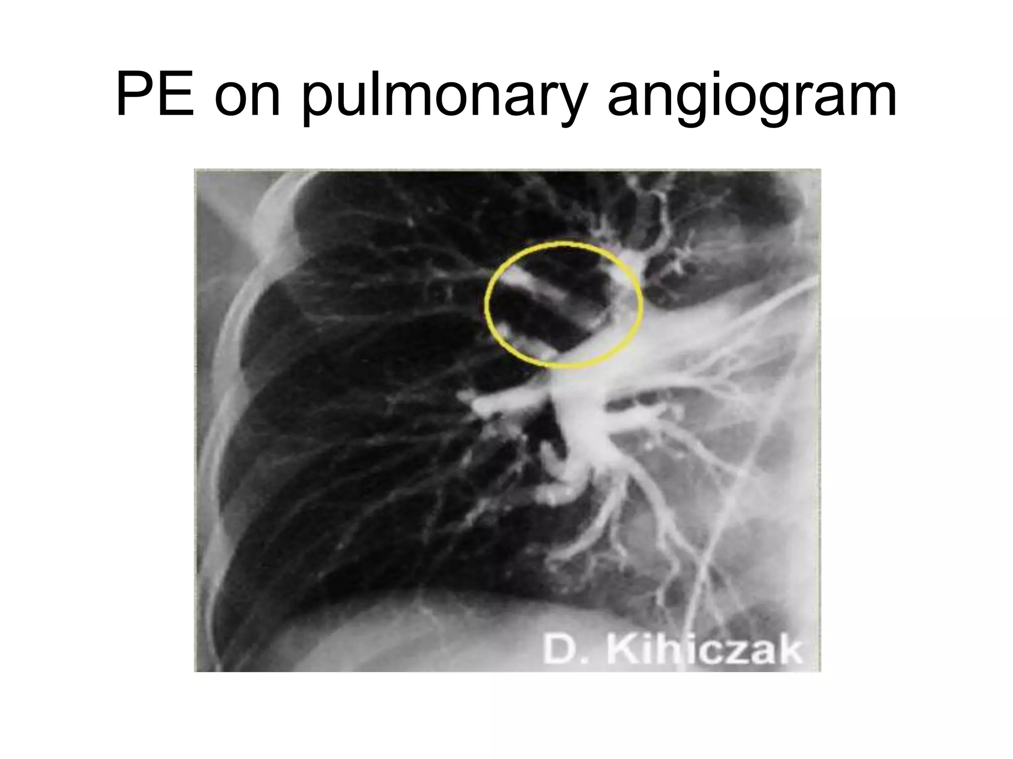
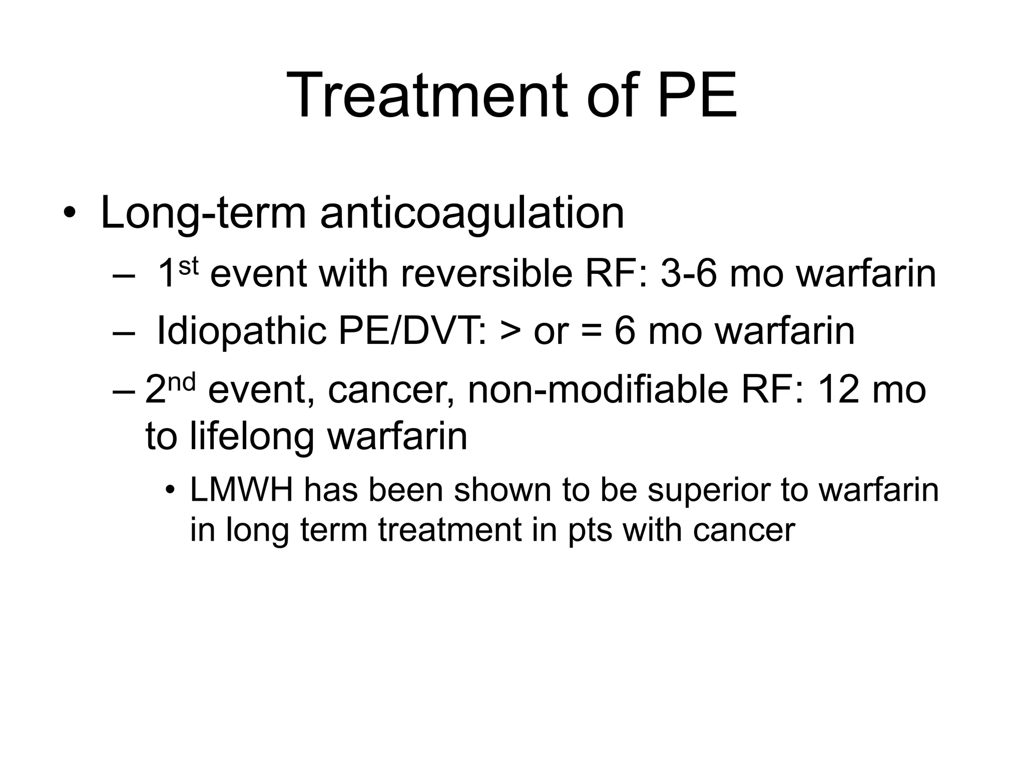
Pulmonary Embolism
Diagnosis, Treatment, and Prevention
By: (Afework A.)
Pulmonary Embolism
• Thrombosis that originates in the venous
system and embolizes to the pulmonary
arterial circulation
– DVT in veins of leg above the knee (>90%)
– DVT elsewhere (pelvic, arm, calf veins, etc.)
– Cardiac thrombi
How Common?
• 650,000 cases in the US each year
• 150,000 – 200,000 US deaths each year
• Most common preventable cause of
hospital death
• 3rd most common acute cardiovascular
emergency (MI and stroke)
Risk Factors (for DVT)
• Venous injury
– Alterations in blood flow (stasis): best rest,
inactivity/immobilization, CHF, paralysis
– Injury to endothelium: trauma, surgery
– Thrombophilia: Factor V Leiden, Protein C or S deficiency, etc.
• Age >50
• History of varicose veins
• History of MI
• History of malignancy
• History of atrial fibrillation
• History of ischemic stroke
• History of diabetes mellitus
• Previous VTE, obesity, pregnancy
5
Pathophysiology
Rudolph Virchow, 1858
Triad:
• Hypercoagulability
• Stasis to flow
• Vessel injury
6
Risk Factors
Hypercoagulability
Malignancy
Nonmalignant thrombophilia
Pregnancy
Postpartum status (<4wk)
Estrogen/ OCP’s
Genetic mutations (Factor V Leiden, Protein C & S deficiency, Factor
VIII, Prothrombin mutations, anti-thrombin III
deficiency)
Venous Statis
Bedrest > 24 hr
Recent cast or external fixator
Long-distance travel or prolong automobile travel
Venous Injury
Recent surgery requiring endotracheal intubation
Recent trauma (especially the lower extremities and pelvis)
7
Clinical Presentation
• The Classic Triad: (Hemoptysis, Dyspnea, Pleuritic
Pain)
• Not very common!
• Occurs in less than 20% of patients with documented
PE
• Three Clinical Presentations
– Pulmonary Infarction
– Submassive Embolism
– Massive Embolism
Clinical Presentation
• Asymptomatic
• Sudden onset of unexplained dyspnea
• Pleuritic chest pain
• Tachypnea
• Tachycardia
• Anxiety/agitation, cough, hemoptysis, syncope,
fever, cyanosis, isolated crackles, pleural friction
rub, loud P2, right-sided S3, pulmonary
insufficiency murmur, elevated JVP, right
ventricular heave, acute worsening of heart
failure or lung disease
Broad Differential
• Pneumothorax
• Myocardial ischemia
• Pericarditis
• Asthma
• Pneumonia
• MI with cardiogenic shock
• Cardiac tamponade
• Aortic dissection
• etc, etc, etc
Nonspecific Workup
• Chest X-ray: abnormal in 88% of acute PE
– Atelectasis (60-70%): most common finding in PE without infarction
– “Classic” findings:
• Westermark sign (increased lucency in area of embolus)
• Hampton Hump (wedge-shaped pleural-based infiltrate)
• Abrupt cutoff of vessel
– Pleural effusion
• EKG
– Most common: sinus tachycardia +/- nonspecific ST-segment and T-
wave changes
– “Classic S1-Q3-T3 pattern”
– Other signs of right heart strain (ie, new RBBB and ST changes in V1,2
• ABG
– Normal does NOT rule out PE
– “Classic” findings:
• Hypoxia, hypocapnia, respiratory alkalosis, increased A-a gradient
11
Chest X-ray Eponyms of PE
• Westermark's sign
– A dilation of the pulmonary vessels proximal to the
embolism along with collapse of distal vessels,
sometimes with a sharp cutoff.
• Hampton’s Hump
– A triangular or rounded pleural-based infiltrate with
the apex toward the hilum, usually located adjacent to
the hilum.
12
Radiographic Eponyms
- Hampton’s Hump, Westermark’s Sign
Westermark’s
Sign
Hampton’s Hump
13
Diagnostic Testing
– EKG’s
• EKG
– Most Common Findings:
• Tachycardia or nonspecific ST/T-wave changes
– Acute cor pulmonale or right strain patterns
• Tall peaked T-waves in lead II (P pulmonale)
• Right axis deviation
• RBBB
• S1-Q3-T3 (occurs in only 20% of PE patients)
EKG Findings
Evaluation and Diagnosis
• Evaluation and imaging
is dependent upon
estimated pretest
probability (Modified
Wells’ Criteria)
• Pretest probability:
– Low (<2 points)
– Intermediate (2-6 points)
– High (>6 points)
VARIABLE POINTS
S/S of DVT 3.0
HR >100 1.5
Immobilization
(bed rest >/= 3d)
OR surgery within
4 weeks
1.5
Prior DVT or PE 1.5
Hemoptysis 1.0
Malignancy
(treated within the
past 6 months or
palliative
1.0
Other diagnoses
less likely than PE
3.0
Preliminary Lab. Testing & Pretest Probability -2
• EKG:unexplained tachycardia:common in
APE but nonspecific
• acute cor pulmonale: S1, Q3, T3 pattern,
RBBB , P-wave pulmonale, or RAD : more
common with massive embolism ---
nonspecific
• CXR: generally nondiagnostic
• arterial oxygen tension may be normal
• A–a oxygen difference may be normal
Preliminary Lab. Testing & Pretest Probability -3
• D-dimer test (+): VTE are possible
diagnoses
• this test is nonspecific
• infection,other inflammatory states, cancer,
& trauma
• D-dimer testing is best considered
together with clinical probability
Preliminary Lab. Testing & Pretest Probability -4
• D-dimer test (-):with a low or moderate
pretest probability, likelihood of VTE is low
• precludes the need for specific imaging
studies
• high pretest probability: imaging should be
performed instead of D-dimer testing
• Other biomarkers: cardiac troponin levels,
plasma levels of brain natriuretic peptide
D-dimer in evaluation of PE
• High sensitivity but poor specificity
• Negative ELISA has >95% negative predictive value and can be
used to r/o PE in low risk patients (less than 2 points)
Low (<2) Intermediate
(2-6)
High (>6)
Overall 3% 20% 60%
(-) D-dimer 2% 6% 20%
(+) D-dimer 7% 36% 75%
Helical CT
• Sensitivity 85% (more sensitive for
proximal emboli)
• Specificity 95%
• Values vary widely in literature
Bilateral PE
V/Q Scan
• Identifies mismatches between areas that are ventilated
but not perfused
• Best initial test in patients with clear CXR
• Scan can be interpreted as High, Intermediate, or Low
probability of PE, or normal
– Normal rules out PE
– High-probability scan is diagnostic of PE if the clinical suspicion
is also high
– Low-probability scan rules out PE only in a pt with low pretest
clinical probability (because PE is found in roughly 15% of pts
with low-probability scans)
– Intermediate-probability scan requires further evaluation (16-
66% chance of PE depending on pretest probability)
V/Q Scan
Duplex US with compression of the
lower extremities
• Non-invasive test that accurately detects
proximal DVT in LE (70-80% of pts with
PE have concomitant proximal DVT)
• Often used in workup of PE before going
to more invasive procedures
Pulmonary Angiography
• “Gold Standard”
• Invasive study
• 5% morbidity
• < 0.5% mortality
• Indicated if the diagnosis remains
uncertain after noninvasive testing
PE on pulmonary angiogram
Treatment of PE
• Acute anticoagulation to therapeutic levels
– IV UFH: 80 U/kg bolus, then 18 U/kg/hr to goal PTT of
46-70 seconds OR
– LMWH: ie) lovenox 1 mg/kg SUBQ BID then start
warfarin (when PTT is therapeutic on UFH or on day 1
of LMWH), overlap x 5 days, titrate to INR 2.0 to 3.0
– Thrombolysis: for massive PE causing
hemodynamic compromise
– IVC Filter: if anticoagulation is contraindicated (ie,
active GI bleed, intracranial neoplasm, know bleeding
diathesis), if thrombus formed despite adequate
anticoagulation, or with a large burden of thrombosis
in the LE that could be fatal if embolized
Treatment of PE
• Long-term anticoagulation
– 1st event with reversible RF: 3-6 mo warfarin
– Idiopathic PE/DVT: > or = 6 mo warfarin
– 2nd event, cancer, non-modifiable RF: 12 mo
to lifelong warfarin
• LMWH has been shown to be superior to warfarin
in long term treatment in pts with cancer

PE afe.ppt

  • 1.
    Pulmonary Embolism Diagnosis, Treatment,and Prevention By: (Afework A.)
  • 2.
    Pulmonary Embolism • Thrombosisthat originates in the venous system and embolizes to the pulmonary arterial circulation – DVT in veins of leg above the knee (>90%) – DVT elsewhere (pelvic, arm, calf veins, etc.) – Cardiac thrombi
  • 3.
    How Common? • 650,000cases in the US each year • 150,000 – 200,000 US deaths each year • Most common preventable cause of hospital death • 3rd most common acute cardiovascular emergency (MI and stroke)
  • 4.
    Risk Factors (forDVT) • Venous injury – Alterations in blood flow (stasis): best rest, inactivity/immobilization, CHF, paralysis – Injury to endothelium: trauma, surgery – Thrombophilia: Factor V Leiden, Protein C or S deficiency, etc. • Age >50 • History of varicose veins • History of MI • History of malignancy • History of atrial fibrillation • History of ischemic stroke • History of diabetes mellitus • Previous VTE, obesity, pregnancy
  • 5.
    5 Pathophysiology Rudolph Virchow, 1858 Triad: •Hypercoagulability • Stasis to flow • Vessel injury
  • 6.
    6 Risk Factors Hypercoagulability Malignancy Nonmalignant thrombophilia Pregnancy Postpartumstatus (<4wk) Estrogen/ OCP’s Genetic mutations (Factor V Leiden, Protein C & S deficiency, Factor VIII, Prothrombin mutations, anti-thrombin III deficiency) Venous Statis Bedrest > 24 hr Recent cast or external fixator Long-distance travel or prolong automobile travel Venous Injury Recent surgery requiring endotracheal intubation Recent trauma (especially the lower extremities and pelvis)
  • 7.
    7 Clinical Presentation • TheClassic Triad: (Hemoptysis, Dyspnea, Pleuritic Pain) • Not very common! • Occurs in less than 20% of patients with documented PE • Three Clinical Presentations – Pulmonary Infarction – Submassive Embolism – Massive Embolism
  • 8.
    Clinical Presentation • Asymptomatic •Sudden onset of unexplained dyspnea • Pleuritic chest pain • Tachypnea • Tachycardia • Anxiety/agitation, cough, hemoptysis, syncope, fever, cyanosis, isolated crackles, pleural friction rub, loud P2, right-sided S3, pulmonary insufficiency murmur, elevated JVP, right ventricular heave, acute worsening of heart failure or lung disease
  • 9.
    Broad Differential • Pneumothorax •Myocardial ischemia • Pericarditis • Asthma • Pneumonia • MI with cardiogenic shock • Cardiac tamponade • Aortic dissection • etc, etc, etc
  • 10.
    Nonspecific Workup • ChestX-ray: abnormal in 88% of acute PE – Atelectasis (60-70%): most common finding in PE without infarction – “Classic” findings: • Westermark sign (increased lucency in area of embolus) • Hampton Hump (wedge-shaped pleural-based infiltrate) • Abrupt cutoff of vessel – Pleural effusion • EKG – Most common: sinus tachycardia +/- nonspecific ST-segment and T- wave changes – “Classic S1-Q3-T3 pattern” – Other signs of right heart strain (ie, new RBBB and ST changes in V1,2 • ABG – Normal does NOT rule out PE – “Classic” findings: • Hypoxia, hypocapnia, respiratory alkalosis, increased A-a gradient
  • 11.
    11 Chest X-ray Eponymsof PE • Westermark's sign – A dilation of the pulmonary vessels proximal to the embolism along with collapse of distal vessels, sometimes with a sharp cutoff. • Hampton’s Hump – A triangular or rounded pleural-based infiltrate with the apex toward the hilum, usually located adjacent to the hilum.
  • 12.
    12 Radiographic Eponyms - Hampton’sHump, Westermark’s Sign Westermark’s Sign Hampton’s Hump
  • 13.
    13 Diagnostic Testing – EKG’s •EKG – Most Common Findings: • Tachycardia or nonspecific ST/T-wave changes – Acute cor pulmonale or right strain patterns • Tall peaked T-waves in lead II (P pulmonale) • Right axis deviation • RBBB • S1-Q3-T3 (occurs in only 20% of PE patients)
  • 14.
  • 15.
    Evaluation and Diagnosis •Evaluation and imaging is dependent upon estimated pretest probability (Modified Wells’ Criteria) • Pretest probability: – Low (<2 points) – Intermediate (2-6 points) – High (>6 points) VARIABLE POINTS S/S of DVT 3.0 HR >100 1.5 Immobilization (bed rest >/= 3d) OR surgery within 4 weeks 1.5 Prior DVT or PE 1.5 Hemoptysis 1.0 Malignancy (treated within the past 6 months or palliative 1.0 Other diagnoses less likely than PE 3.0
  • 16.
    Preliminary Lab. Testing& Pretest Probability -2 • EKG:unexplained tachycardia:common in APE but nonspecific • acute cor pulmonale: S1, Q3, T3 pattern, RBBB , P-wave pulmonale, or RAD : more common with massive embolism --- nonspecific • CXR: generally nondiagnostic • arterial oxygen tension may be normal • A–a oxygen difference may be normal
  • 17.
    Preliminary Lab. Testing& Pretest Probability -3 • D-dimer test (+): VTE are possible diagnoses • this test is nonspecific • infection,other inflammatory states, cancer, & trauma • D-dimer testing is best considered together with clinical probability
  • 18.
    Preliminary Lab. Testing& Pretest Probability -4 • D-dimer test (-):with a low or moderate pretest probability, likelihood of VTE is low • precludes the need for specific imaging studies • high pretest probability: imaging should be performed instead of D-dimer testing • Other biomarkers: cardiac troponin levels, plasma levels of brain natriuretic peptide
  • 19.
    D-dimer in evaluationof PE • High sensitivity but poor specificity • Negative ELISA has >95% negative predictive value and can be used to r/o PE in low risk patients (less than 2 points) Low (<2) Intermediate (2-6) High (>6) Overall 3% 20% 60% (-) D-dimer 2% 6% 20% (+) D-dimer 7% 36% 75%
  • 20.
    Helical CT • Sensitivity85% (more sensitive for proximal emboli) • Specificity 95% • Values vary widely in literature
  • 21.
  • 22.
    V/Q Scan • Identifiesmismatches between areas that are ventilated but not perfused • Best initial test in patients with clear CXR • Scan can be interpreted as High, Intermediate, or Low probability of PE, or normal – Normal rules out PE – High-probability scan is diagnostic of PE if the clinical suspicion is also high – Low-probability scan rules out PE only in a pt with low pretest clinical probability (because PE is found in roughly 15% of pts with low-probability scans) – Intermediate-probability scan requires further evaluation (16- 66% chance of PE depending on pretest probability)
  • 23.
  • 24.
    Duplex US withcompression of the lower extremities • Non-invasive test that accurately detects proximal DVT in LE (70-80% of pts with PE have concomitant proximal DVT) • Often used in workup of PE before going to more invasive procedures
  • 25.
    Pulmonary Angiography • “GoldStandard” • Invasive study • 5% morbidity • < 0.5% mortality • Indicated if the diagnosis remains uncertain after noninvasive testing
  • 26.
    PE on pulmonaryangiogram
  • 27.
    Treatment of PE •Acute anticoagulation to therapeutic levels – IV UFH: 80 U/kg bolus, then 18 U/kg/hr to goal PTT of 46-70 seconds OR – LMWH: ie) lovenox 1 mg/kg SUBQ BID then start warfarin (when PTT is therapeutic on UFH or on day 1 of LMWH), overlap x 5 days, titrate to INR 2.0 to 3.0 – Thrombolysis: for massive PE causing hemodynamic compromise – IVC Filter: if anticoagulation is contraindicated (ie, active GI bleed, intracranial neoplasm, know bleeding diathesis), if thrombus formed despite adequate anticoagulation, or with a large burden of thrombosis in the LE that could be fatal if embolized
  • 28.
    Treatment of PE •Long-term anticoagulation – 1st event with reversible RF: 3-6 mo warfarin – Idiopathic PE/DVT: > or = 6 mo warfarin – 2nd event, cancer, non-modifiable RF: 12 mo to lifelong warfarin • LMWH has been shown to be superior to warfarin in long term treatment in pts with cancer

Editor's Notes