SlideShare a Scribd company logo
Pulmonary Embolism 
By 
Eman mahmoud 
M.D of chest diseases
Pulmonary Embolism 
• Embolism : Impaction of a thrombus or 
foreign matter in the pulmonary vascular 
bed.
Epidemiology 
• 2nd most common cause of unexpected death in 
most age groups. 
• present in 60-80% of patients with DVT, more 
than 50 % them are asymptomatic. 
• Account for 15 % of all postoperative deaths 
• it is estimated that in the USA .100 000 people 
• die each year of pulmonary embolism.
• Thrombotic 
EMBOLUS 
• Non-thrombotic : Fat, Air, Tumour , 
Amniotic fluid.
RISK FACTORS 
• Bed rest 
• Post-operative 
• After severe blood 
loss and trauma 
• COPD 
• CHF 
• Varicose veins 
• Advancing age 
• Obesity 
• Post-partum 
• Malignancy 
• Travel of 4 hours or 
more 
• Smoking 
• 1ry polycythemia 
• Oral contraceptives
THROMBOPHILIA 
• Acquired thrombophilia 
• 
• Inherited thrombophilias
Hereditary factors 
• Antithrombin III deficiency 
• Protein C deficiency 
• Protein S deficiency 
• Factor V Leiden (most common genetic 
risk factor for thrombophilia) 
• Plasminogen abnormality 
• Plasminogen activator abnormality 
• Fibrinogen abnormality 
• Resistance to activated protein C
Acquired thrombophilia 
• This is mostly associated with the 
antiphospholipid syndrome (APS). 
• APS is the combination of (LAC) with 
or without, (ACA), with a history of 
recurrent miscarriage and or 
thrombosis.
Pathophysiology
Clinical Features 
•Size of the embolus and blood 
vessel occluded. 
•State of the lung. 
•Associated disease(s).
PE, Clinical Features 
•dyspnea (75–85%) 
• Pleuritic chest pain (57–87%) 
• cough (40–53% ) 
• Hemoptysis 
• Syncope.
Massive Pulmonary Embolism 
MPE 
•collapse/hypotension 
•unexplained hypoxia 
•engorged neck veins 
•Acut right heart failure
Physical Signs 
•Normal 
•Tachypnea (respiratory rate >16/min) - 96% 
•Rales - 58% 
•Accentuated second heart sound - 53% 
•Tachycardia (heart rate >100/min) - 44% 
•Fever (temperature >37.8°C) - 43% 
•Diaphoresis - 36% 
•S3 or S4 gallop - 34% 
•Clinical signs and symptoms suggesting thrombophlebitis - 
32% 
•Lower extremity edema - 24% 
•Cardiac murmur - 23% 
•Cyanosis - 19%
Clinical Probability of PE
D-Dimer 
•Degradation product produced by plasmin-mediated 
proteases of cross-linked fibrin. 
•Blood D-dimer assay should only be considered following 
assessment of clinical probability. 
•D-dimer assay should not be performed in those 
with high clinical probability of PE.
D-dimer 
.Highly sensitive assays, such rapid ELISA assays, 
have high false positive rates but safely rule out VTE 
in outpatients presenting with a low clinical 
probability of PE. 
•A negative D-dimer test reliably excludes PE in 
patients with low clinical probability; such patients 
do not require imaging for VTE.
Ischemia-Modified Albumin 
levels 
•A potential alternative to D-dimer testing is assessment of the 
ischemia-modified albumin (IMA) level. 
• 93% sensitive and 75% specific for pulmonary embolism 
• The positive predictive value of IMA, in particular, is better than 
D-dimer. However, it should not be used alone.
Chest x-ray 
•14% Normal 
•68% Atelectasis 
•48% Pleural Effusion 
•35% Pleural based opacity 
•24% Elevated diaphragm 
•15% Prominent central pulmonary artery 
•7% Westermark’s sign 
•7% Cardiomegaly 
•5% Pulmonary edema
Elevated L H.Diaph
Decreased vascularity (Westermark sign)
(Hampton hump) 
•The classic radiographic findings of 
pulmonary infarction 
• a wedge-shaped, pleura-based 
triangular opacity with an apex 
pointing toward the hilus
Atelectic band
cardiac troponins 
•Recently, cardiac troponins I and T have been 
shown to be associated with early mortality and a 
complicated hospital course in patients with PE. 
The assessment of cardiac plasma troponin levels 
revealed ventricular injury, especially in patients 
with massive PE who had hypotension 
or shock.
B Natriuretic Peptide (BNP) 
•BNP is a neurohormone secreted from the cardiac 
ventricles in response to dilatation or an increase 
of pressure. 
• BNP levels may increase with right ventricle 
dysfunction when the patients is in bed and 
decrease with treatment. 
•Serum brain natriuretic peptide levels of > 90 pg/mL 
have a sensitivity of 85% and a specificity of 75% for 
predicting adverse clinical outcomes
Arterial blood gases 
•Blood gases may increase the suspicion and 
contribute to the clinical assessment, but they 
are insufficient to permit proof or exclusion of 
PE.
ECG 
•In minor pulmonary embolism, the only finding is sinus 
tachycardia. 
•In massive pulmonary embolism, evidence of right heart 
strain may be seen (rightward shift of the QRS axis, 
transient RBBB, T-wave inversion, SIQIIITIII pattern, P-pulmonale), 
but these signs are non-specific. 
The main value of ECG is exclusion other diagnoses, such 
as MI or pericarditis.
S1 Q3 T3 Pattern
Echocardiography 
•RV dilatation 
•RV size does not change from diastole to systole 
= hypokinesis 
•D-shaped LV 
•40% of pts have RV abnormalities seen by 
ECHO
Echocardiography 
•Transthoracic echocardiography rarely enables direct 
visualisation of the pulmonary embolus but may reveal 
thrombus floating in the right atrium or ventricle. 
• Transoesophageal echocardiography, it is possible to 
visualise massive emboli in the central pulmonary arteries.
Lower extremity venous 
ultrasonography 
Advantages 
Portability 
May avoid further diagnostic imaging if positive 
Limitations 
Low sensitivity and risk of false positives 
No consistent protocol for technique 
Operator dependant
Venous Ultrasonography 
Recommendations of Use 
•First-line if radiographic imaging contraindicated or not 
readily available 
•Not likely required in patient with negative CT-PA. 
•Helpful to rule out DVT in patient with non-diagnostic 
V/Q scan.
Lung scintigraphy 
•The lung scan is an indirect method of diagnosis 
since it does not detect the embolus itself but only 
its consequence, the perfusion abnormality. 
•Used when CT scanning is not available or if the patient 
has a contraindication to CT scanning or intravenous 
contrast material.
The Lung Scan Perfusion 
Perfusion 
• IV injection of human serum albumin labelled w/ technetium-99m 
•Particles are same size as pulmonary capillaries and become trapped 
•Lung peripheral to a clot is not perfused and will show defect 
Ventilation: 
•Inhalation of xenon-133 radioactive gas 
•Degree of ventilation of all lung areas can be assesed 
•Pneumonia, emphysema, tumors can cause defects 
•Pulmonary embolism does not cause ventilation defect 
Patients w/ a perfusion defect w/out a ventilation defect is 
suggestive of a pulmonary embolus.
VQ Scan results 
Presence of several large focal perfusion defects not matched by 
ventilation defects indicates a high probability of PE !!!!! 
Normal scan basically excludes PE and indicates for other 
explanations for the pts condition. 
High probability – start Rx. 
Low probability – withhold Rx – can do CT / angiogram. 
Intermediate probability – can do CT / angio
V/Q Scan 
Advantages 
Excellent negative predictive value (97%) 
Can be used in patients with contraindication to 
contrast medium 
Limitations 
30-50% of patients have non-diagnostic scan 
necessitating further investigation
CTPA 
• CTPA studies using the multislice technique 
showed a high sensitivity (96 to 100%) and 
specificity (97 to 98%), 
•CTPA is now the recommended 
initial lung imaging modality for non-massive 
PE. 
•Patients with a good quality 
negative CTPA do not require further 
invest. or ttt for PE. 
BTS (2003)
Multidetector helical CT 
pulmonary angiography 
Advantages 
Diagnosis of alternative disease entities 
Coverage of entire chest with high spatial 
resolution in one breath hold 
Availability 
Improved depiction of small peripheral 
emboli
Multidetector helical CT pulmonary angiography – 
Limitations 
Reader expertise required 
Expense 
Requires precise timing of contrast bolus 
Radiation exposure 
Not portable 
Contraindications to contrast 
Renal insufficiency 
the radiation dose from 3 to 5 mSv, with an estimated cancer risk 
of 150 excess cancer deaths per million
Multidetector-CT Findings 
Partial or complete filling defects in lumen of pulmonary arteries 
Most reliable sign is filling defect forming acute angle with vessel wall with defect 
outlined by contrast material “polo mint” sign 
“Tram-track sign” Parallel lines of contrast surrounding thrombus in vessel that travels 
in transverse plane 
“Rim sign” Contrast surrounding thrombus in vessel that travels orthogonal to 
transverse plane 
a right-to-left ventricular dimensional ratio of 0.9 or more at MDCTA, had 92% 
sensitivity for right ventricular dysfunction
“railway track” sign on longitudinal images of the vessel
Arterial occlusion with failure to enhance the entire lumen due to a large filling defect
A peripheral intraluminal filling defect that forms acute angles with the arterial wall
A partial filling defect surrounded by contrast material, producing the 
“polo mint” sign on images acquired perpendicular to the long axis of a 
vessel
a peripheral, crescent-shaped intraluminal defect that forms obtuse angles with the 
vessel wall
Pulmonary Angiography 
•Was traditionally regarded as the reference test for PE. 
• Due to the invasive character, including right heart 
catheterization and injection of contrast material. 
•The current availability of noninvasive diagnostic imaging 
catheter pulmonary angiography now has an insignificant role.
Magnetic resonance angiography 
(MRA) 
MRA appears promising & avoids ionising radiation but has poor sensitivity for 
subsegmental clot.
Further Alternative Imaging Tests 
•Dual-energy CTPA 
•Electrocardiographically gated CTPA 
•Three-dimensional images acquired by single-photon 
emission computed tomography (SPECT) using a 
gamma-emitting radioisotope may improve V/Q 
scintigraphy and has a lower radiation dose.
Dual-energy CTPA 
Provides functional and anatomic lung imaging 
Demonstrates perfusion defects beyond obstructive and non-obstructive 
clots 
Diagnostic accuracy requires further research 
Advantages 
Indirect evaluation of peripheral pulmonary arterial bed 
Disadvantages 
Longer data acquisition time 
Increased radiation exposure
Dual-energy CTPA 
[A] Axial reconstruction with color-coded 
dual energy perfusion 
information. Note the large perfusion 
defects in both lungs. 
[B] Coronal reconstruction. 
Only the apical parts show a 
normal perfusion.
Electrocardiographically gated 
CTPA 
•can differentiate between cardiac events and PE 
• may be of use in patients presenting with thoracic pain and 
suspected PE, cardiac events, or aorta dissection. 
• More contrast material is needed, and the radiation dose is 
higher compared with CTPA.
Three-dimensional images 
•Single-photon emission computed tomography (SPECT) using a gamma-emitting 
radioisotope may improve V/Q scintigraphy 
• lower radiation dose.
Low dose MDCT 
using ultra high pitch technique 
Useful in patients who are unable to hold their breath
Treatment 
•For acute PE, the ACCP guidelines recommend starting low– 
molecular weight heparin (LMWH) or fondaparinux, preferred over 
unfractionated heparin (UFH) or subcutaneous heparin 
•Patients should have an oral anticoagulant (warfarin) initiated at 
the time of diagnosis, and they should have UFH, LMWH, or 
fondaparinux discontinued only after the international normalized 
ratio (INR) is 2.0 for at least 24 hours but no sooner than 5 days 
after warfarin therapy has been started
Heparin 
•The initial treatment of PE is LMWH or UHF for at 
least 5 days, followed by warfarin. 
•Dose of UFH: 80 U/kg/h bolus followed by18 U/kg 
. Monitoring by APTT(1.5-3)times normal. 
•It binds to endogenous antithrombin, This heparin - 
antithrombin complex catalyzes the inactivation of 
factor Xa and IIa (thrombin).
Heparin 
•Adverse drug reactions include bleeding, heparin-induced 
thrombocytopenia, and osteoporosis .with 
prolonged use . 
•A platelet count should be obtained weekly for the 
first month. If there is an abrupt decrease in the 
platelet count (approx. 50%) or by skin lesions at 
heparin injection sites or acute systemic reactions 
(eg, chills, cardiorespiratory distress) after IV 
heparin bolus administration consideration must be 
given to the possibility of (HIT).
LMWH 
•It should be used whenever possible for the initial 
inpatient treatment of DVT & PE. 
. 
•Outpatient ttt of DVT, and possibly PE,is safe and 
cost-effective for carefully selected patients.
Oral anticoagulant 
Warfarin inhibits gamma-carboxylation of the 
vitamin K-dependent coagulation factors II, VII, IX, 
and X. 
Warfarin and heparin can be started on the same 
day, but concomitant administration of these drugs 
for 4 or 5 days is recommended.
Oral anticoagulant 
•Warfarin also inhibits carboxylation of natural anticoagulant 
proteins C and S resulting in a rapid decline of their levels. 
This poses a theoretical risk for a venous thromboembolic 
events. 
•Starting the patient on the expected daily dose (eg, 5mg) 
rather than administering a loading dose can minimize 
potential bleeding and avoid an extreme decline in protein C. 
•Target international normalised ratio [INR], 2.0–3.0) for at 
least 3–6 months.
Factor Xa inhibitors 
•Rivaroxaban (Xarelto) is an oral factor Xa inhibitor 
approved by the FDA in November 2012 for the 
treatment of DVT or PE. 
• Reduce risk of recurrent DVT and PE following initial 
treatment. 
•Associated with less bleeding, particularly in elderly 
patients and those with moderate renal impairment.
Factor Xa inhibitors 
•Apixaban was approved for treatment of PE in August 2014. 
•This advance thus offers the prospect of a safe and effective 
regimen of anticoagulation for patients with the advantages of 
simplicity and cost-effectiveness in comparison to current 
management strategies.
Fondaparinux 
• Synthetic polysaccharide derived from the antithrombin binding 
region of heparin. Catalyzes factor Xa inactivation by antithrombin 
without inhibiting thrombin. 
•In patients with acute PE, favored over IV UFH and over SC 
UFH 
• Once-daily fondaparinux was found to be similar as IV UFH.
Direct Thrombin Inhibitors 
Argatroban (Novastan 
) 
approved in 2000: Anticoagulant for pts with HIT needing 
prophylaxis or treatment for thrombosis 
Ximelagatran (Exanta) 
• have shown benefit over warfarin or LMWH
Duration of anticoagulation 
•A patient with a first thromboembolic event occurring in the 
setting of reversible risk factors, should receive warfarin therapy 
for at least 3 months. 
•The current ACCP guidelines recommend that all patients with 
unprovoked PE receive three months of treatment with 
anticoagulation and have an assessment of the risk-benefit ratio 
of extended therapy at the end of three months . 
patients with a second unprovoked episode of venous thromboembolism and 
low or moderate risk of bleeding, extended anticoagulant therapy is 
recommended 
Patients who have pulmonary embolism and preexisting 
irreversible risk factors should be placed on long-term 
anticoagulation.
Heparin-induced 
thrombocytopenia (HIT) 
•Transient prothrombotic disorder initiated by heparin. 
•The main features of HIT are (1) thrombocytopenia (2) 
increased risk of venous and arterial thrombosis. 
•The highest frequency , 5%, has been reported in post– 
orthopedic surgery patients receiving up to 2 weeks of 
unfractionated heparin
Heparin-induced 
thrombocytopenia (HIT) 
•should be suspected whenever the patient's platelet count falls to 
less than 100,000/μL or less than 50% of the baseline value 
•The treatment stop all heparin products, initiate an alternative, 
nonheparin anticoagulant, even when thrombosis is not clinically 
apparent. the use of lepirudin, argatroban, or danaparoid is 
preferred over continued use of heparin, LMWH, or either initiation 
or continuation of a vitamin K antagonist
Thrombolytic Therapy
Thrombolytic Therapy 
•The ACCP guidelines recommend that thrombolytic therapy 
should be used in 
•patients with acute PE associated with hypotension (systolic BP< 
90 mm HG) who do not have a high bleeding risk . 
•select patients with acute PE not associated with hypotension 
and with a low bleeding risk whose initial clinical presentation or 
clinical course after starting anticoagulation suggests a high risk of 
developing hypotension 
•Tissue plasminogen activator (tPA) has a short infusion time 
and has been recommended as the best agent for this 
reason.
Major contraindications 
•A history of intracranial hemorrhage 
•Known intracranial aneurysm or arteriovenous 
malformation 
•Significant head trauma 
•Active internal bleeding 
•Known bleeding diathesis 
•Intracranial or intraspinal surgery within 3 months 
•A cerebrovascular accident within 2 months.
Relative contraindications : 
•Recent internal bleeding 
•Recent surgery or organ 
biopsy 
•Uncontrolled hypertension 
•Pregnancy 
•Age 75 years
Pulmonary Embolectomy 
Surgical embolectomy rarely done. It should 
be reserved for pts with hemodynamic 
instability despite heparin and 
cardiopulmonary support, who either fail 
thrombolytic therapy or have a 
contraindication to it.
Catheter-Based Thrombus Removal 
for the Initial Treatment of Patients 
With PE 
In patients with acute PE associated with hypotension and 
who have 
(i) contraindications to thrombolysis, 
(ii) failed thrombolysis, 
(iii) shock that is likely to cause death before systemic 
thrombolysis can take effect (eg, within hours). 
if appropriate expertise and resources are available, we 
suggest catheter-assisted thrombus removal over no such 
intervention
Vena Cava Filters 
indicated in the following settings: 
•Patients with acute venous thromboembolism who have 
an absolute contraindication to anticoagulant therapy 
•Patients with massive PE who survived but in whom 
recurrent embolism invariably will be fatal
IVC filter
Pulmonary embolism In Pregnancy 
• Pregnancy increases the risk of VTE 4-fold to 5- 
fold over the nonpregnant state. 
• Overall, the prevalence of VTE in pregnancy is 
0.5-2.0 per 1,000 pregnancies . 
• PE is one of the commenest cause of maternal 
death in pregnancy 
• accounts for 1.1 deaths per 100,000 
pregnancies.
Pulmonary embolism In Pregnancy
AETIOLOGY 
• Increase in the levels of coagulation factors 
VII, VIII, IX, and X. 
• Increased fibrinogen levels. 
• Increased platelet activation. 
• increases resistance to the anti-thrombotic 
factors such as protein C and protein S. 
• Venous stasis in the lower limbs due to 
pressure by the gravid uterus
Risk factors for DVT / PE during 
pregnancy 
• Maternal age > 35 years. 
• Pre-pregnancy weight > 80 kg. 
• Pre-existing Thrombophilia. 
• Previous DVT. 
• Severe varicose veins (V.Vs). 
• Prolonged bed rest. 
• Multi foetal pregnancies. 
• Severe pre-eclampsia. 
• Caesarean section delivery.
Laboratory Evaluation 
D-dimer 
• Pregnancy limits the usefulness of this test. 
• Studies indicate that d-dimer values increase 
with gestational age, making the d-dimer test 
less specific. 
• Nonetheless, it remains a test with good 
negative predictive value in pregnancy.
Imaging for PE 
• If at initial presentation a patient has 
concomitant symptoms or signs consistent with 
DVT and PE, compression ultrasonography 
may be considered first. 
• A negative compression ultrasonography of 
lower extremity veins however does not exclude 
PE. 
• If clinical suspicion still exists, further chest 
imaging is necessary.
A chest radiograph 
•Recommended prior to the evaluation for 
PE to define other etiologies that may 
explain the symptoms (eg, pneumonia, 
atelectasis) and define the appropriate next 
imaging test.
•Two alternative radiologic modalities exist for 
diagnosis of PE are spiral CT pulmonary 
angiography (CT-PA) and ventilation-perfusion 
(V/Q) scan. 
•In the pregnant patient with no known pulmonary 
disease and a normal chest 
radiograph,ventilation-perfusion scan is the 
recommended study to evaluate for PE. 
• If the patient has an abnormal chest radiograph 
or known pulmonary disease, spiral CT 
pulmonary angiography is recommended.
CTPA versus V/Q scan 
• Spiral CT scanning and V/Q lung/can be safely performed in all 
trimesters. 
• Fetal radiation exposure for CTPA varies from 0.03 
mGy to 1.3 mGy; increased with gestation and 
enlarged gravid uterus 
• - Fetal radiation dose for V/Q scanning is estimated as 1– 
3.7 mGy 
• The risk of abnormality is considered to be negligible at ≤ 5 
rads (50 mGy) and the risk of malformations is increased at 
doses > 15 rads (150 mGy). 
British Journal of Radiology,2006 (79):441-444 • 
• - ESC (2008): Guidelines on the diagnosis and management of acute pulmonary embolism
CTPA versus V/Q scan 
CTPA 
•More sensitive and specific 
•Short acquisition time, easy access and interpretation 
• Lower radiation dose to the fetus (<10% of that with V/Q 
scanning) 
• Identify other pathology: aortic dissection. 
• Iodinated contrast medium can potentially alter fetal or neonatal 
thyroid function. 
V/Q scan 
- Not suitable if there is X-ray abnormalities or other chest disease 
as asthma or COPD 
- High incidence of indeterminate scans 
- Advantage over CTPA: Less radiation to maternal breast
Treatment 
•Heparin, both low molecular weight and unfractionated does not 
cross the placenta or the breast. It is therefore safe for the fetus 
and for the breast-fed infant. 
• warfarin, cross the placenta In the first trimester warfarin 
•UFH, LMWHs, and warfarin are not secreted in breast milk in 
clinically significant amounts, and so are safe for use in breast-feeding 
patients. So after labour oral anticoagulant can be 
introduced
Duration of therapy 
•A pregnant patient with a DVT or PE should be 
anticoagulated for the duration of the pregnancy and 
at least six weeks postpartum, or for six months, 
whichever is longer 
•Subcutaneous LMWH is preferred over IV UFH or SC 
UFH in most patients . 
•UFH (either IV or SC) is preferred over SC LMWH in 
patients who have severe renal failure.
Labor and delivery 
•Treatment with SC LMWH should be discontinued at least 24 hour 
Prior to delivery if the delivery time is predictable . 
•A period of 24 to 36 hours without anticoagulant therapy may be 
undesirable in pregnant women who are at high risk for recurrent VTE 
(eg, those with an acute PE or proximal DVT that developed within the 
past month). 
•Such patients may benefit from having their SC LMWH or SC UFH 
switched to IV UFH, which can be discontinued 4 to 6 hours prior to 
delivery .
Postpartum 
•Anticoagulation may be restarted with UFH or LMWH 4- 
6 hours following vaginal delivery or 6-12 hours following 
cesarean delivery. 
• If neuraxial blockade was used, prophylactic anti-coagulation 
should not be restarted any sooner than 2 
hours following epidural removal. 
• Although the ideal time to restart therapeutic 
anticoagulation following epidural removal is unclear, 
waiting 12 hours after removal of the epidural may be a 
reasonable approach
Thrombolytic agents in pregnancy 
. Streptokinase (and probably other thrombolytic drugs) does not 
cross the placenta because of its high molecular weight. 
•However in the mother, bleeding is the major side effect, usually 
from the genital tract and often severe.incidence of bleeding is 
about 8%. 
• Because of the risk of bleeding, thrombolysis should not be used 
routinely in pregnancy, 
•Should not be used at the time of delivery unless it appears that 
the patient is likely to die
Malignancy 
• Four- to sevenfold higher risk of thrombosis compared with 
patients without cancer, 
•caused by prothrombotic effects of the tumor and also because 
of treatment, particularly with surgery, use of a central venous 
catheter, and chemotherapy. 
•~20% of patients presenting with VTE have active cancer, 
associated with reduced survival. 
•Value (specificity) of D-dimer testing is reduced, leading to an 
increased need for imaging.
Malignancy 
• initial treatment with heparin and warfarin is given in the standard 
manner. 
• The relative risk of recurrence is 3 and of bleeding is 6 compared 
with other 
•Duration of treatment is arbitrary. 
• For those with recurrence in spite of adequate anticoagulation, 
options include: 
(a) aiming for a higher INR of 3.0–3.5 (which further increases the 
risks of bleeding) 
(b) switching to longterm LMWH while continuing anticoagulation, 
(c) insertingan IVC filter, the value of which is questionable.307
Patients with liver cirrhosis 
• Characterized by decreased synthesis of both pro-and 
anticoagulant factors. 
• Although bleeding is the most common clinical 
manifestation as a result of decreased platelet function 
and number, diminished clotting factors and excessive 
fibrinolysis, 
• Hypercoagulability may play an under recognized but 
important role in many aspects of chronic liver 
disease.
• Particular patients with cirrhosis appear to have a higher 
incidence of DVT and pulmonary embolism(PE) compared with 
the general population. 
• The incidence of DVT/PE ranges from 0.5% to 1.9%, similar to 
patients without comorbidities, but lower than patients with other 
chronic diseases (i.e, renal or heart disease). 
• Serum albumin level was independently associated with the 
occurrence of thrombosis 
•Cirrhotic patient should not be considered as an auto-anticoagulated 
patient.
Liver disease as a risk DVT/PE 
•Patients w autoimmune liver disease have a higher incidence of 
portal vein thrombosis 
•Cholestatic liver disease are characterized by a hypercoagulable 
state . 
• patients with non-cirrhotic liver disease are at greater risk of 
DVT/PE. 
• Non-alcoholic fatty liver disease have been associated with a 
great risk of atherosclerosis and endothelial dysfunction 
•
Liver disease as a risk DVT/PE 
•Patients with hepatocellular carcinoma are known to be 
at greater risk of thrombo embolic complications (i.e. 
portal vein thrombosis and PE), and increased levels of 
thrombin–antithrombin complexes have been 
demonstrated.
Risk factors for DVT/PE 
• Same risk factors as other non-cirrhotic patients such 
as venous stasis, infection, congestive heart failure, 
acute respiratory disease and immobilization.Surgery . 
• liver resection such as for hepatocellular carcinoma 
has been shown to correlate with a higher incidence of 
thrombotic complications.
Thrombotic prophylaxis 
•Current American College of Chest Physicians (ACCP) guidelines 
for thrombo prophylaxis in patients with important risk factors for 
bleedings state: ‘for medical patients with risk factors for VTE, and 
for whom there is a contraindication to anticoagulant 
thromboprophylaxis, we recommend the optimal use of 
mechanical thromboprophylaxis with graduated compression 
stockings or intermittent pneumatic compression. 
•The statement has the maximum grade of evidence and strength 
of recommendation,
•Current guidelines on antithrombotic prophylaxis do 
not specifically comment on the cirrhotic population as 
a result of the perceived risk of bleeding complications 
•Thromboprophylaxis should be recommended in 
patients with liver cirrhosis at least when exposed to 
high-risk conditions for thrombotic complications. 
•Low molecular weight heparins (LWMHs) seem to be 
relatively safe in this group of patients. 
•when important risk factors for bleeding are present, 
graduated compression stockings or intermittent 
pneumatic compression should be considered.
•Disadvantage of the use of LMWH and fondaparinux 
inpatients with cirrhosis, is the unpredictable efficacy, as 
these drugs require antithrombin to exert its 
anticoagulant function, and antithrombin levels are 
frequently decreased in these patients. 
•The use of antithrombotic agents, directly inhibiting 
factor (F)Xa or thrombin, will need very careful 
evaluation in this subset of patients due to the risk of 
haemorrhagic complications.
•The FXa inhibitors rivaroxaban and apixaban are 
metabolized in the liver and they are contraindicated 
in severe hepatic disease 
•Idraparinux, an antithrombin dependent FXa inhibitor, 
has no hepatic clearance,but its long half-life 
(approximately 80 h) and the lack of antidote do 
represent major problems if bleeding occurs. 
•Finally, all these drugs must be used with caution or 
are contraindicated in the presence of renal failure.
Pulmonary embolism,overview

More Related Content

What's hot

Pulmonary Embolism
Pulmonary EmbolismPulmonary Embolism
Pulmonary Embolism
cairo1957
 
DIAGNOSIS & MANAGEMENT OF PULMONARY HYPERTENSION
DIAGNOSIS & MANAGEMENT OF PULMONARY HYPERTENSIONDIAGNOSIS & MANAGEMENT OF PULMONARY HYPERTENSION
DIAGNOSIS & MANAGEMENT OF PULMONARY HYPERTENSION
Kamal Bharathi
 
Pericardial diseases
Pericardial diseasesPericardial diseases
Pericardial diseases
imrana tanvir
 
Pulmonary hypertension
Pulmonary hypertensionPulmonary hypertension
Pulmonary hypertension
Abhay Mange
 
Hypertrophic cardiomyopathy
Hypertrophic cardiomyopathyHypertrophic cardiomyopathy
Hypertrophic cardiomyopathy
Fuad Farooq
 
Supraventricular tachycardias
Supraventricular tachycardiasSupraventricular tachycardias
Supraventricular tachycardias
Praveen Nagula
 

What's hot (20)

Pulmonary embolism
Pulmonary embolism Pulmonary embolism
Pulmonary embolism
 
Chest xray for evaluation of cardiovascular system
Chest xray for evaluation of cardiovascular systemChest xray for evaluation of cardiovascular system
Chest xray for evaluation of cardiovascular system
 
An approach to cardiac xray Dr. Muhammad Bin Zulfiqar
An approach to cardiac xray Dr. Muhammad Bin ZulfiqarAn approach to cardiac xray Dr. Muhammad Bin Zulfiqar
An approach to cardiac xray Dr. Muhammad Bin Zulfiqar
 
Pulmonary Embolism
Pulmonary EmbolismPulmonary Embolism
Pulmonary Embolism
 
Atrial Fibrillation by Dr. Aryan
Atrial Fibrillation by Dr. AryanAtrial Fibrillation by Dr. Aryan
Atrial Fibrillation by Dr. Aryan
 
DIAGNOSIS & MANAGEMENT OF PULMONARY HYPERTENSION
DIAGNOSIS & MANAGEMENT OF PULMONARY HYPERTENSIONDIAGNOSIS & MANAGEMENT OF PULMONARY HYPERTENSION
DIAGNOSIS & MANAGEMENT OF PULMONARY HYPERTENSION
 
Pericardial diseases
Pericardial diseasesPericardial diseases
Pericardial diseases
 
Hypertrophic cardiomyopathy (HCM)
Hypertrophic cardiomyopathy (HCM)Hypertrophic cardiomyopathy (HCM)
Hypertrophic cardiomyopathy (HCM)
 
Pulmonary hypertension
Pulmonary hypertensionPulmonary hypertension
Pulmonary hypertension
 
Total anomalous pulmonary venous connection
Total anomalous pulmonary venous connectionTotal anomalous pulmonary venous connection
Total anomalous pulmonary venous connection
 
pulmonary embolism
pulmonary embolismpulmonary embolism
pulmonary embolism
 
Pulmonary embolism.
Pulmonary embolism.Pulmonary embolism.
Pulmonary embolism.
 
Hypertrophic cardiomyopathy
Hypertrophic cardiomyopathyHypertrophic cardiomyopathy
Hypertrophic cardiomyopathy
 
Congenital heart disease
Congenital heart diseaseCongenital heart disease
Congenital heart disease
 
Pulmonary edema & Management
Pulmonary edema & ManagementPulmonary edema & Management
Pulmonary edema & Management
 
Pulmonary embolism - Diagnosis and management
Pulmonary embolism - Diagnosis and managementPulmonary embolism - Diagnosis and management
Pulmonary embolism - Diagnosis and management
 
Aortic stenosis
Aortic stenosisAortic stenosis
Aortic stenosis
 
dialated cardiomyopathies
dialated cardiomyopathiesdialated cardiomyopathies
dialated cardiomyopathies
 
Cardiology 4.6. Hypertrophic Cardiomyopathy HCM - Dr. Farjad Ikram
Cardiology 4.6. Hypertrophic Cardiomyopathy HCM - Dr. Farjad IkramCardiology 4.6. Hypertrophic Cardiomyopathy HCM - Dr. Farjad Ikram
Cardiology 4.6. Hypertrophic Cardiomyopathy HCM - Dr. Farjad Ikram
 
Supraventricular tachycardias
Supraventricular tachycardiasSupraventricular tachycardias
Supraventricular tachycardias
 

Viewers also liked (9)

Pulmonary embolism
Pulmonary embolismPulmonary embolism
Pulmonary embolism
 
Pulmonary embolism
Pulmonary embolismPulmonary embolism
Pulmonary embolism
 
Pulmonary Embolism and CTEPH
Pulmonary Embolism and CTEPHPulmonary Embolism and CTEPH
Pulmonary Embolism and CTEPH
 
Pulmonary embolism ms
Pulmonary embolism msPulmonary embolism ms
Pulmonary embolism ms
 
Case 001
Case 001Case 001
Case 001
 
Chest Injuries
Chest InjuriesChest Injuries
Chest Injuries
 
Chest Trauma
Chest TraumaChest Trauma
Chest Trauma
 
Thoracic trauma presentation
Thoracic trauma presentationThoracic trauma presentation
Thoracic trauma presentation
 
Copd update 2015
Copd update 2015Copd update 2015
Copd update 2015
 

Similar to Pulmonary embolism,overview

Chronic Thrombo Embolic Pulmonary Hypertension.pptx
Chronic Thrombo Embolic Pulmonary Hypertension.pptxChronic Thrombo Embolic Pulmonary Hypertension.pptx
Chronic Thrombo Embolic Pulmonary Hypertension.pptx
Jaydeep Malakar
 
4.Pe(English终)
4.Pe(English终)4.Pe(English终)
4.Pe(English终)
Deep Deep
 
Pulmonary embolism
Pulmonary embolismPulmonary embolism
Pulmonary embolism
Hossam atef
 
Pulmonaryembolismimaging 190505165517
Pulmonaryembolismimaging 190505165517Pulmonaryembolismimaging 190505165517
Pulmonaryembolismimaging 190505165517
Alzoum
 

Similar to Pulmonary embolism,overview (20)

Pulmonary embolism radiology imaging
Pulmonary embolism radiology imagingPulmonary embolism radiology imaging
Pulmonary embolism radiology imaging
 
deep vein thrombosis and pe
deep vein thrombosis and pedeep vein thrombosis and pe
deep vein thrombosis and pe
 
Pulmonary Embolism
Pulmonary Embolism Pulmonary Embolism
Pulmonary Embolism
 
Diagnosis of Pulmonary Embolism - by Dr KD DELE
Diagnosis of Pulmonary Embolism - by Dr KD DELEDiagnosis of Pulmonary Embolism - by Dr KD DELE
Diagnosis of Pulmonary Embolism - by Dr KD DELE
 
Pulmonary embolisim
Pulmonary embolisimPulmonary embolisim
Pulmonary embolisim
 
Pulmonary embolism - 2019 ESC Guidelines by Dr. Vaibhav Yawalkar MD DM Cardio...
Pulmonary embolism - 2019 ESC Guidelines by Dr. Vaibhav Yawalkar MD DM Cardio...Pulmonary embolism - 2019 ESC Guidelines by Dr. Vaibhav Yawalkar MD DM Cardio...
Pulmonary embolism - 2019 ESC Guidelines by Dr. Vaibhav Yawalkar MD DM Cardio...
 
Pulmonary embolism radiology
Pulmonary embolism radiologyPulmonary embolism radiology
Pulmonary embolism radiology
 
Pulmonary embolism
Pulmonary embolismPulmonary embolism
Pulmonary embolism
 
Pulmonary embolism@ghanem@.2013
Pulmonary embolism@ghanem@.2013Pulmonary embolism@ghanem@.2013
Pulmonary embolism@ghanem@.2013
 
VP pulmonary thmboembolism.pptx
VP pulmonary thmboembolism.pptxVP pulmonary thmboembolism.pptx
VP pulmonary thmboembolism.pptx
 
Venous thromboembolism, THROMBOPROPHYLAXIS and management
Venous thromboembolism, THROMBOPROPHYLAXIS and managementVenous thromboembolism, THROMBOPROPHYLAXIS and management
Venous thromboembolism, THROMBOPROPHYLAXIS and management
 
Chronic Thrombo Embolic Pulmonary Hypertension.pptx
Chronic Thrombo Embolic Pulmonary Hypertension.pptxChronic Thrombo Embolic Pulmonary Hypertension.pptx
Chronic Thrombo Embolic Pulmonary Hypertension.pptx
 
Pulmonaryembolism ....tanmay new
Pulmonaryembolism ....tanmay newPulmonaryembolism ....tanmay new
Pulmonaryembolism ....tanmay new
 
4.Pe(English终)
4.Pe(English终)4.Pe(English终)
4.Pe(English终)
 
Pulmonary embolism
Pulmonary embolismPulmonary embolism
Pulmonary embolism
 
Pulmonary embolism imaging
Pulmonary embolism imagingPulmonary embolism imaging
Pulmonary embolism imaging
 
Pulmonaryembolismimaging 190505165517
Pulmonaryembolismimaging 190505165517Pulmonaryembolismimaging 190505165517
Pulmonaryembolismimaging 190505165517
 
Imaging in haemoptysis
Imaging in haemoptysisImaging in haemoptysis
Imaging in haemoptysis
 
PULMONARY EMBOLISM.pptx
PULMONARY EMBOLISM.pptxPULMONARY EMBOLISM.pptx
PULMONARY EMBOLISM.pptx
 
Update on Pulmonary Embolism
Update on Pulmonary EmbolismUpdate on Pulmonary Embolism
Update on Pulmonary Embolism
 

Recently uploaded

Antibiotic Stewardship by Anushri Srivastava.pptx
Antibiotic Stewardship by Anushri Srivastava.pptxAntibiotic Stewardship by Anushri Srivastava.pptx
Antibiotic Stewardship by Anushri Srivastava.pptx
AnushriSrivastav
 
ASSISTING WITH THE USE OF URINAL BY ANUSHRI SRIVASTAVA.pptx
ASSISTING WITH THE USE OF URINAL BY ANUSHRI SRIVASTAVA.pptxASSISTING WITH THE USE OF URINAL BY ANUSHRI SRIVASTAVA.pptx
ASSISTING WITH THE USE OF URINAL BY ANUSHRI SRIVASTAVA.pptx
AnushriSrivastav
 
GENERAL PHARMACOLOGY - INTRODUCTION DENTAL.ppt
GENERAL PHARMACOLOGY - INTRODUCTION DENTAL.pptGENERAL PHARMACOLOGY - INTRODUCTION DENTAL.ppt
GENERAL PHARMACOLOGY - INTRODUCTION DENTAL.ppt
Mangaiarkkarasi
 

Recently uploaded (20)

Jaipur @ℂall @Girls ꧁❤8901183002❤꧂@ℂall @Girls Service Vip Top Model Safe
Jaipur @ℂall @Girls ꧁❤8901183002❤꧂@ℂall @Girls Service Vip Top Model SafeJaipur @ℂall @Girls ꧁❤8901183002❤꧂@ℂall @Girls Service Vip Top Model Safe
Jaipur @ℂall @Girls ꧁❤8901183002❤꧂@ℂall @Girls Service Vip Top Model Safe
 
CHAPTER- 1 SEMESTER - V NATIONAL HEALTH PROGRAMME RELATED TO CHILD.pdf
CHAPTER- 1 SEMESTER - V NATIONAL HEALTH PROGRAMME RELATED TO CHILD.pdfCHAPTER- 1 SEMESTER - V NATIONAL HEALTH PROGRAMME RELATED TO CHILD.pdf
CHAPTER- 1 SEMESTER - V NATIONAL HEALTH PROGRAMME RELATED TO CHILD.pdf
 
Jesse Jhaj: Building Relationships with Patients as a Doctor or Healthcare Wo...
Jesse Jhaj: Building Relationships with Patients as a Doctor or Healthcare Wo...Jesse Jhaj: Building Relationships with Patients as a Doctor or Healthcare Wo...
Jesse Jhaj: Building Relationships with Patients as a Doctor or Healthcare Wo...
 
Overcome Your Phobias with Hypnotherapy.pptx
Overcome Your Phobias with Hypnotherapy.pptxOvercome Your Phobias with Hypnotherapy.pptx
Overcome Your Phobias with Hypnotherapy.pptx
 
Occupational Therapy Management for Parkinson's Disease - Webinar 2024
Occupational Therapy Management for Parkinson's Disease - Webinar 2024Occupational Therapy Management for Parkinson's Disease - Webinar 2024
Occupational Therapy Management for Parkinson's Disease - Webinar 2024
 
Antibiotic Stewardship by Anushri Srivastava.pptx
Antibiotic Stewardship by Anushri Srivastava.pptxAntibiotic Stewardship by Anushri Srivastava.pptx
Antibiotic Stewardship by Anushri Srivastava.pptx
 
Notify ME 89O1183OO2 #cALL# #gIRLS# In Chhattisgarh By Chhattisgarh #ℂall #gI...
Notify ME 89O1183OO2 #cALL# #gIRLS# In Chhattisgarh By Chhattisgarh #ℂall #gI...Notify ME 89O1183OO2 #cALL# #gIRLS# In Chhattisgarh By Chhattisgarh #ℂall #gI...
Notify ME 89O1183OO2 #cALL# #gIRLS# In Chhattisgarh By Chhattisgarh #ℂall #gI...
 
R3 Stem Cells and Kidney Repair A New Horizon in Nephrology.pptx
R3 Stem Cells and Kidney Repair A New Horizon in Nephrology.pptxR3 Stem Cells and Kidney Repair A New Horizon in Nephrology.pptx
R3 Stem Cells and Kidney Repair A New Horizon in Nephrology.pptx
 
ASSISTING WITH THE USE OF URINAL BY ANUSHRI SRIVASTAVA.pptx
ASSISTING WITH THE USE OF URINAL BY ANUSHRI SRIVASTAVA.pptxASSISTING WITH THE USE OF URINAL BY ANUSHRI SRIVASTAVA.pptx
ASSISTING WITH THE USE OF URINAL BY ANUSHRI SRIVASTAVA.pptx
 
Master the Art of Yoga with Joga Yoga Training
Master the Art of Yoga with Joga Yoga TrainingMaster the Art of Yoga with Joga Yoga Training
Master the Art of Yoga with Joga Yoga Training
 
Importance of Diet on Dental Health.docx
Importance of Diet on Dental Health.docxImportance of Diet on Dental Health.docx
Importance of Diet on Dental Health.docx
 
pathology seminar presentation best ppt by .pptx
pathology seminar presentation best ppt by  .pptxpathology seminar presentation best ppt by  .pptx
pathology seminar presentation best ppt by .pptx
 
The Docs PPG - 30.01.2024.pptx..........
The Docs PPG - 30.01.2024.pptx..........The Docs PPG - 30.01.2024.pptx..........
The Docs PPG - 30.01.2024.pptx..........
 
What can we really do to give meaning and momentum to equality, diversity and...
What can we really do to give meaning and momentum to equality, diversity and...What can we really do to give meaning and momentum to equality, diversity and...
What can we really do to give meaning and momentum to equality, diversity and...
 
Chris Shade BS MEd MS LPC-Associate "Presume" (What Do I Do?)
Chris Shade BS MEd MS LPC-Associate "Presume" (What Do I Do?)Chris Shade BS MEd MS LPC-Associate "Presume" (What Do I Do?)
Chris Shade BS MEd MS LPC-Associate "Presume" (What Do I Do?)
 
Healthcare Companion Robots: Key Features and Functionalities, Benefits, Chal...
Healthcare Companion Robots: Key Features and Functionalities, Benefits, Chal...Healthcare Companion Robots: Key Features and Functionalities, Benefits, Chal...
Healthcare Companion Robots: Key Features and Functionalities, Benefits, Chal...
 
Contact Now 89011**83002 Dehradun ℂall Girls By Full Service ℂall Girl In De...
Contact Now  89011**83002 Dehradun ℂall Girls By Full Service ℂall Girl In De...Contact Now  89011**83002 Dehradun ℂall Girls By Full Service ℂall Girl In De...
Contact Now 89011**83002 Dehradun ℂall Girls By Full Service ℂall Girl In De...
 
GENERAL PHARMACOLOGY - INTRODUCTION DENTAL.ppt
GENERAL PHARMACOLOGY - INTRODUCTION DENTAL.pptGENERAL PHARMACOLOGY - INTRODUCTION DENTAL.ppt
GENERAL PHARMACOLOGY - INTRODUCTION DENTAL.ppt
 
Best Erectile Dysfunction Treatment In Narela
Best Erectile Dysfunction Treatment In NarelaBest Erectile Dysfunction Treatment In Narela
Best Erectile Dysfunction Treatment In Narela
 
💃Joint ❤89011-83002❤ #ℂALL #gIRLS Ludhiana Escorts by ✔️🍑💃Hotel #cALL #gIRLS...
💃Joint ❤89011-83002❤ #ℂALL #gIRLS Ludhiana Escorts  by ✔️🍑💃Hotel #cALL #gIRLS...💃Joint ❤89011-83002❤ #ℂALL #gIRLS Ludhiana Escorts  by ✔️🍑💃Hotel #cALL #gIRLS...
💃Joint ❤89011-83002❤ #ℂALL #gIRLS Ludhiana Escorts by ✔️🍑💃Hotel #cALL #gIRLS...
 

Pulmonary embolism,overview

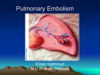
  • 1. Pulmonary Embolism By Eman mahmoud M.D of chest diseases
  • 2. Pulmonary Embolism • Embolism : Impaction of a thrombus or foreign matter in the pulmonary vascular bed.
  • 3. Epidemiology • 2nd most common cause of unexpected death in most age groups. • present in 60-80% of patients with DVT, more than 50 % them are asymptomatic. • Account for 15 % of all postoperative deaths • it is estimated that in the USA .100 000 people • die each year of pulmonary embolism.
  • 4. • Thrombotic EMBOLUS • Non-thrombotic : Fat, Air, Tumour , Amniotic fluid.
  • 5. RISK FACTORS • Bed rest • Post-operative • After severe blood loss and trauma • COPD • CHF • Varicose veins • Advancing age • Obesity • Post-partum • Malignancy • Travel of 4 hours or more • Smoking • 1ry polycythemia • Oral contraceptives
  • 6. THROMBOPHILIA • Acquired thrombophilia • • Inherited thrombophilias
  • 7. Hereditary factors • Antithrombin III deficiency • Protein C deficiency • Protein S deficiency • Factor V Leiden (most common genetic risk factor for thrombophilia) • Plasminogen abnormality • Plasminogen activator abnormality • Fibrinogen abnormality • Resistance to activated protein C
  • 8. Acquired thrombophilia • This is mostly associated with the antiphospholipid syndrome (APS). • APS is the combination of (LAC) with or without, (ACA), with a history of recurrent miscarriage and or thrombosis.
  • 10. Clinical Features •Size of the embolus and blood vessel occluded. •State of the lung. •Associated disease(s).
  • 11. PE, Clinical Features •dyspnea (75–85%) • Pleuritic chest pain (57–87%) • cough (40–53% ) • Hemoptysis • Syncope.
  • 12.
  • 13. Massive Pulmonary Embolism MPE •collapse/hypotension •unexplained hypoxia •engorged neck veins •Acut right heart failure
  • 14. Physical Signs •Normal •Tachypnea (respiratory rate >16/min) - 96% •Rales - 58% •Accentuated second heart sound - 53% •Tachycardia (heart rate >100/min) - 44% •Fever (temperature >37.8°C) - 43% •Diaphoresis - 36% •S3 or S4 gallop - 34% •Clinical signs and symptoms suggesting thrombophlebitis - 32% •Lower extremity edema - 24% •Cardiac murmur - 23% •Cyanosis - 19%
  • 16. D-Dimer •Degradation product produced by plasmin-mediated proteases of cross-linked fibrin. •Blood D-dimer assay should only be considered following assessment of clinical probability. •D-dimer assay should not be performed in those with high clinical probability of PE.
  • 17. D-dimer .Highly sensitive assays, such rapid ELISA assays, have high false positive rates but safely rule out VTE in outpatients presenting with a low clinical probability of PE. •A negative D-dimer test reliably excludes PE in patients with low clinical probability; such patients do not require imaging for VTE.
  • 18. Ischemia-Modified Albumin levels •A potential alternative to D-dimer testing is assessment of the ischemia-modified albumin (IMA) level. • 93% sensitive and 75% specific for pulmonary embolism • The positive predictive value of IMA, in particular, is better than D-dimer. However, it should not be used alone.
  • 19. Chest x-ray •14% Normal •68% Atelectasis •48% Pleural Effusion •35% Pleural based opacity •24% Elevated diaphragm •15% Prominent central pulmonary artery •7% Westermark’s sign •7% Cardiomegaly •5% Pulmonary edema
  • 22. (Hampton hump) •The classic radiographic findings of pulmonary infarction • a wedge-shaped, pleura-based triangular opacity with an apex pointing toward the hilus
  • 24. cardiac troponins •Recently, cardiac troponins I and T have been shown to be associated with early mortality and a complicated hospital course in patients with PE. The assessment of cardiac plasma troponin levels revealed ventricular injury, especially in patients with massive PE who had hypotension or shock.
  • 25. B Natriuretic Peptide (BNP) •BNP is a neurohormone secreted from the cardiac ventricles in response to dilatation or an increase of pressure. • BNP levels may increase with right ventricle dysfunction when the patients is in bed and decrease with treatment. •Serum brain natriuretic peptide levels of > 90 pg/mL have a sensitivity of 85% and a specificity of 75% for predicting adverse clinical outcomes
  • 26. Arterial blood gases •Blood gases may increase the suspicion and contribute to the clinical assessment, but they are insufficient to permit proof or exclusion of PE.
  • 27. ECG •In minor pulmonary embolism, the only finding is sinus tachycardia. •In massive pulmonary embolism, evidence of right heart strain may be seen (rightward shift of the QRS axis, transient RBBB, T-wave inversion, SIQIIITIII pattern, P-pulmonale), but these signs are non-specific. The main value of ECG is exclusion other diagnoses, such as MI or pericarditis.
  • 28. S1 Q3 T3 Pattern
  • 29. Echocardiography •RV dilatation •RV size does not change from diastole to systole = hypokinesis •D-shaped LV •40% of pts have RV abnormalities seen by ECHO
  • 30. Echocardiography •Transthoracic echocardiography rarely enables direct visualisation of the pulmonary embolus but may reveal thrombus floating in the right atrium or ventricle. • Transoesophageal echocardiography, it is possible to visualise massive emboli in the central pulmonary arteries.
  • 31. Lower extremity venous ultrasonography Advantages Portability May avoid further diagnostic imaging if positive Limitations Low sensitivity and risk of false positives No consistent protocol for technique Operator dependant
  • 32. Venous Ultrasonography Recommendations of Use •First-line if radiographic imaging contraindicated or not readily available •Not likely required in patient with negative CT-PA. •Helpful to rule out DVT in patient with non-diagnostic V/Q scan.
  • 33. Lung scintigraphy •The lung scan is an indirect method of diagnosis since it does not detect the embolus itself but only its consequence, the perfusion abnormality. •Used when CT scanning is not available or if the patient has a contraindication to CT scanning or intravenous contrast material.
  • 34. The Lung Scan Perfusion Perfusion • IV injection of human serum albumin labelled w/ technetium-99m •Particles are same size as pulmonary capillaries and become trapped •Lung peripheral to a clot is not perfused and will show defect Ventilation: •Inhalation of xenon-133 radioactive gas •Degree of ventilation of all lung areas can be assesed •Pneumonia, emphysema, tumors can cause defects •Pulmonary embolism does not cause ventilation defect Patients w/ a perfusion defect w/out a ventilation defect is suggestive of a pulmonary embolus.
  • 35.
  • 36. VQ Scan results Presence of several large focal perfusion defects not matched by ventilation defects indicates a high probability of PE !!!!! Normal scan basically excludes PE and indicates for other explanations for the pts condition. High probability – start Rx. Low probability – withhold Rx – can do CT / angiogram. Intermediate probability – can do CT / angio
  • 37. V/Q Scan Advantages Excellent negative predictive value (97%) Can be used in patients with contraindication to contrast medium Limitations 30-50% of patients have non-diagnostic scan necessitating further investigation
  • 38. CTPA • CTPA studies using the multislice technique showed a high sensitivity (96 to 100%) and specificity (97 to 98%), •CTPA is now the recommended initial lung imaging modality for non-massive PE. •Patients with a good quality negative CTPA do not require further invest. or ttt for PE. BTS (2003)
  • 39. Multidetector helical CT pulmonary angiography Advantages Diagnosis of alternative disease entities Coverage of entire chest with high spatial resolution in one breath hold Availability Improved depiction of small peripheral emboli
  • 40. Multidetector helical CT pulmonary angiography – Limitations Reader expertise required Expense Requires precise timing of contrast bolus Radiation exposure Not portable Contraindications to contrast Renal insufficiency the radiation dose from 3 to 5 mSv, with an estimated cancer risk of 150 excess cancer deaths per million
  • 41. Multidetector-CT Findings Partial or complete filling defects in lumen of pulmonary arteries Most reliable sign is filling defect forming acute angle with vessel wall with defect outlined by contrast material “polo mint” sign “Tram-track sign” Parallel lines of contrast surrounding thrombus in vessel that travels in transverse plane “Rim sign” Contrast surrounding thrombus in vessel that travels orthogonal to transverse plane a right-to-left ventricular dimensional ratio of 0.9 or more at MDCTA, had 92% sensitivity for right ventricular dysfunction
  • 42. “railway track” sign on longitudinal images of the vessel
  • 43. Arterial occlusion with failure to enhance the entire lumen due to a large filling defect
  • 44. A peripheral intraluminal filling defect that forms acute angles with the arterial wall
  • 45. A partial filling defect surrounded by contrast material, producing the “polo mint” sign on images acquired perpendicular to the long axis of a vessel
  • 46. a peripheral, crescent-shaped intraluminal defect that forms obtuse angles with the vessel wall
  • 47. Pulmonary Angiography •Was traditionally regarded as the reference test for PE. • Due to the invasive character, including right heart catheterization and injection of contrast material. •The current availability of noninvasive diagnostic imaging catheter pulmonary angiography now has an insignificant role.
  • 48. Magnetic resonance angiography (MRA) MRA appears promising & avoids ionising radiation but has poor sensitivity for subsegmental clot.
  • 49. Further Alternative Imaging Tests •Dual-energy CTPA •Electrocardiographically gated CTPA •Three-dimensional images acquired by single-photon emission computed tomography (SPECT) using a gamma-emitting radioisotope may improve V/Q scintigraphy and has a lower radiation dose.
  • 50. Dual-energy CTPA Provides functional and anatomic lung imaging Demonstrates perfusion defects beyond obstructive and non-obstructive clots Diagnostic accuracy requires further research Advantages Indirect evaluation of peripheral pulmonary arterial bed Disadvantages Longer data acquisition time Increased radiation exposure
  • 51. Dual-energy CTPA [A] Axial reconstruction with color-coded dual energy perfusion information. Note the large perfusion defects in both lungs. [B] Coronal reconstruction. Only the apical parts show a normal perfusion.
  • 52. Electrocardiographically gated CTPA •can differentiate between cardiac events and PE • may be of use in patients presenting with thoracic pain and suspected PE, cardiac events, or aorta dissection. • More contrast material is needed, and the radiation dose is higher compared with CTPA.
  • 53. Three-dimensional images •Single-photon emission computed tomography (SPECT) using a gamma-emitting radioisotope may improve V/Q scintigraphy • lower radiation dose.
  • 54. Low dose MDCT using ultra high pitch technique Useful in patients who are unable to hold their breath
  • 55.
  • 56.
  • 57. Treatment •For acute PE, the ACCP guidelines recommend starting low– molecular weight heparin (LMWH) or fondaparinux, preferred over unfractionated heparin (UFH) or subcutaneous heparin •Patients should have an oral anticoagulant (warfarin) initiated at the time of diagnosis, and they should have UFH, LMWH, or fondaparinux discontinued only after the international normalized ratio (INR) is 2.0 for at least 24 hours but no sooner than 5 days after warfarin therapy has been started
  • 58.
  • 59. Heparin •The initial treatment of PE is LMWH or UHF for at least 5 days, followed by warfarin. •Dose of UFH: 80 U/kg/h bolus followed by18 U/kg . Monitoring by APTT(1.5-3)times normal. •It binds to endogenous antithrombin, This heparin - antithrombin complex catalyzes the inactivation of factor Xa and IIa (thrombin).
  • 60. Heparin •Adverse drug reactions include bleeding, heparin-induced thrombocytopenia, and osteoporosis .with prolonged use . •A platelet count should be obtained weekly for the first month. If there is an abrupt decrease in the platelet count (approx. 50%) or by skin lesions at heparin injection sites or acute systemic reactions (eg, chills, cardiorespiratory distress) after IV heparin bolus administration consideration must be given to the possibility of (HIT).
  • 61. LMWH •It should be used whenever possible for the initial inpatient treatment of DVT & PE. . •Outpatient ttt of DVT, and possibly PE,is safe and cost-effective for carefully selected patients.
  • 62. Oral anticoagulant Warfarin inhibits gamma-carboxylation of the vitamin K-dependent coagulation factors II, VII, IX, and X. Warfarin and heparin can be started on the same day, but concomitant administration of these drugs for 4 or 5 days is recommended.
  • 63. Oral anticoagulant •Warfarin also inhibits carboxylation of natural anticoagulant proteins C and S resulting in a rapid decline of their levels. This poses a theoretical risk for a venous thromboembolic events. •Starting the patient on the expected daily dose (eg, 5mg) rather than administering a loading dose can minimize potential bleeding and avoid an extreme decline in protein C. •Target international normalised ratio [INR], 2.0–3.0) for at least 3–6 months.
  • 64. Factor Xa inhibitors •Rivaroxaban (Xarelto) is an oral factor Xa inhibitor approved by the FDA in November 2012 for the treatment of DVT or PE. • Reduce risk of recurrent DVT and PE following initial treatment. •Associated with less bleeding, particularly in elderly patients and those with moderate renal impairment.
  • 65. Factor Xa inhibitors •Apixaban was approved for treatment of PE in August 2014. •This advance thus offers the prospect of a safe and effective regimen of anticoagulation for patients with the advantages of simplicity and cost-effectiveness in comparison to current management strategies.
  • 66. Fondaparinux • Synthetic polysaccharide derived from the antithrombin binding region of heparin. Catalyzes factor Xa inactivation by antithrombin without inhibiting thrombin. •In patients with acute PE, favored over IV UFH and over SC UFH • Once-daily fondaparinux was found to be similar as IV UFH.
  • 67. Direct Thrombin Inhibitors Argatroban (Novastan ) approved in 2000: Anticoagulant for pts with HIT needing prophylaxis or treatment for thrombosis Ximelagatran (Exanta) • have shown benefit over warfarin or LMWH
  • 68. Duration of anticoagulation •A patient with a first thromboembolic event occurring in the setting of reversible risk factors, should receive warfarin therapy for at least 3 months. •The current ACCP guidelines recommend that all patients with unprovoked PE receive three months of treatment with anticoagulation and have an assessment of the risk-benefit ratio of extended therapy at the end of three months . patients with a second unprovoked episode of venous thromboembolism and low or moderate risk of bleeding, extended anticoagulant therapy is recommended Patients who have pulmonary embolism and preexisting irreversible risk factors should be placed on long-term anticoagulation.
  • 69. Heparin-induced thrombocytopenia (HIT) •Transient prothrombotic disorder initiated by heparin. •The main features of HIT are (1) thrombocytopenia (2) increased risk of venous and arterial thrombosis. •The highest frequency , 5%, has been reported in post– orthopedic surgery patients receiving up to 2 weeks of unfractionated heparin
  • 70. Heparin-induced thrombocytopenia (HIT) •should be suspected whenever the patient's platelet count falls to less than 100,000/μL or less than 50% of the baseline value •The treatment stop all heparin products, initiate an alternative, nonheparin anticoagulant, even when thrombosis is not clinically apparent. the use of lepirudin, argatroban, or danaparoid is preferred over continued use of heparin, LMWH, or either initiation or continuation of a vitamin K antagonist
  • 72. Thrombolytic Therapy •The ACCP guidelines recommend that thrombolytic therapy should be used in •patients with acute PE associated with hypotension (systolic BP< 90 mm HG) who do not have a high bleeding risk . •select patients with acute PE not associated with hypotension and with a low bleeding risk whose initial clinical presentation or clinical course after starting anticoagulation suggests a high risk of developing hypotension •Tissue plasminogen activator (tPA) has a short infusion time and has been recommended as the best agent for this reason.
  • 73. Major contraindications •A history of intracranial hemorrhage •Known intracranial aneurysm or arteriovenous malformation •Significant head trauma •Active internal bleeding •Known bleeding diathesis •Intracranial or intraspinal surgery within 3 months •A cerebrovascular accident within 2 months.
  • 74. Relative contraindications : •Recent internal bleeding •Recent surgery or organ biopsy •Uncontrolled hypertension •Pregnancy •Age 75 years
  • 75.
  • 76.
  • 77. Pulmonary Embolectomy Surgical embolectomy rarely done. It should be reserved for pts with hemodynamic instability despite heparin and cardiopulmonary support, who either fail thrombolytic therapy or have a contraindication to it.
  • 78. Catheter-Based Thrombus Removal for the Initial Treatment of Patients With PE In patients with acute PE associated with hypotension and who have (i) contraindications to thrombolysis, (ii) failed thrombolysis, (iii) shock that is likely to cause death before systemic thrombolysis can take effect (eg, within hours). if appropriate expertise and resources are available, we suggest catheter-assisted thrombus removal over no such intervention
  • 79. Vena Cava Filters indicated in the following settings: •Patients with acute venous thromboembolism who have an absolute contraindication to anticoagulant therapy •Patients with massive PE who survived but in whom recurrent embolism invariably will be fatal
  • 81. Pulmonary embolism In Pregnancy • Pregnancy increases the risk of VTE 4-fold to 5- fold over the nonpregnant state. • Overall, the prevalence of VTE in pregnancy is 0.5-2.0 per 1,000 pregnancies . • PE is one of the commenest cause of maternal death in pregnancy • accounts for 1.1 deaths per 100,000 pregnancies.
  • 83. AETIOLOGY • Increase in the levels of coagulation factors VII, VIII, IX, and X. • Increased fibrinogen levels. • Increased platelet activation. • increases resistance to the anti-thrombotic factors such as protein C and protein S. • Venous stasis in the lower limbs due to pressure by the gravid uterus
  • 84. Risk factors for DVT / PE during pregnancy • Maternal age > 35 years. • Pre-pregnancy weight > 80 kg. • Pre-existing Thrombophilia. • Previous DVT. • Severe varicose veins (V.Vs). • Prolonged bed rest. • Multi foetal pregnancies. • Severe pre-eclampsia. • Caesarean section delivery.
  • 85. Laboratory Evaluation D-dimer • Pregnancy limits the usefulness of this test. • Studies indicate that d-dimer values increase with gestational age, making the d-dimer test less specific. • Nonetheless, it remains a test with good negative predictive value in pregnancy.
  • 86. Imaging for PE • If at initial presentation a patient has concomitant symptoms or signs consistent with DVT and PE, compression ultrasonography may be considered first. • A negative compression ultrasonography of lower extremity veins however does not exclude PE. • If clinical suspicion still exists, further chest imaging is necessary.
  • 87. A chest radiograph •Recommended prior to the evaluation for PE to define other etiologies that may explain the symptoms (eg, pneumonia, atelectasis) and define the appropriate next imaging test.
  • 88. •Two alternative radiologic modalities exist for diagnosis of PE are spiral CT pulmonary angiography (CT-PA) and ventilation-perfusion (V/Q) scan. •In the pregnant patient with no known pulmonary disease and a normal chest radiograph,ventilation-perfusion scan is the recommended study to evaluate for PE. • If the patient has an abnormal chest radiograph or known pulmonary disease, spiral CT pulmonary angiography is recommended.
  • 89. CTPA versus V/Q scan • Spiral CT scanning and V/Q lung/can be safely performed in all trimesters. • Fetal radiation exposure for CTPA varies from 0.03 mGy to 1.3 mGy; increased with gestation and enlarged gravid uterus • - Fetal radiation dose for V/Q scanning is estimated as 1– 3.7 mGy • The risk of abnormality is considered to be negligible at ≤ 5 rads (50 mGy) and the risk of malformations is increased at doses > 15 rads (150 mGy). British Journal of Radiology,2006 (79):441-444 • • - ESC (2008): Guidelines on the diagnosis and management of acute pulmonary embolism
  • 90. CTPA versus V/Q scan CTPA •More sensitive and specific •Short acquisition time, easy access and interpretation • Lower radiation dose to the fetus (<10% of that with V/Q scanning) • Identify other pathology: aortic dissection. • Iodinated contrast medium can potentially alter fetal or neonatal thyroid function. V/Q scan - Not suitable if there is X-ray abnormalities or other chest disease as asthma or COPD - High incidence of indeterminate scans - Advantage over CTPA: Less radiation to maternal breast
  • 91.
  • 92. Treatment •Heparin, both low molecular weight and unfractionated does not cross the placenta or the breast. It is therefore safe for the fetus and for the breast-fed infant. • warfarin, cross the placenta In the first trimester warfarin •UFH, LMWHs, and warfarin are not secreted in breast milk in clinically significant amounts, and so are safe for use in breast-feeding patients. So after labour oral anticoagulant can be introduced
  • 93. Duration of therapy •A pregnant patient with a DVT or PE should be anticoagulated for the duration of the pregnancy and at least six weeks postpartum, or for six months, whichever is longer •Subcutaneous LMWH is preferred over IV UFH or SC UFH in most patients . •UFH (either IV or SC) is preferred over SC LMWH in patients who have severe renal failure.
  • 94. Labor and delivery •Treatment with SC LMWH should be discontinued at least 24 hour Prior to delivery if the delivery time is predictable . •A period of 24 to 36 hours without anticoagulant therapy may be undesirable in pregnant women who are at high risk for recurrent VTE (eg, those with an acute PE or proximal DVT that developed within the past month). •Such patients may benefit from having their SC LMWH or SC UFH switched to IV UFH, which can be discontinued 4 to 6 hours prior to delivery .
  • 95. Postpartum •Anticoagulation may be restarted with UFH or LMWH 4- 6 hours following vaginal delivery or 6-12 hours following cesarean delivery. • If neuraxial blockade was used, prophylactic anti-coagulation should not be restarted any sooner than 2 hours following epidural removal. • Although the ideal time to restart therapeutic anticoagulation following epidural removal is unclear, waiting 12 hours after removal of the epidural may be a reasonable approach
  • 96. Thrombolytic agents in pregnancy . Streptokinase (and probably other thrombolytic drugs) does not cross the placenta because of its high molecular weight. •However in the mother, bleeding is the major side effect, usually from the genital tract and often severe.incidence of bleeding is about 8%. • Because of the risk of bleeding, thrombolysis should not be used routinely in pregnancy, •Should not be used at the time of delivery unless it appears that the patient is likely to die
  • 97. Malignancy • Four- to sevenfold higher risk of thrombosis compared with patients without cancer, •caused by prothrombotic effects of the tumor and also because of treatment, particularly with surgery, use of a central venous catheter, and chemotherapy. •~20% of patients presenting with VTE have active cancer, associated with reduced survival. •Value (specificity) of D-dimer testing is reduced, leading to an increased need for imaging.
  • 98. Malignancy • initial treatment with heparin and warfarin is given in the standard manner. • The relative risk of recurrence is 3 and of bleeding is 6 compared with other •Duration of treatment is arbitrary. • For those with recurrence in spite of adequate anticoagulation, options include: (a) aiming for a higher INR of 3.0–3.5 (which further increases the risks of bleeding) (b) switching to longterm LMWH while continuing anticoagulation, (c) insertingan IVC filter, the value of which is questionable.307
  • 99. Patients with liver cirrhosis • Characterized by decreased synthesis of both pro-and anticoagulant factors. • Although bleeding is the most common clinical manifestation as a result of decreased platelet function and number, diminished clotting factors and excessive fibrinolysis, • Hypercoagulability may play an under recognized but important role in many aspects of chronic liver disease.
  • 100. • Particular patients with cirrhosis appear to have a higher incidence of DVT and pulmonary embolism(PE) compared with the general population. • The incidence of DVT/PE ranges from 0.5% to 1.9%, similar to patients without comorbidities, but lower than patients with other chronic diseases (i.e, renal or heart disease). • Serum albumin level was independently associated with the occurrence of thrombosis •Cirrhotic patient should not be considered as an auto-anticoagulated patient.
  • 101. Liver disease as a risk DVT/PE •Patients w autoimmune liver disease have a higher incidence of portal vein thrombosis •Cholestatic liver disease are characterized by a hypercoagulable state . • patients with non-cirrhotic liver disease are at greater risk of DVT/PE. • Non-alcoholic fatty liver disease have been associated with a great risk of atherosclerosis and endothelial dysfunction •
  • 102. Liver disease as a risk DVT/PE •Patients with hepatocellular carcinoma are known to be at greater risk of thrombo embolic complications (i.e. portal vein thrombosis and PE), and increased levels of thrombin–antithrombin complexes have been demonstrated.
  • 103. Risk factors for DVT/PE • Same risk factors as other non-cirrhotic patients such as venous stasis, infection, congestive heart failure, acute respiratory disease and immobilization.Surgery . • liver resection such as for hepatocellular carcinoma has been shown to correlate with a higher incidence of thrombotic complications.
  • 104. Thrombotic prophylaxis •Current American College of Chest Physicians (ACCP) guidelines for thrombo prophylaxis in patients with important risk factors for bleedings state: ‘for medical patients with risk factors for VTE, and for whom there is a contraindication to anticoagulant thromboprophylaxis, we recommend the optimal use of mechanical thromboprophylaxis with graduated compression stockings or intermittent pneumatic compression. •The statement has the maximum grade of evidence and strength of recommendation,
  • 105. •Current guidelines on antithrombotic prophylaxis do not specifically comment on the cirrhotic population as a result of the perceived risk of bleeding complications •Thromboprophylaxis should be recommended in patients with liver cirrhosis at least when exposed to high-risk conditions for thrombotic complications. •Low molecular weight heparins (LWMHs) seem to be relatively safe in this group of patients. •when important risk factors for bleeding are present, graduated compression stockings or intermittent pneumatic compression should be considered.
  • 106. •Disadvantage of the use of LMWH and fondaparinux inpatients with cirrhosis, is the unpredictable efficacy, as these drugs require antithrombin to exert its anticoagulant function, and antithrombin levels are frequently decreased in these patients. •The use of antithrombotic agents, directly inhibiting factor (F)Xa or thrombin, will need very careful evaluation in this subset of patients due to the risk of haemorrhagic complications.
  • 107. •The FXa inhibitors rivaroxaban and apixaban are metabolized in the liver and they are contraindicated in severe hepatic disease •Idraparinux, an antithrombin dependent FXa inhibitor, has no hepatic clearance,but its long half-life (approximately 80 h) and the lack of antidote do represent major problems if bleeding occurs. •Finally, all these drugs must be used with caution or are contraindicated in the presence of renal failure.