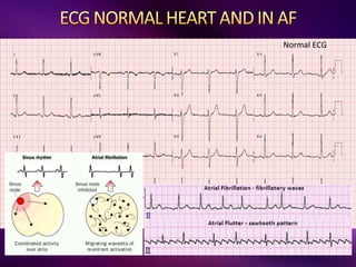
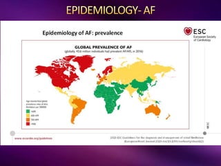
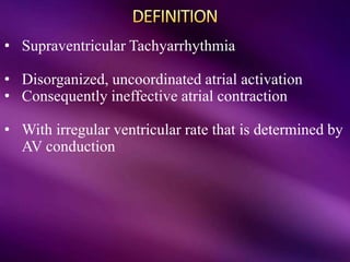
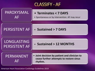
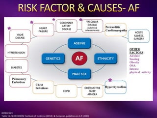
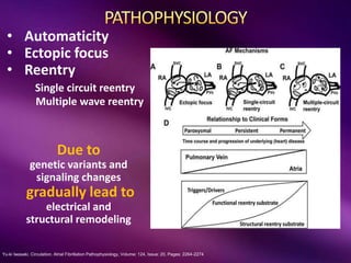
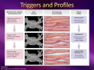
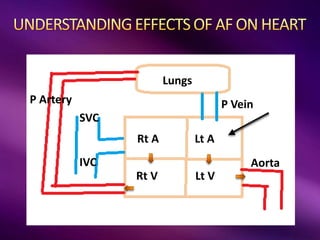
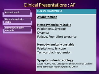
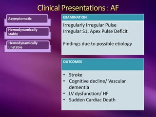
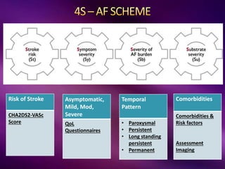
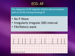
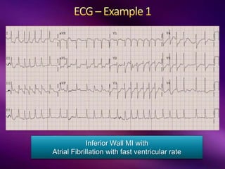
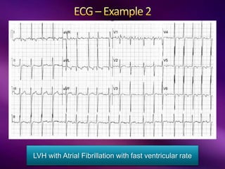
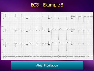
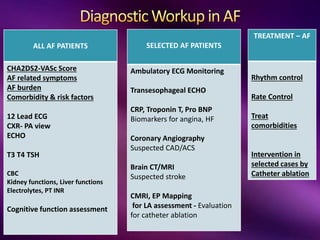
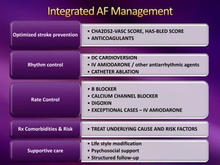
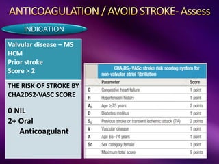
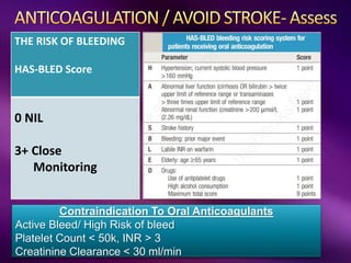
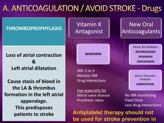
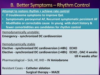
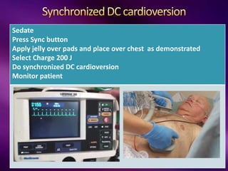
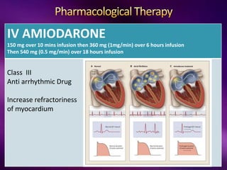
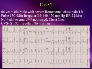
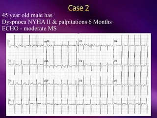
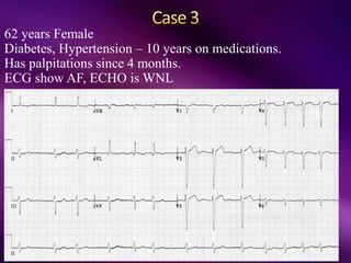
Col Bharat Malhotra
Senior Advisor (Medicine)
REFERENCE
Harrison’s Principles of internal medicine Harrisons 20th Ed
Davidson’s Principles and practice of Medicine (2018)
2020 European Cardiology guidelines on Atrial Fibrillation 2019 American Heart Association Guidelines on AF
LEFT ATRIA
LEFT ATRIA
Normal
Electrical Activity
• 60 – 100 /min
• Sinus origin
• In Conduction system
• Defined Velocity
BRADYARRTHYMIAS
• Sinus Bradycardia
• Sick Sinus Syndrome
• Sinus node arrest
• AV block
• First degree
• Second degree
• Third degree
TACHYARRHYTHMIAS
• Sinus tachycardia
• Atrial flutter
• Atrial Fibrillation
• Atrial Tachycardia
• Focal
• Multifocal
• AV Junction
• AVRT
• AVNRT
• Ventricular Tachycardia
• Ventricular Fibrillation
ECTOPICS
• Atrial ectopic
• Ventricular Ectopic
BUNDLE BRANCH BLOCK
• Rt BBB
• Lt BBB
LEFT ATRIA
Normal ECG
• Supraventricular Tachyarrhythmia
• Disorganized, uncoordinated atrial activation
• Consequently ineffective atrial contraction
• With irregular ventricular rate that is determined by
AV conduction
American Heart Association Cardiology Guidelines 2019
• Terminates < 7 DAYS
• Spontaneous or by Intervention. AF may recur
PAROXYSMAL
AF
• Sustained > 7 DAYS
PERSISTENT AF
• Sustained > 12 MONTHS
LONGLASTING
PERSISTENT AF
• Joint decision by patient and clinician to
cease further attempts to restore sinus
rhythm.
PERMANENT
AF
REFERENCE
Table 16.21 DAVIDSON Textbook of medicine (2018) & European guidelines on A F (2020)
Yu-ki Iwasaki. Circulation. Atrial Fibrillation Pathophysiology, Volume: 124, Issue: 20, Pages: 2264-2274
• Automaticity
• Ectopic focus
• Reentry
Single circuit reentry
Multiple wave reentry
Due to
genetic variants and
signaling changes
gradually lead to
electrical and
structural remodeling
REFERENCE
New England Journal of Medicine JAN 2021
SVC
IVC
P Artery P Vein
Aorta
Lungs
Lt A
Rt A
Rt V Lt V
Asymptomatic
Hemodynamically
stable
Hemodynamically
unstable
CLINICAL PRSENTATIONS
Asymptomatic
Hemodynamically Stable
Palpitations, Syncope
Dyspnea
Fatigue, Poor effort tolerance
Hemodynamically unstable
Palpitations, Syncope
Tachycardia, Hypotension
Symptoms due to etiology
Acute HF, LVF, ACS, Cardiogenic Shock, Valvular Disease
Lung pathology, Hyperthyroidism, Others
Asymptomatic
Hemodynamically
stable
Hemodynamically
unstable
EXAMINATION
Irregularly Irregular Pulse
Irregular S1, Apex Pulse Deficit
Findings due to possible etiology
OUTCOMES
• Stroke
• Cognitive decline/ Vascular
dementia
• LV dysfunction/ HF
• Sudden Cardiac Death
Risk of Stroke
CHA2DS2-VASc
Score
Asymptomatic,
Mild, Mod,
Severe
QoL
Questionnaires
Temporal
Pattern
• Paroxysmal
• Persistent
• Long standing
persistent
• Permanent
Comorbidities
Comorbidities &
Risk factors
Assessment
Imaging
The diagnosis of AF requires rhythm documentation
with an (ECG) tracing showing AF
• No P Wave
• Irregularly Irregular QRS interval
• Fibrillatory wave
Inferior Wall MI with
Atrial Fibrillation with fast ventricular rate
LVH with Atrial Fibrillation with fast ventricular rate
Atrial Fibrillation
ALL AF PATIENTS
CHA2DS2-VASc Score
AF related symptoms
AF burden
Comorbidity & risk factors
12 Lead ECG
CXR- PA view
ECHO
T3 T4 TSH
CBC
Kidney functions, Liver functions
Electrolytes, PT INR
Cognitive function assessment
TREATMENT – AF
Rhythm control
Rate Control
Treat
comorbidities
Intervention in
selected cases by
Catheter ablation
SELECTED AF PATIENTS
Ambulatory ECG Monitoring
Transesophageal ECHO
CRP, Troponin T, Pro BNP
Biomarkers for angina, HF
Coronary Angiography
Suspected CAD/ACS
Brain CT/MRI
Suspected stroke
CMRI, EP Mapping
for LA assessment - Evaluation
for catheter ablation
• CHA2DS2-VASC SCORE, HAS-BLED SCORE
• ANTICOAGULANTS
Optimized stroke prevention
• DC CARDIOVERSION
• IV AMIODARONE / other antiarrhythmic agents
• CATHETER ABLATION
Rhythm control
• B BLOCKER
• CALCIUM CHANNEL BLOCKER
• DIGOXIN
• EXCEPTIONAL CASES – IV AMIODARONE
Rate Control
• TREAT UNDERLYING CAUSE AND RISK FACTORS
Rx Comorbidities & Risk
• Life style modification
• Psychosocial support
• Structured follow-up
Supportive care
Valvular disease – MS
HCM
Prior stroke
Score > 2
THE RISK OF STROKE BY
CHA2DS2-VASC SCORE
0 NIL
2+ Oral
Anticoagulant
INDICATION
THE RISK OF BLEEDING
HAS-BLED Score
0 NIL
3+ Close
Monitoring
Contraindication To Oral Anticoagulants
Active Bleed/ High Risk of bleed
Platelet Count < 50k, INR > 3
Creatinine Clearance < 30 ml/min
THROMBOPROPHYLAXIS
Loss of atrial contraction
&
Left atrial dilatation
Cause stasis of blood in
the LA & thrombus
formation in the left atrial
appendage.
This predisposes
patients to stroke Antiplatelet therapy should not
be used for stroke prevention in
Vitamin K
Antagonist
WARFARIN
New Oral
Anticoagulants
Factor Xa Inhibitors
RIVOROXABAN
APIXABAN
ENDOXABAN
Direct Thrombin
Inhibitor
DABIGATRAN
No INR monitoring
Fixed Dose
Less drug interactions
INR: 2 to 3
Monitor INR
Drug Interactions
Use especially for
Mitral valve disease
Prosthetic Valve
Attempt to restore rhythm + achieve rate control
• If troublesome symptoms to improve QoL
• Symptomatic paroxysmal AF, Recurrent symptomatic persistent AF
• Modifiable or correctable cause in young with short history &
fewer comorbidities are candidates for rhythm control
Hemodynamically unstable:
Emergency - synchronized DC cardioversion
Hemodynamically stable
Elective - synchronized DC cardioversion (<48h) ECHO
Elective - synchronized DC cardioversion (>48h) ECHO , OAC 4 weeks
till 4 weeks after
Pharmacological – Sick, HF, IHD – IV Amiodarone
Resistant Cases – Catheter ablation
Surgical therapy – MAZE
Sedate
Press Sync button
Apply jelly over pads and place over chest as demonstrated
Select Charge 200 J
Do synchronized DC cardioversion
Monitor patient
IV AMIODARONE
150 mg over 10 mins infusion then 360 mg (1mg/min) over 6 hours infusion
Then 540 mg (0.5 mg/min) over 18 hours infusion
Class III
Anti arrhythmic Drug
Increase refractoriness
of myocardium
Prepare – TEE, ECG, CT Angio of Heart
Ablation Cath Lab
Electrical mapping
Ablate around pulmonary veins
Attempt to restore rhythm + achieve rate control
Accept presence of AF but achieve rate control
B Blocker (IV Metoprolol, IV Esmolol)
(Metoprolol, Bisoprolol, Nebivolol, Carvedilol)
Calcium Channel Blocker – (Diltiazem, Verapamil)
Digoxin
Exceptional Cases- Implant a permanent pacemaker
Plus Complete AV node block with catheter ablation
Last Resort - IV Amiodarone
<110/min
< 80/Min
Treat the coexisting illness & Risk Factors
66 years old Male with severe Retrosternal chest pain 1 h
Pulse 150 /Min Irregular BP 140 / 70 mmHg RR 22/Min
No Pedal enema, JVP not raised, Chest Clear
CVS- S1 S2 irregular, No murmur
45 year old male has
Dyspnoea NYHA II & palpitations 6 Months
ECHO - moderate MS
62 years Female
Diabetes, Hypertension – 10 years on medications.
Has palpitations since 4 months.
ECG show AF, ECHO is WNL
ATRIAL FIBRILLATION

ATRIAL FIBRILLATION

  • 1.
    Col Bharat Malhotra SeniorAdvisor (Medicine) REFERENCE Harrison’s Principles of internal medicine Harrisons 20th Ed Davidson’s Principles and practice of Medicine (2018) 2020 European Cardiology guidelines on Atrial Fibrillation 2019 American Heart Association Guidelines on AF
  • 3.
  • 4.
    LEFT ATRIA Normal Electrical Activity •60 – 100 /min • Sinus origin • In Conduction system • Defined Velocity
  • 5.
    BRADYARRTHYMIAS • Sinus Bradycardia •Sick Sinus Syndrome • Sinus node arrest • AV block • First degree • Second degree • Third degree TACHYARRHYTHMIAS • Sinus tachycardia • Atrial flutter • Atrial Fibrillation • Atrial Tachycardia • Focal • Multifocal • AV Junction • AVRT • AVNRT • Ventricular Tachycardia • Ventricular Fibrillation ECTOPICS • Atrial ectopic • Ventricular Ectopic BUNDLE BRANCH BLOCK • Rt BBB • Lt BBB
  • 6.
  • 9.
    • Supraventricular Tachyarrhythmia •Disorganized, uncoordinated atrial activation • Consequently ineffective atrial contraction • With irregular ventricular rate that is determined by AV conduction
  • 10.
    American Heart AssociationCardiology Guidelines 2019 • Terminates < 7 DAYS • Spontaneous or by Intervention. AF may recur PAROXYSMAL AF • Sustained > 7 DAYS PERSISTENT AF • Sustained > 12 MONTHS LONGLASTING PERSISTENT AF • Joint decision by patient and clinician to cease further attempts to restore sinus rhythm. PERMANENT AF
  • 11.
    REFERENCE Table 16.21 DAVIDSONTextbook of medicine (2018) & European guidelines on A F (2020)
  • 12.
    Yu-ki Iwasaki. Circulation.Atrial Fibrillation Pathophysiology, Volume: 124, Issue: 20, Pages: 2264-2274 • Automaticity • Ectopic focus • Reentry Single circuit reentry Multiple wave reentry Due to genetic variants and signaling changes gradually lead to electrical and structural remodeling
  • 13.
    REFERENCE New England Journalof Medicine JAN 2021
  • 14.
    SVC IVC P Artery PVein Aorta Lungs Lt A Rt A Rt V Lt V
  • 15.
    Asymptomatic Hemodynamically stable Hemodynamically unstable CLINICAL PRSENTATIONS Asymptomatic Hemodynamically Stable Palpitations,Syncope Dyspnea Fatigue, Poor effort tolerance Hemodynamically unstable Palpitations, Syncope Tachycardia, Hypotension Symptoms due to etiology Acute HF, LVF, ACS, Cardiogenic Shock, Valvular Disease Lung pathology, Hyperthyroidism, Others
  • 16.
    Asymptomatic Hemodynamically stable Hemodynamically unstable EXAMINATION Irregularly Irregular Pulse IrregularS1, Apex Pulse Deficit Findings due to possible etiology OUTCOMES • Stroke • Cognitive decline/ Vascular dementia • LV dysfunction/ HF • Sudden Cardiac Death
  • 17.
    Risk of Stroke CHA2DS2-VASc Score Asymptomatic, Mild,Mod, Severe QoL Questionnaires Temporal Pattern • Paroxysmal • Persistent • Long standing persistent • Permanent Comorbidities Comorbidities & Risk factors Assessment Imaging
  • 18.
    The diagnosis ofAF requires rhythm documentation with an (ECG) tracing showing AF • No P Wave • Irregularly Irregular QRS interval • Fibrillatory wave
  • 19.
    Inferior Wall MIwith Atrial Fibrillation with fast ventricular rate
  • 20.
    LVH with AtrialFibrillation with fast ventricular rate
  • 21.
  • 22.
    ALL AF PATIENTS CHA2DS2-VAScScore AF related symptoms AF burden Comorbidity & risk factors 12 Lead ECG CXR- PA view ECHO T3 T4 TSH CBC Kidney functions, Liver functions Electrolytes, PT INR Cognitive function assessment TREATMENT – AF Rhythm control Rate Control Treat comorbidities Intervention in selected cases by Catheter ablation SELECTED AF PATIENTS Ambulatory ECG Monitoring Transesophageal ECHO CRP, Troponin T, Pro BNP Biomarkers for angina, HF Coronary Angiography Suspected CAD/ACS Brain CT/MRI Suspected stroke CMRI, EP Mapping for LA assessment - Evaluation for catheter ablation
  • 23.
    • CHA2DS2-VASC SCORE,HAS-BLED SCORE • ANTICOAGULANTS Optimized stroke prevention • DC CARDIOVERSION • IV AMIODARONE / other antiarrhythmic agents • CATHETER ABLATION Rhythm control • B BLOCKER • CALCIUM CHANNEL BLOCKER • DIGOXIN • EXCEPTIONAL CASES – IV AMIODARONE Rate Control • TREAT UNDERLYING CAUSE AND RISK FACTORS Rx Comorbidities & Risk • Life style modification • Psychosocial support • Structured follow-up Supportive care
  • 24.
    Valvular disease –MS HCM Prior stroke Score > 2 THE RISK OF STROKE BY CHA2DS2-VASC SCORE 0 NIL 2+ Oral Anticoagulant INDICATION
  • 25.
    THE RISK OFBLEEDING HAS-BLED Score 0 NIL 3+ Close Monitoring Contraindication To Oral Anticoagulants Active Bleed/ High Risk of bleed Platelet Count < 50k, INR > 3 Creatinine Clearance < 30 ml/min
  • 26.
    THROMBOPROPHYLAXIS Loss of atrialcontraction & Left atrial dilatation Cause stasis of blood in the LA & thrombus formation in the left atrial appendage. This predisposes patients to stroke Antiplatelet therapy should not be used for stroke prevention in Vitamin K Antagonist WARFARIN New Oral Anticoagulants Factor Xa Inhibitors RIVOROXABAN APIXABAN ENDOXABAN Direct Thrombin Inhibitor DABIGATRAN No INR monitoring Fixed Dose Less drug interactions INR: 2 to 3 Monitor INR Drug Interactions Use especially for Mitral valve disease Prosthetic Valve
  • 27.
    Attempt to restorerhythm + achieve rate control • If troublesome symptoms to improve QoL • Symptomatic paroxysmal AF, Recurrent symptomatic persistent AF • Modifiable or correctable cause in young with short history & fewer comorbidities are candidates for rhythm control Hemodynamically unstable: Emergency - synchronized DC cardioversion Hemodynamically stable Elective - synchronized DC cardioversion (<48h) ECHO Elective - synchronized DC cardioversion (>48h) ECHO , OAC 4 weeks till 4 weeks after Pharmacological – Sick, HF, IHD – IV Amiodarone Resistant Cases – Catheter ablation Surgical therapy – MAZE
  • 28.
    Sedate Press Sync button Applyjelly over pads and place over chest as demonstrated Select Charge 200 J Do synchronized DC cardioversion Monitor patient
  • 29.
    IV AMIODARONE 150 mgover 10 mins infusion then 360 mg (1mg/min) over 6 hours infusion Then 540 mg (0.5 mg/min) over 18 hours infusion Class III Anti arrhythmic Drug Increase refractoriness of myocardium
  • 30.
    Prepare – TEE,ECG, CT Angio of Heart Ablation Cath Lab Electrical mapping Ablate around pulmonary veins
  • 32.
    Attempt to restorerhythm + achieve rate control Accept presence of AF but achieve rate control B Blocker (IV Metoprolol, IV Esmolol) (Metoprolol, Bisoprolol, Nebivolol, Carvedilol) Calcium Channel Blocker – (Diltiazem, Verapamil) Digoxin Exceptional Cases- Implant a permanent pacemaker Plus Complete AV node block with catheter ablation Last Resort - IV Amiodarone <110/min < 80/Min
  • 33.
    Treat the coexistingillness & Risk Factors
  • 34.
    66 years oldMale with severe Retrosternal chest pain 1 h Pulse 150 /Min Irregular BP 140 / 70 mmHg RR 22/Min No Pedal enema, JVP not raised, Chest Clear CVS- S1 S2 irregular, No murmur
  • 35.
    45 year oldmale has Dyspnoea NYHA II & palpitations 6 Months ECHO - moderate MS
  • 36.
    62 years Female Diabetes,Hypertension – 10 years on medications. Has palpitations since 4 months. ECG show AF, ECHO is WNL