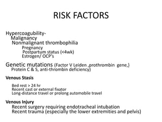
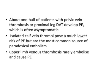
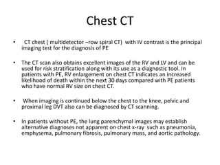
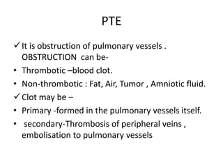
1) Pulmonary thromboembolism (PTE) is caused by obstruction of pulmonary vessels, usually by blood clots. Risk factors include hypercoagulability, recent surgery or trauma, pregnancy, genetic mutations.
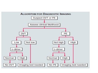
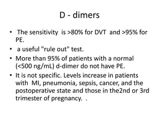
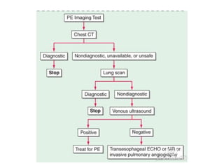
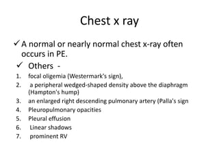
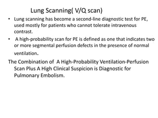
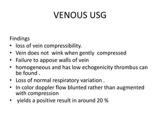
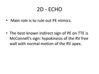
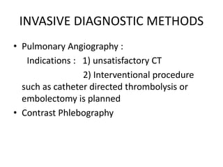
2) Diagnostic tests include D-dimer, chest CT, lung scan, echocardiogram. Chest CT has replaced lung scan as the primary imaging test.
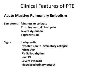
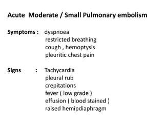
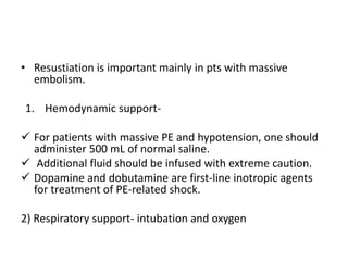
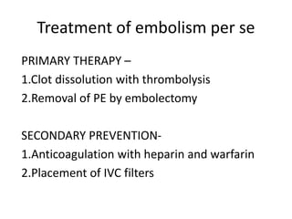
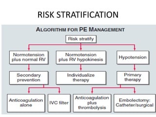
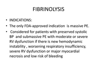
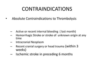
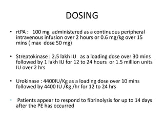
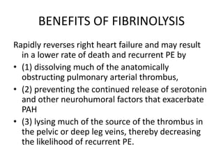
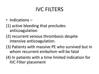
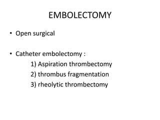
3) Treatment involves resuscitation for massive PTE, anticoagulation with heparin or low molecular weight heparin initially followed by warfarin for at least 3 months, and potentially thrombolysis for selected high-risk cases or inferior vena cava filters for recurrent clots despite
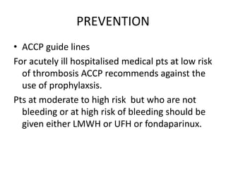
Acute pulmonary embolism and its management.Puja Gupta
Critical Care Nursing (CCN).Respiratory disorders. Critical Care Nursing (CCN).Critical Care Nursing (CCN).Critical Care Nursing (CCN).Critical Care Nursing (CCN).Critical Care Nursing (CCN).Critical Care Nursing (CCN).Critical Care Nursing (CCN).Critical Care Nursing (CCN).Critical Care Nursing (CCN).Critical Care Nursing (CCN).Critical Care Nursing (CCN).Critical Care Nursing (CCN).
Deep vein thrombosis (DVT) and Pulmonary embolism (PE)Aminul Haque
Deep vein thrombosis (DVT) and pulmonary embolism (PE), collectively referred to as venous thromboembolism (VTE), constitute a major global burden of disease.
Pulmonary embolism - Diagnosis and managementDr Vivek Baliga
Pulmonary embolism is a common problem seen in medical practice. This presentation by Dr Vivek Baliga discusses the basic aspects and evidence behind current management.
Pulmonary embolism - Diagnosis and managementDr Vivek Baliga
Pulmonary embolism is a common problem seen in medical practice. This presentation by Dr Vivek Baliga discusses the basic aspects and evidence behind current management.
Physician should have a high suspicion to diagnose patient with pulmonary Embolism, this slides will give you precise Diagnosis, Investigation and guideline directed Treatment.
Diagnosis of Pulmonary Embolism is often difficult. This presentation highlights step-wise and practical approach to the diagnosis of PE in short and precise fashion.
Students, digital devices and success - Andreas Schleicher - 27 May 2024..pptxEduSkills OECD
Andreas Schleicher presents at the OECD webinar ‘Digital devices in schools: detrimental distraction or secret to success?’ on 27 May 2024. The presentation was based on findings from PISA 2022 results and the webinar helped launch the PISA in Focus ‘Managing screen time: How to protect and equip students against distraction’ https://www.oecd-ilibrary.org/education/managing-screen-time_7c225af4-en and the OECD Education Policy Perspective ‘Students, digital devices and success’ can be found here - https://oe.cd/il/5yV
Synthetic Fiber Construction in lab .pptxPavel ( NSTU)
Synthetic fiber production is a fascinating and complex field that blends chemistry, engineering, and environmental science. By understanding these aspects, students can gain a comprehensive view of synthetic fiber production, its impact on society and the environment, and the potential for future innovations. Synthetic fibers play a crucial role in modern society, impacting various aspects of daily life, industry, and the environment. ynthetic fibers are integral to modern life, offering a range of benefits from cost-effectiveness and versatility to innovative applications and performance characteristics. While they pose environmental challenges, ongoing research and development aim to create more sustainable and eco-friendly alternatives. Understanding the importance of synthetic fibers helps in appreciating their role in the economy, industry, and daily life, while also emphasizing the need for sustainable practices and innovation.
We all have good and bad thoughts from time to time and situation to situation. We are bombarded daily with spiraling thoughts(both negative and positive) creating all-consuming feel , making us difficult to manage with associated suffering. Good thoughts are like our Mob Signal (Positive thought) amidst noise(negative thought) in the atmosphere. Negative thoughts like noise outweigh positive thoughts. These thoughts often create unwanted confusion, trouble, stress and frustration in our mind as well as chaos in our physical world. Negative thoughts are also known as “distorted thinking”.
2024.06.01 Introducing a competency framework for languag learning materials ...Sandy Millin
http://sandymillin.wordpress.com/iateflwebinar2024
Published classroom materials form the basis of syllabuses, drive teacher professional development, and have a potentially huge influence on learners, teachers and education systems. All teachers also create their own materials, whether a few sentences on a blackboard, a highly-structured fully-realised online course, or anything in between. Despite this, the knowledge and skills needed to create effective language learning materials are rarely part of teacher training, and are mostly learnt by trial and error.
Knowledge and skills frameworks, generally called competency frameworks, for ELT teachers, trainers and managers have existed for a few years now. However, until I created one for my MA dissertation, there wasn’t one drawing together what we need to know and do to be able to effectively produce language learning materials.
This webinar will introduce you to my framework, highlighting the key competencies I identified from my research. It will also show how anybody involved in language teaching (any language, not just English!), teacher training, managing schools or developing language learning materials can benefit from using the framework.
This is a presentation by Dada Robert in a Your Skill Boost masterclass organised by the Excellence Foundation for South Sudan (EFSS) on Saturday, the 25th and Sunday, the 26th of May 2024.
He discussed the concept of quality improvement, emphasizing its applicability to various aspects of life, including personal, project, and program improvements. He defined quality as doing the right thing at the right time in the right way to achieve the best possible results and discussed the concept of the "gap" between what we know and what we do, and how this gap represents the areas we need to improve. He explained the scientific approach to quality improvement, which involves systematic performance analysis, testing and learning, and implementing change ideas. He also highlighted the importance of client focus and a team approach to quality improvement.
Read| The latest issue of The Challenger is here! We are thrilled to announce that our school paper has qualified for the NATIONAL SCHOOLS PRESS CONFERENCE (NSPC) 2024. Thank you for your unwavering support and trust. Dive into the stories that made us stand out!
4. RISK FACTORS
Hypercoagubility-
Malignancy
Nonmalignant thrombophilia
Pregnancy
Postpartum status (<4wk)
Estrogen/ OCP’s
Genetic mutations (Factor V Leiden ,prothrombin gene,}
Protein C & S, anti-thrombin deficiency)
Venous Stasis
Bed rest > 24 hr
Recent cast or external fixator
Long-distance travel or prolong automobile travel
Venous Injury
Recent surgery requiring endotracheal intubation
Recent trauma (especially the lower extremities and pelvis)
5. • About one-half of patients with pelvic vein
thrombosis or proximal leg DVT develop PE,
which is often asymptomatic.
• Isolated calf vein thrombi pose a much lower
risk of PE but are the most common source of
paradoxical embolism.
• upper limb venous thrombosis rarely embolise
and cause PE.
6. Clinical Features of PTE
Acute Massive Pulmonary Embolism
Symptoms : faintness or collapse
Crushing central chest pain
severe dyspnoea
apprehension
Signs : tachycardia
hypotension to circulatory collapse
raised JVP
RV Gallop rhythm
loud P2
Severe cyanosis
decreased urinary output
8. Principal markers used for risk stratification
1) RV Dysfunction : hypotension
RV dilation ,hypokinesis or overload
RV dilation on spiral CT
BNP or NT –Pro BNP
elevated right heart pressure at RHC
2) Myocardial injury : Cardiac troponin T or I positive
Patients with massive PE : Rv dysfunction + hypotension
Moderate to large PE : RV dysfunction + normotension
Small to moderate PE : Normal RV function + normotension
10. evaluating patients with possible VTE
• The initial task is to decide on the clinical
likelihood of the disorder.
• For this we have various scores like
wells,geneva etc
11. Well’s Criteria
Clinical Signs and Symptoms of DVT?
(Calf tenderness, swelling >3cm, errythema, pitting
edema affected leg only)
+3
PE Is #1 Diagnosis, or Equally Likely +3
Heart Rate > 100 +1.5
Immobilization at least 3 days, or Surgery in the
Previous 4 weeks
+1.5
Previous, objectively diagnosed PE or DVT? +1.5
Hemoptysis +1
Malignancy w/ Rx within 6 mo, or palliative? +1
>6 : High Risk
2 to 6 : Moderate Risk
< 2 : Low Risk
Adapted with permission from Wells PS, Anderson DR, Rodger M, Ginsberg JS, Kearon C, Gent M, et al. Derivation of a simple
clinical model to categorize patients probability of pulmonary embolism: increasing the models utility with the SimpliRED d-dimer.
Thromb Haemost 2000;83:416-20.
12.
13. D - dimers
• The sensitivity is >80% for DVT and >95% for
PE.
• a useful "rule out" test.
• More than 95% of patients with a normal
(<500 ng/mL) d-dimer do not have PE.
• It is not specific. Levels increase in patients
with MI, pneumonia, sepsis, cancer, and the
postoperative state and those in the2nd or 3rd
trimester of pregnancy. .
14.
15. Chest x ray
A normal or nearly normal chest x-ray often
occurs in PE.
Others -
1. focal oligemia (Westermark's sign),
2. a peripheral wedged-shaped density above the diaphragm
(Hampton's hump)
3. an enlarged right descending pulmonary artery (Palla's sign
4. Pleuropulmonary opacities
5. Pleural effusion
6. Linear shadows
7. prominent RV
19. • ECG
– 2 Most Common finding on EKG:
• Nonspecific ST-segment and T-wave changes
• Sinus Tachycardia
– Historical abnormality suggestive of PE
• S1Q3T3
• Right ventricular strain
• New incomplete RBBB
20. Chest CT
• CT chest ( multidetector –row spiral CT) with IV contrast is the principal
imaging test for the diagnosis of PE
• The CT scan also obtains excellent images of the RV and LV and can be
used for risk stratification along with its use as a diagnostic tool. In
patients with PE, RV enlargement on chest CT indicates an increased
likelihood of death within the next 30 days compared with PE patients
who have normal RV size on chest CT.
• When imaging is continued below the chest to the knee, pelvic and
proximal leg DVT also can be diagnosed by CT scanning.
• In patients without PE, the lung parenchymal images may establish
alternative diagnoses not apparent on chest x-ray such as pneumonia,
emphysema, pulmonary fibrosis, pulmonary mass, and aortic pathology.
21. Lung Scanning( V/Q scan)
• Lung scanning has become a second-line diagnostic test for PE,
used mostly for patients who cannot tolerate intravenous
contrast.
• A high-probability scan for PE is defined as one that indicates two
or more segmental perfusion defects in the presence of normal
ventilation.
The Combination of A High-Probability Ventilation-Perfusion
Scan Plus A High Clinical Suspicion is Diagnostic for
Pulmonary Embolism.
22. VENOUS USG
Findings
• loss of vein compressibility.
• Vein does not wink when gently compressed
• Failure to appose walls of vein
• homogeneous and has low echogenicity thrombus can
be found .
• Loss of normal respiratory variation .
• In color doppler flow blunted rather than augmented
with compression
• yields a positive result in around 20 %
23. 2D - ECHO
• Main role is to rule out PE mimics.
• The best-known indirect sign of PE on TTE is
McConnell's sign: hypokinesis of the RV free
wall with normal motion of the RV apex.
24. INVASIVE DIAGNOSTIC METHODS
• Pulmonary Angiography :
Indications : 1) unsatisfactory CT
2) Interventional procedure
such as catheter directed thrombolysis or
embolectomy is planned
• Contrast Phlebography
26. • Resustiation is important mainly in pts with massive
embolism.
1. Hemodynamic support-
For patients with massive PE and hypotension, one should
administer 500 mL of normal saline.
Additional fluid should be infused with extreme caution.
Dopamine and dobutamine are first-line inotropic agents
for treatment of PE-related shock.
2) Respiratory support- intubation and oxygen
27. Treatment of embolism per se
PRIMARY THERAPY –
1.Clot dissolution with thrombolysis
2.Removal of PE by embolectomy
SECONDARY PREVENTION-
1.Anticoagulation with heparin and warfarin
2.Placement of IVC filters
30. What anticoagulants to give?
• Parenteral - 1. IV or SC UFH
2.LMWH(enoxaparin )
3. fondaparinux
Oral - 1.warfarin
2. rivaroxaban
3. dabigatran
31. If patient is having proven or suspected HIT
use a direct thrombin inhibitor like
argatroban,lepirudin,or bivalirudin
32. Prefered anticoagulant ?
• ACCP suggests LMWH or fondaparinux instead
of UFH
EXCEPTION –
• Pt in whom SC absorption is inadequate
• Pts who are being considered for thrombolytic
therapy
33. Why LMWH is prefered?
• Better subcutaneous bioavailability.
• Longer and more consistent monoexponential
t1/2 once daily dose.
• Since aPTT is not prolonged no need of lab
monitoring .
• Incidence of HIT is less
34. How to start the pt on anticoagulation
?
• Once the diagnosis is confirmed ,begin t/t
with parenteral form and also start oral form
on the same day or next day.
• Continue the parenteral form for atleast 5
days ( even if INR reaches 2 earlier) or until
the INR is atleast 2 for 24 hrs or more
35. Both the parenteral and oral forms are started
simultaneously because
Warfarin takes 5-7 days to achieve a
therapeutic effect .
If warfarin is initiated as a montherapy during
an acute thrombotic illness, paradoxical
exacerbation of hypercoagulability can
increase the likelihood of thrombosis rather
than prevent it.
36. For UFH
• Initial bolus 80units/kg followed by 18/kg/hr
ie bolus of 5000-10000 units followed by
infusion of 1000-1500 units /hr.
For LMWH
Enoxaparin : 1 mg /kg twice daily with normal
renal function
Dalteparin: 200 u/kg once daily or 100 u/kg
twice daily
Tinzaparin : 175 u/kg once a day
Fondaparinux : weight based once daily
37.
38. Dose of warfarin
• strating dose of warfarin is 10 mg daily for 2
days then dose by INR
• Target INR is 2.5 range is 2-3
40. FIBRINOLYSIS
• INDICATIONS:
• The only FDA-approved indication is massive PE.
• Considered for patients with preserved systolic
BP and submassive PE with moderate or severe
RV dysfunction if there is new hemodynamic
instability , worsening respiratory insufficiency,
severe RV dysfunction or major myocardial
necrosis and low risk of bleeding
41. CONTRAINDICATIONS
• Absolute Contraindications to Thrombolysis
– Active or recent internal bleeding ( last month)
– Hemorrhagic Stroke or stroke of unknown origin at any
time
– Intracranial Neoplasm
– Recent cranial surgery or head trauma (within 3
weeks)
– Ischemic stroke in preceding 6 months
42. DOSING
• rtPA : 100 mg administered as a continuous peripheral
intravenous infusion over 2 hours or 0.6 mg/kg over 15
mins ( max dose 50 mg)
• Streptokinase : 2.5 lakh IU as a loading dose over 30 mins
followed by 1 lakh IU for 12 to 24 hours or 1.5 million units
IU over 2 hrs
• Urokinase : 4400IU/Kg as a loading dose over 10 mins
followed by 4400 IU /Kg /hr for 12 to 24 hrs
• Patients appear to respond to fibrinolysis for up to 14 days
after the PE has occurred
43. BENEFITS OF FIBRINOLYSIS
Rapidly reverses right heart failure and may result
in a lower rate of death and recurrent PE by
• (1) dissolving much of the anatomically
obstructing pulmonary arterial thrombus,
• (2) preventing the continued release of serotonin
and other neurohumoral factors that exacerbate
PAH
• (3) lysing much of the source of the thrombus in
the pelvic or deep leg veins, thereby decreasing
the likelihood of recurrent PE.
44. IVC FILTERS
• Indications –
(1) active bleeding that precludes
anticoagulation
(2) recurrent venous thrombosis despite
intensive anticoagulation.
(3) Patients with massive PE who survived but in
whom recurrent embolism will be fatal
(4) In patients with a time limited indication for
IVC Filter placement
46. PREVENTION
• ACCP guide lines
For acutely ill hospitalised medical pts at low risk
of thrombosis ACCP recommends against the
use of prophylaxsis.
Pts at moderate to high risk but who are not
bleeding or at high risk of bleeding should be
given either LMWH or UFH or fondaparinux.
47. PAUDA SCORE
• RISK FACTOR
1) Active cancer
2) Previous VTE
3) Reduced mobility
4) Thrombophilic condition
5) Recent <1 month trauma or surgery
6) Age > 70 yrs
7) Heart or respiratory failure
8) Acute MI or Ischemic stroke
9) Active infection and /or rheumatological disease
10) BMI > 30
11) Ongoing hormonal treatment
• SCORE
3
3
3
3
2
1
1
1
1
1
1
48. • Patients with score more than 4 requires
prophylactic therapy .
• For pts who are bleeding or at risk of bleeding
use leg compression devices only.
• Pts are considered to be at high risk of
bleeding if they meet any of the following
criteria
active gastroduodenal ulcer
Bleeding in 3 months prior to admission
Platelet count <50,000
49. • Or if they had multiple risk factors for bleeding
of lesser predictive strengthlike age >84
yrs,severe renal failure , hepatic failure with
INR > 1.5 , male ,current cancer, ICU
admission.