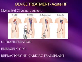
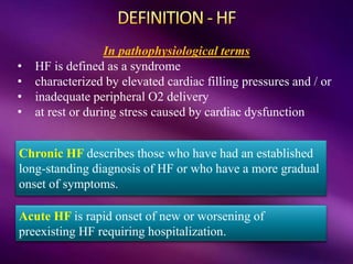
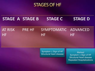
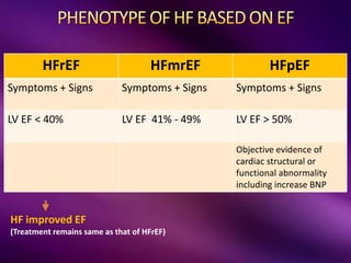
Col Bharat Malhotra
Senior Advisor (Medicine)
REFERENCE
Harrison’s Principles of internal medicine Harrison's 21st Ed (2022)
Davidson’s Principles and practice of Medicine (2018)
AHA & European Guidelines on management of Acute and Chronic Heart Failure (2021 & 2022)
Incidence and Prevalence of HF worldwide
PREVALENCE
Developed Countries: 2%
Asia : 0.1 -7%
India : 0.12- 0.44%
40-60 yrs: 1-2%
> 80 years: 12%
• Complex clinical syndrome
• that results from structural or functional impairment
• of ventricular filling or ejection of blood
• leads to cardinal manifestation of dyspnea and
fatigue and fluid retention
American Heart Association Cardiology Guidelines (2022)
Chronic HF describes those who have had an established
long-standing diagnosis of HF or who have a more gradual
onset of symptoms.
Acute HF is rapid onset of new or worsening of
preexisting HF requiring hospitalization.
In pathophysiological terms
• HF is defined as a syndrome
• characterized by elevated cardiac filling pressures and / or
• inadequate peripheral O2 delivery
• at rest or during stress caused by cardiac dysfunction
STAGE A STAGE B STAGE C STAGE D
AT RISK
HF
PRE HF SYMPTOMATIC
HF
ADVANCED
HF
Symptom + Sign of HF
Structural heart disease
Marked
Symptom + Sign of HF
Structural heart disease
Repeated Hospitalizations
HFrEF HFmrEF HFpEF
Symptoms + Signs Symptoms + Signs Symptoms + Signs
LV EF < 40% LV EF 41% - 49% LV EF > 50%
Objective evidence of
cardiac structural or
functional abnormality
including increase BNP
HF improved EF
(Treatment remains same as that of HFrEF)
DILATED
CARDIOMYOPATHY
CORONARY ARTERY
DISEASE
TACHYARRHYTHMIAS
Drug Induced
Alcohol
Metabolic
Endocrinal
Autoimmune
Viral - Myocarditis
VALVULAR DISEASE
Lt –Rt SHUNTING
HYPERTENSION
HYPERTROPHIC
CARDIOMYOPATHY
RESTRICTIVE
CARDIOMYOPATHY
Amylodosis
Sarcoidosis
Haemochromatosis
STENOTIC
VALVULAR DISEASE
HYPERTENSION
ENDOMYOCARDIAL
DISORDERS
CONSTRICTIVE
PERICARDITIS
HIGH OUTPUT STATES
AV Fistula
Chronic Anemia
Thyrotoxicosis
Beri Beri
OTHER FACTORS
Coronary Ischemia
Arrhythmias
Uncontrolled hypertension
Pulmonary embolism
Systemic infection/ sepsis
Patient related factors
Provider related factors
Na & Water
RETENTION
INCREASE PRELOAD
VASOCONSTRICTION
INCREASE AFTERLOAD
DECREASES MYOCARDIAL
CONTRACTILITY
DECREASES CARDIAC OUTPUT
SVC
IVC
P Artery P Vein
Aorta
Lungs
Lt A
Rt A
Rt V Lt V
Chronic HF describes those who have had an established
long-standing diagnosis of HF or who have a more gradual
onset of symptoms.
The diagnosis of CHF requires the presence of symptoms and/or signs of HF
and objective evidence of cardiac dysfunction
PRO
BNP
BNP
ECHO
Chronic Symptoms & signs
of HF
Risk factors for HF
Abnormal ECG/ CXR
Confirm Heart Failure
Define EF & Etiology
EF
Structure
> 125 pg/mL
> 35 pg/mL
Dyspnoea during exercise  on less strenuous activity 
ultimately may occur at rest
Paroxysmal nocturnal dyspnoea, Orthopnoea
Fatigue, Poor effort tolerance
Ankle edema
Nocturnal cough
Anorexia, nausea, early satiety, Rt upper quadrant
abdominal pain & fullness
Confusion, Disorientation, Oliguria
CONGESTION
RAISED JVP
Prominent P2
Hepatomegaly
Hepatojugular
reflux
Ascites
Pedal edema
Cardiomegaly
S3, S4
Pulmonary crackles
Pleural Effusions
LOW CARDIAC OUTPUT
Lethargy, fatigue, Alter mental state
Cool Extremities
Sinus tachycardia
Narrow pulse pressure
Lower BP
• AKI – Due to poor renal perfusion
• Impair liver function
• Hypokalemia, hyperkalemia
• Hyponatremia
• Thromboembolism
• Atrial or ventricular arrhythmias
• Sudden cardiac death
12 lead ECG
CXR PA View
BNP/ NT Pro BNP test
ECHO
Test for Comorbidities Test for Etiology
CBC
Urea Creatinine
Electrolytes
Fasting Glucose, HBA1c
Lipid Profile
TSH
Iron status – Serum Iron, TIBC, Ferritin
High pretest probability of CAD –PCI
Intermediate probability of CAD Stress
ECHO, Exercise testing
Specific disease workup if required
Cardiac – MRI [Infiltrative disorders)
Endomyocardial biopsy
Rt Heart catheterization – done rarely in
evaluation for transplant
ECG – May by abnormal due to ACS, tachyarrhythmias, LBBB, Chamber enlargement
BNP [ >35 pg/mL CHF] [ >100 pg/dLAHF ]
NT Pro BNP [ > 125 pg/mL CHF] [ >400 pg/dLAHF ]
Released from failing heart (sensitive marker for HF)
• Support diagnosis of HF
• Prognosis
• GDMT
• Falsely elevated in acute cardiac illness,
critical illness, Sepsis, PE, AKI
BIOMARKERS: Troponin T (Ischemia)
New diagnosis of HF
Clinical change in patient with known diagnosis of HF
Assess structure, chambers, Valves,
flow dynamics, function, EF
ACEI/ARB/ARNI
B Blocker
MRA
Dapagliflozin/Empagliflozin
Diuretics for fluid retention
LV EF < 35%
QRS WNL
ICD
LV EF > 35%
Device not indicated
If symptom persist
(go to Class II
recommendation)
LV EF < 35%
SR, QRS BROAD
CRT
AIM of Rx
Improve survival
Improve clinical status
ACEI
• Enalapril
• Ramipril
• Lisinopril
ARB
• Losartan
• Valsartan
ARNI
• Sacubitril+
Valsartan
•(Combination)
B Blocker MRA SGLT2 Diuretics
Bisoprolol
Metoprolol
Nebivolol
Carvedilol
Spironolactone
Eplerenone
Dapagliflozin
Empagliflozin
Furosemide
Torsemide
Metolazone
ARNI
Reduce HR
further
In Blacks
only
In AF cases Trials Stage
IVABRADINE Hydralazine &
Isosorbide
Digoxin Vericiguat
• Dyslipidemia, Hypertension, diabetes, obesity
Treat risk factors
• CABG/ PCI
CAD
• Anticoagulation, Digoxin
AF
• Assess and treat if required
Sleep Disorders
• IV Ferric Carboxy maltose
Iron deficiency
• Limited Exercise and weight reduction
• Low salt diet, Low fat diet, smoking cessation
Advise
• Influenza
• Pneumococcal
Vaccine
Reduce risk of SCD
LV EF < 35%
QRS WNL
 Ischemic origin after 40 d
if survival is > 40 days
 Post cardiac arrest
 VT with hypotension
Reduce mortality and morbidity in selected patients and
improves cardiac function
LV EF < 35%
QRS wide with LBBB
Sinus rhythm
 Symptomatic
HF patients
CABG/
PCI
Valve
Repair
Acute HF is rapid onset of new or worsening of
preexisting HF requiring hospitalization.
PRO
BNP
BNP
ECHO
Acute Symptoms & signs
of HF
Risk factors for HF
Abnormal ECG/ CXR/
SpO2
Confirm Heart Failure
Define EF & Etiology
EF
Structure
> 400 pg/mL
> 100 pg/mL
The diagnosis of AHF requires the presence of Rapid onset of HF
or acute decompensation of existing HF
HIGH OUTPUT STATES
Chronic Anemia
Thyrotoxicosis
Beri Beri
OTHER FACTORS
Coronary Ischemia
Arrhythmias
Uncontrolled hypertension
Valvular heart disease
Pulmonary embolism
Systemic infection/ sepsis
Patient related factors
Provider related factors
• Volume overload
Acute Decompensation
(Typical)
• Volume overload
• Hypoperfusion with End organ dysfunction
Acute Decompensation
(Low Output)
• Severe Pulmonary congestion
• Hypoxia
Acute Pulmonary
Edema
• Hypotension with low cardiac output
• End Organ Failure
Cardiogenic Shock
CONGESTION
Orthopnea
Pedal edema
Pleural Effusions
Ascites
Cardiomegaly
S3
Tachypnoea
Pulmonary
crackles
Raised JVP
LOW PERFUSION
Lethargy, fatigue,
Alter mental state
Cool Extremities
Sinus tachycardia
Narrow pulse pressure
Lower BP
End organ dysfunction
Low
P
E
R
F
U
S
I
O
N
CONGESTION
NO YES
NO Warm
Dry
Warm
Wet
YES Cold
Dry
Cold
Wet
Hospitalized
12 lead ECG
CXR PA View, SpO2, ABG
BNP/ NT Pro BNP test
ECHO
Test for Comorbidities Test for Etiology
CBC
Urea Creatinine
Electrolytes
Fasting Glucose, HBA1c
Lipid Profile
Evaluate – coexisting infections
TSH
Iron status – Serum Iron, TIBC, Ferritin
High pretest probability of CAD –PCI
SpO2 monitor
ABG
Swan Ganz Catheter
(Assess PA pressures)
In cardiogenic shock
O2, NIV, Ventilatory support
Congestion/ Fluid overload
• IV Loop Diuretics
• Increase dose - IV loop diuretic,
• Loop diuretic + metolazone
• Renal replacement therapy
Hypoperfusion
• Inotropes – Dobutamine, Dopamine (Milrinone, Levosimendan)
• Vasopressors - Norepinephrine
• Mechanical circulatory support, Emergency PCI
Vasodilators- IV Nitrates , IV Nitroprusside
Other drugs :IV opioids (in acute pulm edema), Anticoagulants (in AF)
DIURETICS
• Furosemide
• Continuous
• vs Bolus
Add on
• Chlorothiazide
or Metolazone
Refractory or in
Cardiorenal Syndrome
• RRT
Vasodilator Inotropes Vasopressor Other Rx
Nitroglycerine
Na
Nitroprusside
(Reserve drug)
Nesiritide
(Utilization waned)
Dobutamine
Dopamine
Milrinone, Levosimendan
(Utilization waned)
Norepinephrine ACS
Arrhythmia
Infection
Anemia
Switch to oral Rx
as for HFrEF
Mechanical Circulatory support
ULTRAFILTERATION
EMERGENCY PCI
REFRACTORY HF- CARDIAC TRANSPLANT
69 years old Female
Dyspnoea on exertion - 3 month
Anorexia
Vital Stable
Pedal edema present
Pro BNP 544 pg/mL
ECHO – EF 40% , MR +
65 years Male smoker, diabetes
Progressive dyspnoea 3 days and now dyspnoeic at rest 1 day.
Temp 100 F, Pallor
Pulse 104/min
RR 28/min, Spo2 90%
BP 122/80 mmHg
Warm peripheries
JVP raised
Pedal edema present
Tender hepatomegaly.
Pro BNP 544 pg/mL
ECHO – EF 25% , MR + RWMA +
Trop T positive
52 years Male
Progressive dyspnoea 1 year associated with wheeze
On treatment with bronchodilators from Pvt Nursing home
reported to our hospital due to no improvement in symptoms.
Don’t mistake
cardiac asthma for
bronchial asthma
EXAMINE
PATIENT
HAD SIGNIFICANT MURMUR
Heart Failure Approach class.pptx

Heart Failure Approach class.pptx

  • 1.
    Col Bharat Malhotra SeniorAdvisor (Medicine) REFERENCE Harrison’s Principles of internal medicine Harrison's 21st Ed (2022) Davidson’s Principles and practice of Medicine (2018) AHA & European Guidelines on management of Acute and Chronic Heart Failure (2021 & 2022)
  • 2.
    Incidence and Prevalenceof HF worldwide PREVALENCE Developed Countries: 2% Asia : 0.1 -7% India : 0.12- 0.44% 40-60 yrs: 1-2% > 80 years: 12%
  • 3.
    • Complex clinicalsyndrome • that results from structural or functional impairment • of ventricular filling or ejection of blood • leads to cardinal manifestation of dyspnea and fatigue and fluid retention American Heart Association Cardiology Guidelines (2022)
  • 4.
    Chronic HF describesthose who have had an established long-standing diagnosis of HF or who have a more gradual onset of symptoms. Acute HF is rapid onset of new or worsening of preexisting HF requiring hospitalization. In pathophysiological terms • HF is defined as a syndrome • characterized by elevated cardiac filling pressures and / or • inadequate peripheral O2 delivery • at rest or during stress caused by cardiac dysfunction
  • 5.
    STAGE A STAGEB STAGE C STAGE D AT RISK HF PRE HF SYMPTOMATIC HF ADVANCED HF Symptom + Sign of HF Structural heart disease Marked Symptom + Sign of HF Structural heart disease Repeated Hospitalizations
  • 6.
    HFrEF HFmrEF HFpEF Symptoms+ Signs Symptoms + Signs Symptoms + Signs LV EF < 40% LV EF 41% - 49% LV EF > 50% Objective evidence of cardiac structural or functional abnormality including increase BNP HF improved EF (Treatment remains same as that of HFrEF)
  • 7.
  • 8.
  • 9.
    HIGH OUTPUT STATES AVFistula Chronic Anemia Thyrotoxicosis Beri Beri OTHER FACTORS Coronary Ischemia Arrhythmias Uncontrolled hypertension Pulmonary embolism Systemic infection/ sepsis Patient related factors Provider related factors
  • 10.
    Na & Water RETENTION INCREASEPRELOAD VASOCONSTRICTION INCREASE AFTERLOAD DECREASES MYOCARDIAL CONTRACTILITY DECREASES CARDIAC OUTPUT
  • 11.
    SVC IVC P Artery PVein Aorta Lungs Lt A Rt A Rt V Lt V
  • 12.
    Chronic HF describesthose who have had an established long-standing diagnosis of HF or who have a more gradual onset of symptoms.
  • 13.
    The diagnosis ofCHF requires the presence of symptoms and/or signs of HF and objective evidence of cardiac dysfunction PRO BNP BNP ECHO Chronic Symptoms & signs of HF Risk factors for HF Abnormal ECG/ CXR Confirm Heart Failure Define EF & Etiology EF Structure > 125 pg/mL > 35 pg/mL
  • 14.
    Dyspnoea during exercise on less strenuous activity  ultimately may occur at rest Paroxysmal nocturnal dyspnoea, Orthopnoea Fatigue, Poor effort tolerance Ankle edema Nocturnal cough Anorexia, nausea, early satiety, Rt upper quadrant abdominal pain & fullness Confusion, Disorientation, Oliguria
  • 15.
    CONGESTION RAISED JVP Prominent P2 Hepatomegaly Hepatojugular reflux Ascites Pedaledema Cardiomegaly S3, S4 Pulmonary crackles Pleural Effusions LOW CARDIAC OUTPUT Lethargy, fatigue, Alter mental state Cool Extremities Sinus tachycardia Narrow pulse pressure Lower BP
  • 16.
    • AKI –Due to poor renal perfusion • Impair liver function • Hypokalemia, hyperkalemia • Hyponatremia • Thromboembolism • Atrial or ventricular arrhythmias • Sudden cardiac death
  • 17.
    12 lead ECG CXRPA View BNP/ NT Pro BNP test ECHO Test for Comorbidities Test for Etiology CBC Urea Creatinine Electrolytes Fasting Glucose, HBA1c Lipid Profile TSH Iron status – Serum Iron, TIBC, Ferritin High pretest probability of CAD –PCI Intermediate probability of CAD Stress ECHO, Exercise testing Specific disease workup if required Cardiac – MRI [Infiltrative disorders) Endomyocardial biopsy Rt Heart catheterization – done rarely in evaluation for transplant
  • 18.
    ECG – Mayby abnormal due to ACS, tachyarrhythmias, LBBB, Chamber enlargement
  • 20.
    BNP [ >35pg/mL CHF] [ >100 pg/dLAHF ] NT Pro BNP [ > 125 pg/mL CHF] [ >400 pg/dLAHF ] Released from failing heart (sensitive marker for HF) • Support diagnosis of HF • Prognosis • GDMT • Falsely elevated in acute cardiac illness, critical illness, Sepsis, PE, AKI BIOMARKERS: Troponin T (Ischemia)
  • 21.
    New diagnosis ofHF Clinical change in patient with known diagnosis of HF Assess structure, chambers, Valves, flow dynamics, function, EF
  • 22.
    ACEI/ARB/ARNI B Blocker MRA Dapagliflozin/Empagliflozin Diuretics forfluid retention LV EF < 35% QRS WNL ICD LV EF > 35% Device not indicated If symptom persist (go to Class II recommendation) LV EF < 35% SR, QRS BROAD CRT AIM of Rx Improve survival Improve clinical status
  • 23.
    ACEI • Enalapril • Ramipril •Lisinopril ARB • Losartan • Valsartan ARNI • Sacubitril+ Valsartan •(Combination) B Blocker MRA SGLT2 Diuretics Bisoprolol Metoprolol Nebivolol Carvedilol Spironolactone Eplerenone Dapagliflozin Empagliflozin Furosemide Torsemide Metolazone
  • 24.
  • 25.
    Reduce HR further In Blacks only InAF cases Trials Stage IVABRADINE Hydralazine & Isosorbide Digoxin Vericiguat
  • 26.
    • Dyslipidemia, Hypertension,diabetes, obesity Treat risk factors • CABG/ PCI CAD • Anticoagulation, Digoxin AF • Assess and treat if required Sleep Disorders • IV Ferric Carboxy maltose Iron deficiency • Limited Exercise and weight reduction • Low salt diet, Low fat diet, smoking cessation Advise • Influenza • Pneumococcal Vaccine
  • 27.
    Reduce risk ofSCD LV EF < 35% QRS WNL  Ischemic origin after 40 d if survival is > 40 days  Post cardiac arrest  VT with hypotension
  • 28.
    Reduce mortality andmorbidity in selected patients and improves cardiac function LV EF < 35% QRS wide with LBBB Sinus rhythm  Symptomatic HF patients
  • 29.
  • 30.
    Acute HF israpid onset of new or worsening of preexisting HF requiring hospitalization.
  • 31.
    PRO BNP BNP ECHO Acute Symptoms &signs of HF Risk factors for HF Abnormal ECG/ CXR/ SpO2 Confirm Heart Failure Define EF & Etiology EF Structure > 400 pg/mL > 100 pg/mL The diagnosis of AHF requires the presence of Rapid onset of HF or acute decompensation of existing HF
  • 32.
    HIGH OUTPUT STATES ChronicAnemia Thyrotoxicosis Beri Beri OTHER FACTORS Coronary Ischemia Arrhythmias Uncontrolled hypertension Valvular heart disease Pulmonary embolism Systemic infection/ sepsis Patient related factors Provider related factors
  • 33.
    • Volume overload AcuteDecompensation (Typical) • Volume overload • Hypoperfusion with End organ dysfunction Acute Decompensation (Low Output) • Severe Pulmonary congestion • Hypoxia Acute Pulmonary Edema • Hypotension with low cardiac output • End Organ Failure Cardiogenic Shock
  • 34.
    CONGESTION Orthopnea Pedal edema Pleural Effusions Ascites Cardiomegaly S3 Tachypnoea Pulmonary crackles RaisedJVP LOW PERFUSION Lethargy, fatigue, Alter mental state Cool Extremities Sinus tachycardia Narrow pulse pressure Lower BP End organ dysfunction Low P E R F U S I O N CONGESTION NO YES NO Warm Dry Warm Wet YES Cold Dry Cold Wet Hospitalized
  • 35.
    12 lead ECG CXRPA View, SpO2, ABG BNP/ NT Pro BNP test ECHO Test for Comorbidities Test for Etiology CBC Urea Creatinine Electrolytes Fasting Glucose, HBA1c Lipid Profile Evaluate – coexisting infections TSH Iron status – Serum Iron, TIBC, Ferritin High pretest probability of CAD –PCI SpO2 monitor ABG Swan Ganz Catheter (Assess PA pressures) In cardiogenic shock
  • 36.
    O2, NIV, Ventilatorysupport Congestion/ Fluid overload • IV Loop Diuretics • Increase dose - IV loop diuretic, • Loop diuretic + metolazone • Renal replacement therapy Hypoperfusion • Inotropes – Dobutamine, Dopamine (Milrinone, Levosimendan) • Vasopressors - Norepinephrine • Mechanical circulatory support, Emergency PCI Vasodilators- IV Nitrates , IV Nitroprusside Other drugs :IV opioids (in acute pulm edema), Anticoagulants (in AF)
  • 37.
    DIURETICS • Furosemide • Continuous •vs Bolus Add on • Chlorothiazide or Metolazone Refractory or in Cardiorenal Syndrome • RRT Vasodilator Inotropes Vasopressor Other Rx Nitroglycerine Na Nitroprusside (Reserve drug) Nesiritide (Utilization waned) Dobutamine Dopamine Milrinone, Levosimendan (Utilization waned) Norepinephrine ACS Arrhythmia Infection Anemia Switch to oral Rx as for HFrEF
  • 38.
    Mechanical Circulatory support ULTRAFILTERATION EMERGENCYPCI REFRACTORY HF- CARDIAC TRANSPLANT
  • 40.
    69 years oldFemale Dyspnoea on exertion - 3 month Anorexia Vital Stable Pedal edema present Pro BNP 544 pg/mL ECHO – EF 40% , MR +
  • 41.
    65 years Malesmoker, diabetes Progressive dyspnoea 3 days and now dyspnoeic at rest 1 day. Temp 100 F, Pallor Pulse 104/min RR 28/min, Spo2 90% BP 122/80 mmHg Warm peripheries JVP raised Pedal edema present Tender hepatomegaly. Pro BNP 544 pg/mL ECHO – EF 25% , MR + RWMA + Trop T positive
  • 42.
    52 years Male Progressivedyspnoea 1 year associated with wheeze On treatment with bronchodilators from Pvt Nursing home reported to our hospital due to no improvement in symptoms. Don’t mistake cardiac asthma for bronchial asthma EXAMINE PATIENT HAD SIGNIFICANT MURMUR