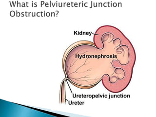
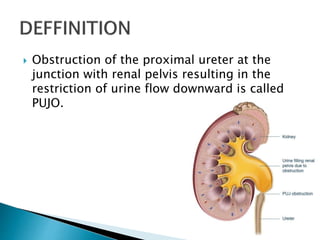
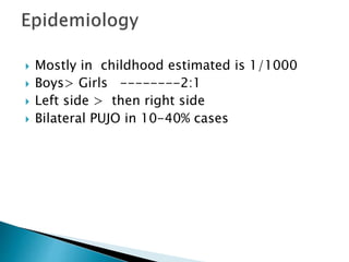
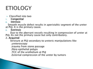
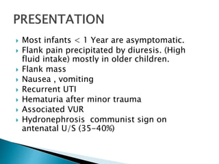
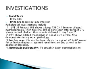
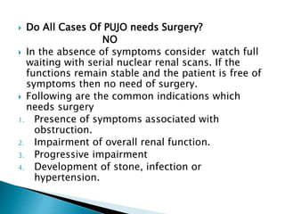
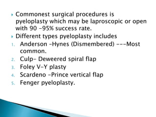
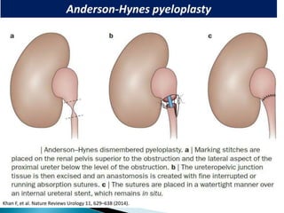
Prepared By
Dr. Muhammad Idrees Khan
PGR IKD HMC
 Definition
 Epidemiology and etiology
 Presentation
 Investigations
 Management
 Obstruction of the proximal ureter at the
junction with renal pelvis resulting in the
restriction of urine flow downward is called
PUJO.
 Mostly in childhood estimated is 1/1000
 Boys> Girls --------2:1
 Left side > then right side
 Bilateral PUJO in 10-40% cases
 Classified into two
1. Congenital
A. Intrinsic
Smooth muscle defect results in aperistaltic segment of the ureter
at PUJ. It is the primary cause.
A. Extrinsic
Due to the aberrant vessels resulting in compression of ureter at
PUJ. Its not the primary cause but only contributory.
2. Acquired
Stricture at PUJ secondary to ureteric manipulations like
.ureteroscopy
.trauma from stone passage
.fibro epithelial polyps
.TCC of the urothelium at PUJ
.External compression of the ureter by tumors
 Most infants < 1 Year are asymptomatic.
 Flank pain precipitated by diuresis. (High
fluid intake) mostly in older children.
 Flank mass
 Nausea , vomiting
 Recurrent UTI
 Hematuria after minor trauma
 Associated VUR
 Hydronephrosis communist sign on
antenatal U/S (35-40%)
 Blood Tests
RFTS, CBC
 Urine R/E to rule out any infection
Radiological investigations include
1. U/S : If Prenatal U/S shows a large TAPD> 15mm or bilateral
hydronephrosis. Then f/u renal U/S is done soon after birth. If U/S
shows normal bladder then scan is deferred to day 3 and 7.
2. CT : shows dilated renal pelvis in non dilated ureter. Also
demonstrates in any other pathology.
3. Nuclear scan: this can be done above the age of 4th to 6th weeks
for definitive diagnosis, splitted renal function and as well as for
degree of blockage.
4. Retrograde pyelography : To establish exact obstruction site.
 Do All Cases Of PUJO needs Surgery?
NO
 In the absence of symptoms consider watch full
waiting with serial nuclear renal scans. If the
functions remain stable and the patient is free of
symptoms then no need of surgery.
 Following are the common indications which
needs surgery
1. Presence of symptoms associated with
obstruction.
2. Impairment of overall renal function.
3. Progressive impairment
4. Development of stone, infection or
hypertension.
 Commonest surgical procedures is
pyeloplasty which may be laproscopic or open
with 90 -95% success rate.
 Different types pyeloplasty includes
1. Anderson –Hynes (Dismembered) ---Most
common.
2. Culp- Deweered spiral flap
3. Foley V-Y plasty
4. Scardeno -Prince vertical flap
5. Fenger pyeloplasty.
PUJO.pptx
PUJO.pptx
PUJO.pptx
PUJO.pptx
PUJO.pptx

PUJO.pptx

  • 2.
    Prepared By Dr. MuhammadIdrees Khan PGR IKD HMC
  • 3.
     Definition  Epidemiologyand etiology  Presentation  Investigations  Management
  • 5.
     Obstruction ofthe proximal ureter at the junction with renal pelvis resulting in the restriction of urine flow downward is called PUJO.
  • 6.
     Mostly inchildhood estimated is 1/1000  Boys> Girls --------2:1  Left side > then right side  Bilateral PUJO in 10-40% cases
  • 7.
     Classified intotwo 1. Congenital A. Intrinsic Smooth muscle defect results in aperistaltic segment of the ureter at PUJ. It is the primary cause. A. Extrinsic Due to the aberrant vessels resulting in compression of ureter at PUJ. Its not the primary cause but only contributory. 2. Acquired Stricture at PUJ secondary to ureteric manipulations like .ureteroscopy .trauma from stone passage .fibro epithelial polyps .TCC of the urothelium at PUJ .External compression of the ureter by tumors
  • 8.
     Most infants< 1 Year are asymptomatic.  Flank pain precipitated by diuresis. (High fluid intake) mostly in older children.  Flank mass  Nausea , vomiting  Recurrent UTI  Hematuria after minor trauma  Associated VUR  Hydronephrosis communist sign on antenatal U/S (35-40%)
  • 10.
     Blood Tests RFTS,CBC  Urine R/E to rule out any infection Radiological investigations include 1. U/S : If Prenatal U/S shows a large TAPD> 15mm or bilateral hydronephrosis. Then f/u renal U/S is done soon after birth. If U/S shows normal bladder then scan is deferred to day 3 and 7. 2. CT : shows dilated renal pelvis in non dilated ureter. Also demonstrates in any other pathology. 3. Nuclear scan: this can be done above the age of 4th to 6th weeks for definitive diagnosis, splitted renal function and as well as for degree of blockage. 4. Retrograde pyelography : To establish exact obstruction site.
  • 11.
     Do AllCases Of PUJO needs Surgery? NO  In the absence of symptoms consider watch full waiting with serial nuclear renal scans. If the functions remain stable and the patient is free of symptoms then no need of surgery.  Following are the common indications which needs surgery 1. Presence of symptoms associated with obstruction. 2. Impairment of overall renal function. 3. Progressive impairment 4. Development of stone, infection or hypertension.
  • 12.
     Commonest surgicalprocedures is pyeloplasty which may be laproscopic or open with 90 -95% success rate.  Different types pyeloplasty includes 1. Anderson –Hynes (Dismembered) ---Most common. 2. Culp- Deweered spiral flap 3. Foley V-Y plasty 4. Scardeno -Prince vertical flap 5. Fenger pyeloplasty.