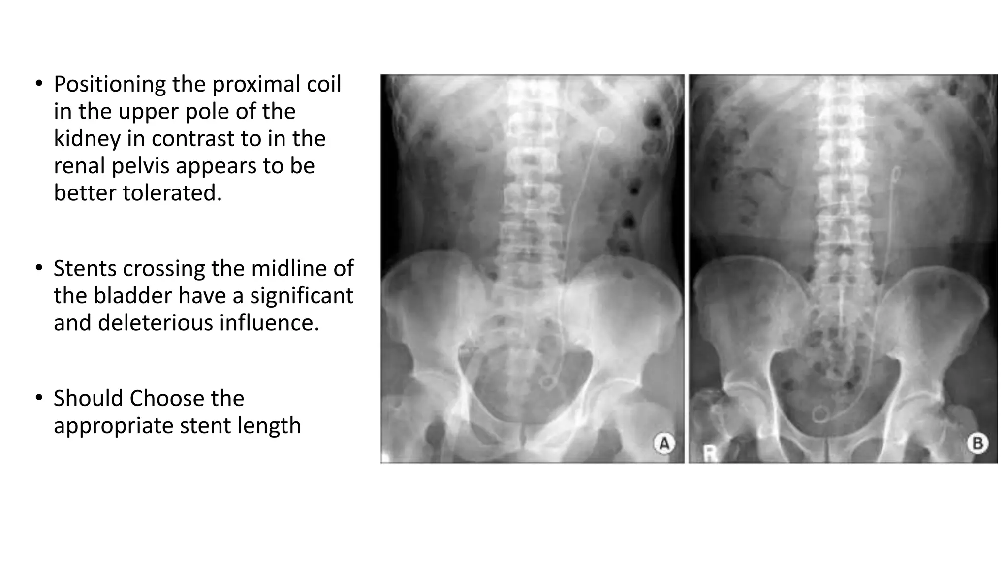
STENTS IN UROLOGY
Dr. Deepesh Kalra
Institute Of Urology
MMC,Chennai
• Zimskind (1967) - first to describe the cystoscopic placement of
indwelling ureteral stents for obstructed ureters.
• Gibbons - first to patent stent as a self-retaining mechanism.
• The first “double-J” (DJ) or double pigtail stent was developed by
Finney and Hepperlen
Ideal stent -
• Easy to insert
• Ability to relieve intraluminal and
extraluminal obstruction
• Excellent flow characteristics
• Resistant to encrustation and
infection.
• Chemically stable after implantation
in a urinary environment
• Does not induce patient symptoms
• High tensile strength
• A low friction coefficient
• Memory
• A self-retainment mechanism
• Should be both biocompatible and
affordable.
Biomaterials
 Silicone -
• Most biocompatible material tested to date
• Least encrustation
• High friction coefficient and flexibility
• more difficult to navigate through a tortuous or obstructed ureter
 Polyethylene -
• First plastic polymer
• Become brittle after prolonged exposure to the urinary environment
• Prone to encrustation, blockage, and fragmentation
Currently used stents are commonly composed of
 Polyurethane
 Silicone
 Proprietary copolymers
Silitek
C-Flex (stiffest) max. Resistance
Black silicone (softest, long duro)
Percuflex
Tecoflex
q The self-expanding
metallic Wallstent -
ü primary patency rates are low,
29% to 54% at 3 to 12
months mainly because of
hyperplastic tissue ingrowth.
q The Resonance metallic ureteral stent-
ü Tightly coiled spirals of a corrosion-
resistant nickel-cobalt-chromium-
molybdenum alloy wire.
ü Resist encrustation and tissue
overgrowth.
 Indwelling stents lasting up to 12
months or longer
ü Inferior overall flow
ü superior in withstanding compression
forces
The Silhouette stent -
Soft, coil-reinforced
 hydrophilic coating.
Less prone to kinking
Can resist higher
compressional forces than the Resonance stent
The Passage and Snake stents -
Open-ended and less tightly coiled than the Resonance and Silhouette stents,
Allowing for more flexibility.
Sustain higher extrinsic radial compression forces than the Silhouette stent
 The Memokath 051
ureteral stent -
Nickel-titanium alloy (NITINOL)
stent with a thermo-expandable
anchoring mechanism.
Long-term patency in ureteral
obstruction
Better tolerated than
conventional ureteral stents in
terms of urinary symptoms, pain.
q The Uventa stent -
ü nickel-titanium alloy, Segmental,
Thermally expandable stent.
ü Meshed
ü A PTFE coating is positioned
between the outer and inner mesh.
ü The outer mesh provides extra
friction to prevent stent migration,
and the PTFE coating prevents
hyperplastic stent ingrowth
 The Allium stent -
large-caliber (24 Fr or 30 Fr) nickel-
titanium alloy, expandable
Meshed
coated with a biocompatible polymer
to prevent stent ingrowth.
The Allium stent was specifically
developed for use in the distal ureter
and has an intravesical anchor to
facilitate removal.
• The development of a biodegradable stent could theoretically eliminate the need
for cystoscopic stent removal and could help prevent the occurrence of forgotten
stents.
• Biodegradable materials are composed of high-molecular-weight polymers such
as polylactide or polyglycolide.
q Uriprene -
 A biodegradable copolymer composed of L-
glycolic acid, polyethylene glycol, and barium
sulfate
 Reliably achieve degradation after 4 weeks
 Induced a lower degree of ureteral
inflammatory change when compared with
conventional stents in a porcine model
Coatings
 Drug-eluting and antiadhesive stent coatings -
• Goals -
Improving stent handling
Reducing biofilm formation
Preventing encrustation
Improving patient comfort
 Hydrogel -
Hydrophilic polymers.
This added surface water reduces friction and increases elasticity
Easier to insert and theoretically more biocompatible.
Pentosan polysulfate (PPS), phosphorylcholine (PC) copolymer, and
polyvinylpyrrolidone (PVP)
Reduces -
 Inflammatory response
 encrustation,
biofilm formation.
q Polyvinylpyrrolidone-iodine (PVP-I) complex modified polyurethane Tecoflex stents
ü Highly hydrophilic and to reduce encrustation deposits and adherence of Pseudomonas
aeruginosa and Staphylococcus aureus by 80% to 86% in in vitro tests
Diamond-like carbon (DLC) coating -
Ultrasmooth smooth
Decreasing friction
Improving biocompatibility
Silicone coated with Oxalobacter formigenes derived oxalate degrading enzymes -
demonstrated a modest reduction in encrustation
 Zotarolimus-eluting metal stent
induced a significantly lower hyperplastic reaction without influencing
inflammation
 Paclitaxel drug-eluting metal mesh stent
 Triclosan-eluting stents (Triumph) -
significantly reduced stent-related pain and urinary symptoms
ü reduced symptomatic UTI rate
 Ketorolac-eluting stent - Reduced stent-induced pain
 Heparin-coated stents - Reduced encrustation rate
 mPEGDOPA3, is a novel antifouling coating resistance to bacterial attachment
and biofilm formation
 Sustained-release varnish containing chlorhexidine (CHX-SRV) – coated stents
significantly reduce bacterial growth
• Applying silver coatings on ureteral stents appears to be an effective strategy in
reducing biofilm adherence without the risk of inducing resistance
Stent Design -
Simple variations to the initial DJ stent
developed by Finney
 3F Microstent -
 Uses a film anchor as a proximal
retaining mechanism.
 Flow characteristics are equivalent
to those of a 4.7-Fr DJ stent
 Grooved stent -
better extraluminal and total flow
post-lithotripsy treatment option, in order to
improve the stone clearance by introducing
multiple pathways for urine drainage
The Towers stent
The LithoStent (olympus)
 Dual-lumen stent -
Significantly improved the flow
Similar flow rates compared with two ipsilateral 7-
Fr stents
 The Spirastent -
a DJ stent with helical metal ridges
superior ability to conform to the ureter shape
Open-Pass ureteral stent -
Has 15 to 17 radially expanding baskets along its length.
Was developed for dilation of the ureter up to 20 Fr and stone fragment
entrapment after SWL.
 Stents equipped with an antireflux valve mechanism at the intravesical portion of
the stent demonstrate a significant decrease in reflux rate compared with a
conventional DJ stent, resulting in less flank and bladder pain and thus improved
patient comfort.
 Tail stents or buoy stents -
composed of a 7-Fr or 10-Fr upper body that
tapers down to a 3-Fr distal tail.
significantly better drainage,
reduced bladder inflammation
reduced irritative bladder symptoms
 Dual durometer stents -
composed of a conventional upper body and a
softer biomaterial at the distal segment
 Magnetip stent -
To avoid cystoscopic removal of the stent.
It has a metallic bead at the distal tip and can
be removed with a magnetic-tipped urethral
catheter
Indications
 Most commonly performed to relieve Ureteral obstruction.
 Intrinsic obstruction - stones, tumors, or strictures
 extrinsic obstruction - compression by tumor, overlying vessels, retroperitoneal
fibrosis, or lymphadenopathies.
Absolute and usually emergent indications for drainage of the kidney(s) are
 B/L obstruction
U/L obstruction in the single functional kidney
Ureteral obstruction with hydronephrosis and urinary infection or sepsis
ESWL
• EAU and the AUA indicate that routine use of DJ stenting before SWL for kidney
or ureteral stones does not improve stone-free rates.
Solitary kidney
Stone > 1.5 cm
Steinstrasse – post-ESWL
URSL
• Routine stenting has no beneficial effect on stone-free rate or ureteral stricture
formation. The procedure takes longer and costs more.
• EAU stone guidelines advise that stenting is not routinely required after
uncomplicated URSL.
• Stenting a ureter post-URSL is, still advised-
Sizeable residual fragments
Presence of an anatomically or functionally solitary kidney
Ureter has been balloon dilated
 UTI
 Complication such as bleeding or perforation has occurred.
• Indwelling time shorter than 14 days was associated with fewer adverse effects
• Placing a ureteral stent after an un-successful URSL for secondary URSL is
beneficial
PCNL
• Routine placement of an internal stent after uncomplicated PCNL is not
necessarily required.
• Stenting is, however, advised in the presence of
residual stone burden
migration of residual fragments to the ureter
 extensive edema,
perforation of the collecting system
 high tract placement with risk of hydrothorax
tubeless PCNL
persistent urinary leakage after nephrostomy tube removal.
 Urologic reconstructive surgery for splinting the ureter
Scaffolding the tissue to improve organized healing
Allow urine to flow unhindered past the operated field
Ureteral trauma treatment
Ureteral realignment
Pyeloplasty
Ureteral re-implantation
Uretero-ureterostomy
Transplant
• Routine prophylactic stenting significantly reduces the incidence of major
urologic complications (Wilson et al, 2013).
• Removing the stent after 8 days as opposed to after 15 days reduces UTI rate
(40% vs. 73%) and is more cost-effective
Prophylactically before gynecologic, urologic, or abdominal surgery.
This facilitates identification of the ureter during surgery and theoretically may
reduce iatrogenic ureteral trauma.
No significant difference in ureteral injury
Easier to identify ureteric trauma with a stent in situ
Hydronephrosis of pregnancy -
Intracractable pain
Pyelonephritis
Rising creatinine
Calculi of pregnancy (after 22 wks) (increase encrustation)
 Drainage modalities for pyonephrosis,hydronephrosis
 Treatment of malignant pathology of the upper urinary tract with, for instance,
BCG or Mitomycin C.
Single-J ureteral catheter, via reflux through DJ stent.
 Persistent urinary extravasation after blunt renal trauma
Technique
Endoscopic retrograde
Antegrade placement
during open or laparoscopic surgery.
• Antibiotic prophylaxis before endoscopic stent placement is recommended in AUA
guidelines ( FQ )
Supine position with flexible cystoscopy
Lithotomy position when a rigid cystoscope is used.
Fluoroscopic guidance
Ultrasound guidance ( pregnant woman )
• As stent diameter does not seem to influence stent symptoms, one
should choose the largest fitting stent available for optimal drainage
• Hydrophilic Nitinol guidewires -
optimal characteristics to
Easily overcome obstruction
To follow the course of a
tortuous ureter
 With a minimal risk of
perforation
• Teflon-containing wires - higher
resistance against bending
• PTFE coated guidewires
• Cystoscopic placement of guidewire in renal pelvis
• Check the position fluoroscopically
• Stent over the guidewire with pusher
• When pusher is at neck retract guidewire
• Check coiling in pelvis fluoroscopically
• Check coiling in bladder cystoscopically
Place a stent by primarily relying on fluoroscopic guidance.
Place the guidewire in the renal pelvis
Cystoscope is removed
8-Fr to 10-Fr coaxial Amplatz dilator is advanced over the guidewire
under fluoroscopic guidance until the 10-Fr component is at the
urethral meatus.
Remove the 8-Fr component, the 10-Fr sheath will allow a 7-Fr stent
to be passed through it over the guidewire
Complications
 Stent Symptoms -
Hematuria
Urgency
Frequency
 Dysuria
 Bladder and flank pain
• Joshi and colleagues developed the ureteral stent symptom questionnaire
(USSQ) to evaluate symptoms and impact on quality of life of ureteral stents.
• 80% - quality of life was influenced
• 58% of patients had reduced work capacity
• 50% patients sought medical professional help
• 33% patients required early removal of ureteral stents because of stent
discomfort
• 42% to 82% of male patients and 30% to 86% of female patients – sexual
dysfunction
• The pathophysiologic explanation for such stent-related symptoms is not
yet fully understood.
 Irritation of the bladder mucosa and especially the trigone
 Reflux of urine
Smooth muscle spasm
• Vesicoureteral reflux - 56% to 62%
• Fluoroscopic imaging revealed positional changes of the stent in relation to
standing, sitting, and bending (physical activity can influence stent
discomfort )
• Positioning the proximal coil
in the upper pole of the
kidney in contrast to in the
renal pelvis appears to be
better tolerated.
• Stents crossing the midline of
the bladder have a significant
and deleterious influence.
• Should Choose the
appropriate stent length
Shorter than 5 feet 10 inches 22-cm stent
5 feet 10 inches to 6 feet 4 inches 24-cm stent
Taller than 6 feet 4 inches 26-cm stent
• Straight linear measurement from PUJ to VUJ on preoperative IVP
• Actual ureteric length to correlate better with CT–measured length than with any
other imaging-based or anthropomorphic measurement
• Ideal stent length for children has been formulated as “child's age + 10” cm
• The use of α-blocker - recommended by EAU guidelines
• Anticholinergic ( solifenacin / tolterodine )
• α-blocker & Anticholinergic better than monotherapy
• Intravesical instillation with ketorolac - short-lived but significant
• Periureteral injection of botulinum toxin A - up to 1 week after stent
placement
• Injection of ropivacaine in proximity to the ureteric orifice and at the
bladder neck demonstrated
• Appropriate stent position with the distal coil not crossing over the
midline of the bladder appeared to have more effect on stent-related
symptoms than α-blockers
Stent of appropriate length
allows reconstitution of the
proximal
pigtail loop in the renal
pelvis and the
distal loop above the
bladder base to
prevent migration and
reduce the occurrence
of irritative bladder
symptoms
(6) Fish reeling. (a) proximal pigtail loop of the stent is not completely reconstituted. (b) reconstitution of the proximal loop with resultant
retraction of the distal loop into the lower ureter. (7) Stent malposition. Persistent pericatheter leakage distal stent tip in the proximal urethra
Stent Migration
 Proximal stent migration
into the ureter - 1% to
8%
• Prevention -
Sufficiently long stent
Adequate loop both in the
renal pelvis and in the
bladder
Inadequate Relief of Obstruction
• Most patent stents will
demonstrate reflux at
the time of voiding
cystography, and this can
be used to assess
patency
• Ultrasonography (US) for
assessment of stent jets
has also been used
• Diuretic renography has
been reported to be the
most sensitive test for
determining ureteral stent
patency
Colonization
• Factors influencing colonization -
ü Female sex
ü Diabetes
ü CKD
ü Indwelling time
ü Routine screening for bacteriuria and treatment of asymptomatic bacteriuria is
not recommended.
ü Antibiotics are recommended only in instances of symptomatic UTI
Urinary Tract Infection
• Inherently subject to bacterial colonization and then UTI
• In Short-term ureteral stent placement (3 weeks)
UTI - 4.8%
Asymptomatic bacteriuria - 6.5%
Bacterial colonisation – 50%
• In chronically stented patients, bacterial colonization reaches 100%
Encrustation
• The duration of indwelling time of ureteral stents is the most important risk
factor for development of encrustation.
9.2% to 26.8% - less than 6 weeks
47.5% to 56.9% - 6 to 12 weeks
75% - longer than 12 weeks
• Stents smaller than 6 Fr are significantly more likely to encrust than stents
7 Fr or larger.
• Complete obstruction in 8.6% of stents after more than 12 weeks.
• Additional risk factors for stent encrustation include
Pregnancy
 UTI or urosepsis
History of stone disease
Metabolic or congenital abnormalities
Urinary diversion
Chronic renal failure
• It occurs in association with the presence of bacteria (such as Proteus mirabilis), which are
known to produce urease.
• These bacteria cause an increase of urine pH, leading to crystals’ formation
• Calcium oxalate appears to be the major component of stent encrustation in the absence of UTI,
pH values below 5.5, and hyperuricosuria.
• Timely stent removal or exchange is the most important preventive measure.
• Recommend stent removal or exchange within 4 months of placement.
• In patients with additional risk factors for encrustation; 6 to 8 weeks
• Pregnant patients are prone to stent encrustation; every 4 to 6 weeks
Peristaltic
“wiping”
• Encrustation and inability to extract a stent are usually diagnosed in an
office setting at a trial of stent removal
• Applying excessive force -
Ureteral damage/avulsion
Stent fragmentation
• placement of an additional stent for 1 to 2 weeks
friction disrupts the encrustation
ureteral dilation
• SWL, URS, cystolitholapaxy, and PCNL
Stent Fracture
• Urine is a hostile environment
• Polyethylene was abandoned
• Most fractures occur at
fenestration sites
• Encrustation is also likely to
play a role in stent
fragmentation
Forgotten or Neglected Stents
Poor patient compliance
Health system issues related to patient
follow-up
The surgeon responsible for stent
insertion is also accountable for its
timely removal.
• The cost of forgetting a stent is on
average sevenfold higher than the cost
of cystoscopic timely removal.
Ureteral Erosion or Fistulization
• The rarest, most feared complication
is erosion of the stent into adjacent
structures, especially the arterial
system.
• massive hematuria to the point of
circulatory collapse
• Extensive pelvic surgery and
irradiation appear to be contributing
factors to the development of this
complication because both may lead
to ureteral ischemia.
• Can develop
severe
encrustation.
• In the presence
of a large
encrustation
burden, nuclear
imaging to
quantify the
renal function is
advised for
planning stent
removal
Urethral Stents
• Titanium stent
• Expandable stent made by
Nitinol (nickel-titanium)
• Permanently Implantable
(UroLume) (Alloy)
 Indications -
 BPH -
EAU and the AUA guidelines on BPH recommend urethral stent placement in high risk
patients who are poor surgical candidate
Post TUMT
q Urethral stricture -
Post OIU or dilation
Post reconstruction
ü Prevent the process of epithelialization from incorporating the stent into the urethral
wall
ü 6 months to 1 year
Removable – Nitinol
Permanently Implantable (UroLume) –
 Alloy
Incorporated into the wall of the urethra and corpus spongiosum.
Relatively short strictures of the bulbous urethra associated with
minimal spongio-fibrosis
• Longer bulbous strictures require two stents that are overlapped.
• These stents can migrate away from each other, leaving a gap between them where
recurrence of stricture is inevitable.
• When this occurs, the stricture recurrence is excised, and a third stent is placed to span
the gap.
Contraindications -
• Prior substitution urethral reconstruction, particularly where skin has been incorporated.
As it is associated with a virulent hypertrophic reaction
• Strictures associated with deep spongiofibrosis (urethral distraction injuries and straddle
injuries)
 As a guide during posterior urethral and bladder neck reconstruction but is
removed after the repair is completed.
 Cystoscopic placement of transurethral stent in a fetus with urethral stenosis
 Neurogenic bladder (to facilitate emptying)
 Post urethro-vesical anastomotic stenoses
 Bladder-neck stenosis
• Complications -
 hyperplastic tissue growth
 stent separation
 stent encrustation
 recurrent urinary tract infections
 recurrent urethral obstruction
Stent misplacement
Stent migration
• Proximal stents implanted for BPH and DSD were more likely to require removal
in comparison to stents placed for bulbous urethral strictures.
• Stent misplacement and stent migration were among the main causes of stent
failure within proximal urethra
 Endoscopic Removal - For patients with urethral stents placed for BPH and/or
DSD, endoscopic management was recommended with resection of the overlying
urothelium, advancement of the stent into the bladder, and then en bloc removal
of the stent through a larger sheath
 Open Removal & reconstruction
THANK YOU

Stents in urology

  • 1.
    STENTS IN UROLOGY Dr.Deepesh Kalra Institute Of Urology MMC,Chennai
  • 2.
    • Zimskind (1967)- first to describe the cystoscopic placement of indwelling ureteral stents for obstructed ureters. • Gibbons - first to patent stent as a self-retaining mechanism. • The first “double-J” (DJ) or double pigtail stent was developed by Finney and Hepperlen
  • 3.
    Ideal stent - •Easy to insert • Ability to relieve intraluminal and extraluminal obstruction • Excellent flow characteristics • Resistant to encrustation and infection. • Chemically stable after implantation in a urinary environment • Does not induce patient symptoms • High tensile strength • A low friction coefficient • Memory • A self-retainment mechanism • Should be both biocompatible and affordable.
  • 4.
    Biomaterials  Silicone - •Most biocompatible material tested to date • Least encrustation • High friction coefficient and flexibility • more difficult to navigate through a tortuous or obstructed ureter  Polyethylene - • First plastic polymer • Become brittle after prolonged exposure to the urinary environment • Prone to encrustation, blockage, and fragmentation
  • 5.
    Currently used stentsare commonly composed of  Polyurethane  Silicone  Proprietary copolymers Silitek C-Flex (stiffest) max. Resistance Black silicone (softest, long duro) Percuflex Tecoflex
  • 6.
    q The self-expanding metallicWallstent - ü primary patency rates are low, 29% to 54% at 3 to 12 months mainly because of hyperplastic tissue ingrowth.
  • 7.
    q The Resonancemetallic ureteral stent- ü Tightly coiled spirals of a corrosion- resistant nickel-cobalt-chromium- molybdenum alloy wire. ü Resist encrustation and tissue overgrowth.  Indwelling stents lasting up to 12 months or longer ü Inferior overall flow ü superior in withstanding compression forces
  • 8.
    The Silhouette stent- Soft, coil-reinforced  hydrophilic coating. Less prone to kinking Can resist higher compressional forces than the Resonance stent The Passage and Snake stents - Open-ended and less tightly coiled than the Resonance and Silhouette stents, Allowing for more flexibility. Sustain higher extrinsic radial compression forces than the Silhouette stent
  • 9.
     The Memokath051 ureteral stent - Nickel-titanium alloy (NITINOL) stent with a thermo-expandable anchoring mechanism. Long-term patency in ureteral obstruction Better tolerated than conventional ureteral stents in terms of urinary symptoms, pain.
  • 10.
    q The Uventastent - ü nickel-titanium alloy, Segmental, Thermally expandable stent. ü Meshed ü A PTFE coating is positioned between the outer and inner mesh. ü The outer mesh provides extra friction to prevent stent migration, and the PTFE coating prevents hyperplastic stent ingrowth
  • 11.
     The Alliumstent - large-caliber (24 Fr or 30 Fr) nickel- titanium alloy, expandable Meshed coated with a biocompatible polymer to prevent stent ingrowth. The Allium stent was specifically developed for use in the distal ureter and has an intravesical anchor to facilitate removal.
  • 12.
    • The developmentof a biodegradable stent could theoretically eliminate the need for cystoscopic stent removal and could help prevent the occurrence of forgotten stents. • Biodegradable materials are composed of high-molecular-weight polymers such as polylactide or polyglycolide.
  • 13.
    q Uriprene - A biodegradable copolymer composed of L- glycolic acid, polyethylene glycol, and barium sulfate  Reliably achieve degradation after 4 weeks  Induced a lower degree of ureteral inflammatory change when compared with conventional stents in a porcine model
  • 14.
    Coatings  Drug-eluting andantiadhesive stent coatings - • Goals - Improving stent handling Reducing biofilm formation Preventing encrustation Improving patient comfort
  • 15.
     Hydrogel - Hydrophilicpolymers. This added surface water reduces friction and increases elasticity Easier to insert and theoretically more biocompatible. Pentosan polysulfate (PPS), phosphorylcholine (PC) copolymer, and polyvinylpyrrolidone (PVP) Reduces -  Inflammatory response  encrustation, biofilm formation.
  • 16.
    q Polyvinylpyrrolidone-iodine (PVP-I)complex modified polyurethane Tecoflex stents ü Highly hydrophilic and to reduce encrustation deposits and adherence of Pseudomonas aeruginosa and Staphylococcus aureus by 80% to 86% in in vitro tests Diamond-like carbon (DLC) coating - Ultrasmooth smooth Decreasing friction Improving biocompatibility Silicone coated with Oxalobacter formigenes derived oxalate degrading enzymes - demonstrated a modest reduction in encrustation
  • 17.
     Zotarolimus-eluting metalstent induced a significantly lower hyperplastic reaction without influencing inflammation  Paclitaxel drug-eluting metal mesh stent  Triclosan-eluting stents (Triumph) - significantly reduced stent-related pain and urinary symptoms ü reduced symptomatic UTI rate
  • 18.
     Ketorolac-eluting stent- Reduced stent-induced pain  Heparin-coated stents - Reduced encrustation rate  mPEGDOPA3, is a novel antifouling coating resistance to bacterial attachment and biofilm formation  Sustained-release varnish containing chlorhexidine (CHX-SRV) – coated stents significantly reduce bacterial growth
  • 19.
    • Applying silvercoatings on ureteral stents appears to be an effective strategy in reducing biofilm adherence without the risk of inducing resistance
  • 20.
    Stent Design - Simplevariations to the initial DJ stent developed by Finney  3F Microstent -  Uses a film anchor as a proximal retaining mechanism.  Flow characteristics are equivalent to those of a 4.7-Fr DJ stent
  • 21.
     Grooved stent- better extraluminal and total flow post-lithotripsy treatment option, in order to improve the stone clearance by introducing multiple pathways for urine drainage The Towers stent The LithoStent (olympus)  Dual-lumen stent - Significantly improved the flow Similar flow rates compared with two ipsilateral 7- Fr stents
  • 22.
     The Spirastent- a DJ stent with helical metal ridges superior ability to conform to the ureter shape Open-Pass ureteral stent - Has 15 to 17 radially expanding baskets along its length. Was developed for dilation of the ureter up to 20 Fr and stone fragment entrapment after SWL.  Stents equipped with an antireflux valve mechanism at the intravesical portion of the stent demonstrate a significant decrease in reflux rate compared with a conventional DJ stent, resulting in less flank and bladder pain and thus improved patient comfort.
  • 23.
     Tail stentsor buoy stents - composed of a 7-Fr or 10-Fr upper body that tapers down to a 3-Fr distal tail. significantly better drainage, reduced bladder inflammation reduced irritative bladder symptoms
  • 24.
     Dual durometerstents - composed of a conventional upper body and a softer biomaterial at the distal segment  Magnetip stent - To avoid cystoscopic removal of the stent. It has a metallic bead at the distal tip and can be removed with a magnetic-tipped urethral catheter
  • 25.
    Indications  Most commonlyperformed to relieve Ureteral obstruction.  Intrinsic obstruction - stones, tumors, or strictures  extrinsic obstruction - compression by tumor, overlying vessels, retroperitoneal fibrosis, or lymphadenopathies. Absolute and usually emergent indications for drainage of the kidney(s) are  B/L obstruction U/L obstruction in the single functional kidney Ureteral obstruction with hydronephrosis and urinary infection or sepsis
  • 26.
    ESWL • EAU andthe AUA indicate that routine use of DJ stenting before SWL for kidney or ureteral stones does not improve stone-free rates. Solitary kidney Stone > 1.5 cm Steinstrasse – post-ESWL
  • 27.
    URSL • Routine stentinghas no beneficial effect on stone-free rate or ureteral stricture formation. The procedure takes longer and costs more. • EAU stone guidelines advise that stenting is not routinely required after uncomplicated URSL. • Stenting a ureter post-URSL is, still advised- Sizeable residual fragments Presence of an anatomically or functionally solitary kidney Ureter has been balloon dilated  UTI  Complication such as bleeding or perforation has occurred.
  • 28.
    • Indwelling timeshorter than 14 days was associated with fewer adverse effects • Placing a ureteral stent after an un-successful URSL for secondary URSL is beneficial
  • 29.
    PCNL • Routine placementof an internal stent after uncomplicated PCNL is not necessarily required. • Stenting is, however, advised in the presence of residual stone burden migration of residual fragments to the ureter  extensive edema, perforation of the collecting system  high tract placement with risk of hydrothorax tubeless PCNL persistent urinary leakage after nephrostomy tube removal.
  • 30.
     Urologic reconstructivesurgery for splinting the ureter Scaffolding the tissue to improve organized healing Allow urine to flow unhindered past the operated field Ureteral trauma treatment Ureteral realignment Pyeloplasty Ureteral re-implantation Uretero-ureterostomy
  • 31.
    Transplant • Routine prophylacticstenting significantly reduces the incidence of major urologic complications (Wilson et al, 2013). • Removing the stent after 8 days as opposed to after 15 days reduces UTI rate (40% vs. 73%) and is more cost-effective
  • 32.
    Prophylactically before gynecologic,urologic, or abdominal surgery. This facilitates identification of the ureter during surgery and theoretically may reduce iatrogenic ureteral trauma. No significant difference in ureteral injury Easier to identify ureteric trauma with a stent in situ
  • 33.
    Hydronephrosis of pregnancy- Intracractable pain Pyelonephritis Rising creatinine Calculi of pregnancy (after 22 wks) (increase encrustation)
  • 34.
     Drainage modalitiesfor pyonephrosis,hydronephrosis  Treatment of malignant pathology of the upper urinary tract with, for instance, BCG or Mitomycin C. Single-J ureteral catheter, via reflux through DJ stent.  Persistent urinary extravasation after blunt renal trauma
  • 35.
    Technique Endoscopic retrograde Antegrade placement duringopen or laparoscopic surgery. • Antibiotic prophylaxis before endoscopic stent placement is recommended in AUA guidelines ( FQ ) Supine position with flexible cystoscopy Lithotomy position when a rigid cystoscope is used. Fluoroscopic guidance Ultrasound guidance ( pregnant woman )
  • 36.
    • As stentdiameter does not seem to influence stent symptoms, one should choose the largest fitting stent available for optimal drainage
  • 37.
    • Hydrophilic Nitinolguidewires - optimal characteristics to Easily overcome obstruction To follow the course of a tortuous ureter  With a minimal risk of perforation • Teflon-containing wires - higher resistance against bending • PTFE coated guidewires
  • 38.
    • Cystoscopic placementof guidewire in renal pelvis • Check the position fluoroscopically • Stent over the guidewire with pusher • When pusher is at neck retract guidewire • Check coiling in pelvis fluoroscopically • Check coiling in bladder cystoscopically
  • 39.
    Place a stentby primarily relying on fluoroscopic guidance. Place the guidewire in the renal pelvis Cystoscope is removed 8-Fr to 10-Fr coaxial Amplatz dilator is advanced over the guidewire under fluoroscopic guidance until the 10-Fr component is at the urethral meatus. Remove the 8-Fr component, the 10-Fr sheath will allow a 7-Fr stent to be passed through it over the guidewire
  • 43.
    Complications  Stent Symptoms- Hematuria Urgency Frequency  Dysuria  Bladder and flank pain
  • 44.
    • Joshi andcolleagues developed the ureteral stent symptom questionnaire (USSQ) to evaluate symptoms and impact on quality of life of ureteral stents. • 80% - quality of life was influenced • 58% of patients had reduced work capacity • 50% patients sought medical professional help • 33% patients required early removal of ureteral stents because of stent discomfort • 42% to 82% of male patients and 30% to 86% of female patients – sexual dysfunction
  • 45.
    • The pathophysiologicexplanation for such stent-related symptoms is not yet fully understood.  Irritation of the bladder mucosa and especially the trigone  Reflux of urine Smooth muscle spasm • Vesicoureteral reflux - 56% to 62% • Fluoroscopic imaging revealed positional changes of the stent in relation to standing, sitting, and bending (physical activity can influence stent discomfort )
  • 46.
    • Positioning theproximal coil in the upper pole of the kidney in contrast to in the renal pelvis appears to be better tolerated. • Stents crossing the midline of the bladder have a significant and deleterious influence. • Should Choose the appropriate stent length
  • 47.
    Shorter than 5feet 10 inches 22-cm stent 5 feet 10 inches to 6 feet 4 inches 24-cm stent Taller than 6 feet 4 inches 26-cm stent • Straight linear measurement from PUJ to VUJ on preoperative IVP • Actual ureteric length to correlate better with CT–measured length than with any other imaging-based or anthropomorphic measurement • Ideal stent length for children has been formulated as “child's age + 10” cm
  • 48.
    • The useof α-blocker - recommended by EAU guidelines • Anticholinergic ( solifenacin / tolterodine ) • α-blocker & Anticholinergic better than monotherapy
  • 49.
    • Intravesical instillationwith ketorolac - short-lived but significant • Periureteral injection of botulinum toxin A - up to 1 week after stent placement • Injection of ropivacaine in proximity to the ureteric orifice and at the bladder neck demonstrated
  • 50.
    • Appropriate stentposition with the distal coil not crossing over the midline of the bladder appeared to have more effect on stent-related symptoms than α-blockers
  • 51.
    Stent of appropriatelength allows reconstitution of the proximal pigtail loop in the renal pelvis and the distal loop above the bladder base to prevent migration and reduce the occurrence of irritative bladder symptoms
  • 53.
    (6) Fish reeling.(a) proximal pigtail loop of the stent is not completely reconstituted. (b) reconstitution of the proximal loop with resultant retraction of the distal loop into the lower ureter. (7) Stent malposition. Persistent pericatheter leakage distal stent tip in the proximal urethra
  • 55.
    Stent Migration  Proximalstent migration into the ureter - 1% to 8% • Prevention - Sufficiently long stent Adequate loop both in the renal pelvis and in the bladder
  • 57.
    Inadequate Relief ofObstruction • Most patent stents will demonstrate reflux at the time of voiding cystography, and this can be used to assess patency
  • 58.
    • Ultrasonography (US)for assessment of stent jets has also been used • Diuretic renography has been reported to be the most sensitive test for determining ureteral stent patency
  • 59.
    Colonization • Factors influencingcolonization - ü Female sex ü Diabetes ü CKD ü Indwelling time ü Routine screening for bacteriuria and treatment of asymptomatic bacteriuria is not recommended. ü Antibiotics are recommended only in instances of symptomatic UTI
  • 60.
    Urinary Tract Infection •Inherently subject to bacterial colonization and then UTI • In Short-term ureteral stent placement (3 weeks) UTI - 4.8% Asymptomatic bacteriuria - 6.5% Bacterial colonisation – 50% • In chronically stented patients, bacterial colonization reaches 100%
  • 61.
    Encrustation • The durationof indwelling time of ureteral stents is the most important risk factor for development of encrustation. 9.2% to 26.8% - less than 6 weeks 47.5% to 56.9% - 6 to 12 weeks 75% - longer than 12 weeks • Stents smaller than 6 Fr are significantly more likely to encrust than stents 7 Fr or larger. • Complete obstruction in 8.6% of stents after more than 12 weeks.
  • 62.
    • Additional riskfactors for stent encrustation include Pregnancy  UTI or urosepsis History of stone disease Metabolic or congenital abnormalities Urinary diversion Chronic renal failure • It occurs in association with the presence of bacteria (such as Proteus mirabilis), which are known to produce urease. • These bacteria cause an increase of urine pH, leading to crystals’ formation • Calcium oxalate appears to be the major component of stent encrustation in the absence of UTI, pH values below 5.5, and hyperuricosuria.
  • 63.
    • Timely stentremoval or exchange is the most important preventive measure. • Recommend stent removal or exchange within 4 months of placement. • In patients with additional risk factors for encrustation; 6 to 8 weeks • Pregnant patients are prone to stent encrustation; every 4 to 6 weeks
  • 64.
  • 65.
    • Encrustation andinability to extract a stent are usually diagnosed in an office setting at a trial of stent removal • Applying excessive force - Ureteral damage/avulsion Stent fragmentation • placement of an additional stent for 1 to 2 weeks friction disrupts the encrustation ureteral dilation • SWL, URS, cystolitholapaxy, and PCNL
  • 66.
    Stent Fracture • Urineis a hostile environment • Polyethylene was abandoned • Most fractures occur at fenestration sites • Encrustation is also likely to play a role in stent fragmentation
  • 67.
    Forgotten or NeglectedStents Poor patient compliance Health system issues related to patient follow-up The surgeon responsible for stent insertion is also accountable for its timely removal. • The cost of forgetting a stent is on average sevenfold higher than the cost of cystoscopic timely removal.
  • 68.
    Ureteral Erosion orFistulization • The rarest, most feared complication is erosion of the stent into adjacent structures, especially the arterial system. • massive hematuria to the point of circulatory collapse • Extensive pelvic surgery and irradiation appear to be contributing factors to the development of this complication because both may lead to ureteral ischemia.
  • 69.
    • Can develop severe encrustation. •In the presence of a large encrustation burden, nuclear imaging to quantify the renal function is advised for planning stent removal
  • 70.
    Urethral Stents • Titaniumstent • Expandable stent made by Nitinol (nickel-titanium) • Permanently Implantable (UroLume) (Alloy)
  • 71.
     Indications - BPH - EAU and the AUA guidelines on BPH recommend urethral stent placement in high risk patients who are poor surgical candidate Post TUMT q Urethral stricture - Post OIU or dilation Post reconstruction ü Prevent the process of epithelialization from incorporating the stent into the urethral wall ü 6 months to 1 year
  • 72.
    Removable – Nitinol PermanentlyImplantable (UroLume) –  Alloy Incorporated into the wall of the urethra and corpus spongiosum. Relatively short strictures of the bulbous urethra associated with minimal spongio-fibrosis
  • 73.
    • Longer bulbousstrictures require two stents that are overlapped. • These stents can migrate away from each other, leaving a gap between them where recurrence of stricture is inevitable. • When this occurs, the stricture recurrence is excised, and a third stent is placed to span the gap. Contraindications - • Prior substitution urethral reconstruction, particularly where skin has been incorporated. As it is associated with a virulent hypertrophic reaction • Strictures associated with deep spongiofibrosis (urethral distraction injuries and straddle injuries)
  • 74.
     As aguide during posterior urethral and bladder neck reconstruction but is removed after the repair is completed.  Cystoscopic placement of transurethral stent in a fetus with urethral stenosis  Neurogenic bladder (to facilitate emptying)  Post urethro-vesical anastomotic stenoses  Bladder-neck stenosis
  • 75.
    • Complications - hyperplastic tissue growth  stent separation  stent encrustation  recurrent urinary tract infections  recurrent urethral obstruction Stent misplacement Stent migration
  • 76.
    • Proximal stentsimplanted for BPH and DSD were more likely to require removal in comparison to stents placed for bulbous urethral strictures. • Stent misplacement and stent migration were among the main causes of stent failure within proximal urethra  Endoscopic Removal - For patients with urethral stents placed for BPH and/or DSD, endoscopic management was recommended with resection of the overlying urothelium, advancement of the stent into the bladder, and then en bloc removal of the stent through a larger sheath  Open Removal & reconstruction
  • 77.