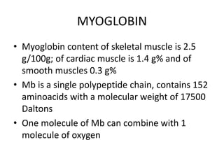
HAEMOGLOBIN &
MYOGLOBIN
STRUCTURE
HAEMOGLOBIN
• Normal level of haemoglobin
14-16 g/dl (males)
13-15 g/dl (females)
• Globular in shape
• Molecular weight is 67,000 Daltons
• is conjugated protein containing globin –apoprotein and
heme - non-protein part
• Tetrameric allosteric protein
• Globin consists of four polypeptide chains of two different
primary structures
• Adult haemoglobulin (HbA) has 2 alpha chains and 2 beta
chains
Alpha & beta chains
Hb A Hb A2 Hb F
structure a2b2 a2d2 a2g2
Normal % 96-98 % 1.5-3.2 % 0.5-0.8 %
Adult haemoblobin
• Hb F (fetal Hb) is made up of 2 alpha and 2 gamma
chains, Hb A2 has 2 alpha and 2 delta chains
• Normal adult blood contains 97% HbA, about 2%
HbA2 and about 1% HbF
• Alpha chain gene is on chromosome 16 while beta,
gamma and delta chains are in chromosome 11
• Each alpha chain has 141 aminoacids residue and
beta, gamma and delta chains have 146 aminoacids
• Alpha and beta subunits are connected by relatively
weak non-covalent bonds
• Other varieties are Embryonic Hb , Glycosylated Hb
• There are 4 heme residues per Hb
molecules, one for each subunit in
Hb. The 4 heme groups account for
about 4% of the whole mass of Hb.
The heme is located in a
hydrophobic cleft of globin chain.
• Heme contains a porphyrin
molecule namely protoporphyrin IX,
with iron at its center
• Heme is common prosthetic group
present in cytochromes, in certain
enzymes such as catalase,
tryptophan pyrolase, and
chlorophyll
Function of Haemoglobin
Haemoglobin performs two important biological
functions concerned with respiration
Delivery of O2 from the lungs to the tissues
Transport of CO2 and protons from tissues to
lungs for excretion
(R)
relaxed state
(T)
tense state
The Transportation of Blood Oxygen
Hemoglobin
Lung
O2
Myoglobin
Muscle
Vein
Artery
When environmental [O2]
increases, Hb binds oxygen
efficiently
When environmental [O2]
decreases, Hb releases
oxygen to Mb
Any one subunit receives an oxygen molecule will increase the
oxygen-binding affinity of the others
Juang RH (2004) BCbasics
Combination of haemoglobin with gas
• Oxy-Haemoglobin
• Oxygenation not oxidation
• One Hb can bind to four O2 molecules
• Less than .01 sec required for oxygenation
• b chain move closer when oxygenated
• When oxygenated 2,3-BPG is pushed out
• b chains are pulled apart when O2 is unloaded,
permitting entry of 2,3-BPG resulting in lower affinity of
O2
Oxy & deoxyhaemoglobin
• Carboxy-Hb
– CO combines with heme portion of Hb
– Affinity of Hb to CO is 210 times more
than O2
– Lethal action due to inhibition of
cytochrome oxidase
• Combination with CO2
– Hb combine with CO2 to form
carbaminohaemoglobin
– Combination on globin rather than with
heme
• Sulfhaemoglobin
– Formed by the action of H2S on oxy-Hb
• Action with cyanide
– Cyanides do not react directly with haemoglobin
but they react with methaemoglobin to form
cyanmethaemoglobin which is not toxic
Abnormal haemoglobin
• Two types
• Mutation affects on structural gene
– E.g. HbS, HbM, HbC, HbD and others
• Mutation affects the regulator gene
– E.g. α-chain thalassaemias and β-chain
thalassaemias
MYOGLOBIN
• Myoglobin content of skeletal muscle is 2.5
g/100g; of cardiac muscle is 1.4 g% and of
smooth muscles 0.3 g%
• Mb is a single polypeptide chain, contains 152
aminoacids with a molecular weight of 17500
Daltons
• One molecule of Mb can combine with 1
molecule of oxygen
• Myoglobin functions as a reservoir for oxygen
• Also serves as oxygen carrier that promotes
the transport of oxygen to the rapidly
respiring muscle cells
Binding of O2 to haemoglobin
• Oxygen dissociation curve: the binding ability of
hemoglobin with O2 at different partial pressures
of oxygen (pO2) is shown by the oxygen
dissociation curve
• In pulmonary alveoli, Hb is 97% saturated with
oxygen where pO2 is 100 mmHg and in tissue
capillaries, where pO2 is 40 mmHg,theoretically,
Hb saturation is 75% but practically about 60%
saturated. So physiologically, 40% of oxygen is
released
0 20 40 60 80 100 120
100
80
60
40
20
0
Percent
O
2
saturation
Partial pressure of oxygen (pO2, mmHg)
Muscle in
exercising
Relaxing
muscle Myoglobin
Hemoglobin
Artery
[O2]
Vein
[O2]
Environmental Oxygen Effects Binding Affinity
Adapted
from
Garrett
&
Grisham
(1999)
Biochemistry
(2e)
p.480
• The sigmoid shape of the oxygen dissociation
curve is due to the allosteric effect or
cooperativity
• The binding of oxygen to one heme residue
increases the affinity of remaining heme
residues for oxygen
• Each successive addition of O2 increases the
affinity of Hb to oxygen synergistically
• The quaternary structure of oxy-Hb is
described as R (relaxed) form; and that of
deoxy-Hb is T (tight) form
• These two forms are interconvertible and each
form has its own equilibrium constant (KR &
KT) for binding of O2
Hb-oxygen dissociation curve
• The normal position of curve depends on
• Concentration of 2,3-BPG
• H+ ion concentration (pH)
• CO2 in red blood cells
• Structure of Hb
Hb-oxygen dissociation curve
• Right shift (easy oxygen delivery)
• High 2,3-BPG
• High H+
• High CO2
• HbS
• Left shift (give up oxygen less readily)
• Low 2,3-BPG
• HbF
Hb-oxygen dissociation curve
BOHR EFFECT
• The binding of oxygen to hemoglobin
decreases with increasing H+ concentration or
when the hemoglobin is exposed to
increased partial pressure of CO2
• Bohr effect causes a shift in the oxygen
dissociation curve to the right
• Bohr effect is primarily responsible for the
release of O2 from the oxyhemoglobin to the
tissue
• Any increase in protons and/or lower pO2 shifts the
equilibrium to the right to produce
deoxyhemoglobin as happens in the tissues
• When CO2 binds to hemoglobin, carbamyl
hemoglobin is produced which causes the removal
of protons from the terminal NH2 group and
stabilizes the structure of Hb in the T form.
Haldane effect
• The carbon dioxide
equilibrium curve is nearly a
straight-line function of
PCO2 in the normal arterial
CO2 range
• The higher PO2 will shift the
curve downward and to the
right (Haldane effect)
• Its advantage is that it
allows the blood to load
more CO2 in the tissues and
unload more CO2 in the
lungs

HAEMOGLOBIN & MYOGLOBIN STRUCTURE.pptx

  • 1.
  • 2.
    HAEMOGLOBIN • Normal levelof haemoglobin 14-16 g/dl (males) 13-15 g/dl (females) • Globular in shape • Molecular weight is 67,000 Daltons • is conjugated protein containing globin –apoprotein and heme - non-protein part • Tetrameric allosteric protein • Globin consists of four polypeptide chains of two different primary structures • Adult haemoglobulin (HbA) has 2 alpha chains and 2 beta chains
  • 3.
  • 4.
    Hb A HbA2 Hb F structure a2b2 a2d2 a2g2 Normal % 96-98 % 1.5-3.2 % 0.5-0.8 % Adult haemoblobin
  • 5.
    • Hb F(fetal Hb) is made up of 2 alpha and 2 gamma chains, Hb A2 has 2 alpha and 2 delta chains • Normal adult blood contains 97% HbA, about 2% HbA2 and about 1% HbF • Alpha chain gene is on chromosome 16 while beta, gamma and delta chains are in chromosome 11 • Each alpha chain has 141 aminoacids residue and beta, gamma and delta chains have 146 aminoacids • Alpha and beta subunits are connected by relatively weak non-covalent bonds • Other varieties are Embryonic Hb , Glycosylated Hb
  • 6.
    • There are4 heme residues per Hb molecules, one for each subunit in Hb. The 4 heme groups account for about 4% of the whole mass of Hb. The heme is located in a hydrophobic cleft of globin chain. • Heme contains a porphyrin molecule namely protoporphyrin IX, with iron at its center • Heme is common prosthetic group present in cytochromes, in certain enzymes such as catalase, tryptophan pyrolase, and chlorophyll
  • 7.
    Function of Haemoglobin Haemoglobinperforms two important biological functions concerned with respiration Delivery of O2 from the lungs to the tissues Transport of CO2 and protons from tissues to lungs for excretion
  • 8.
    (R) relaxed state (T) tense state TheTransportation of Blood Oxygen Hemoglobin Lung O2 Myoglobin Muscle Vein Artery When environmental [O2] increases, Hb binds oxygen efficiently When environmental [O2] decreases, Hb releases oxygen to Mb Any one subunit receives an oxygen molecule will increase the oxygen-binding affinity of the others Juang RH (2004) BCbasics
  • 9.
    Combination of haemoglobinwith gas • Oxy-Haemoglobin • Oxygenation not oxidation • One Hb can bind to four O2 molecules • Less than .01 sec required for oxygenation • b chain move closer when oxygenated • When oxygenated 2,3-BPG is pushed out • b chains are pulled apart when O2 is unloaded, permitting entry of 2,3-BPG resulting in lower affinity of O2
  • 10.
  • 11.
    • Carboxy-Hb – COcombines with heme portion of Hb – Affinity of Hb to CO is 210 times more than O2 – Lethal action due to inhibition of cytochrome oxidase • Combination with CO2 – Hb combine with CO2 to form carbaminohaemoglobin – Combination on globin rather than with heme
  • 12.
    • Sulfhaemoglobin – Formedby the action of H2S on oxy-Hb • Action with cyanide – Cyanides do not react directly with haemoglobin but they react with methaemoglobin to form cyanmethaemoglobin which is not toxic
  • 13.
    Abnormal haemoglobin • Twotypes • Mutation affects on structural gene – E.g. HbS, HbM, HbC, HbD and others • Mutation affects the regulator gene – E.g. α-chain thalassaemias and β-chain thalassaemias
  • 14.
    MYOGLOBIN • Myoglobin contentof skeletal muscle is 2.5 g/100g; of cardiac muscle is 1.4 g% and of smooth muscles 0.3 g% • Mb is a single polypeptide chain, contains 152 aminoacids with a molecular weight of 17500 Daltons • One molecule of Mb can combine with 1 molecule of oxygen
  • 15.
    • Myoglobin functionsas a reservoir for oxygen • Also serves as oxygen carrier that promotes the transport of oxygen to the rapidly respiring muscle cells
  • 16.
    Binding of O2to haemoglobin • Oxygen dissociation curve: the binding ability of hemoglobin with O2 at different partial pressures of oxygen (pO2) is shown by the oxygen dissociation curve • In pulmonary alveoli, Hb is 97% saturated with oxygen where pO2 is 100 mmHg and in tissue capillaries, where pO2 is 40 mmHg,theoretically, Hb saturation is 75% but practically about 60% saturated. So physiologically, 40% of oxygen is released
  • 17.
    0 20 4060 80 100 120 100 80 60 40 20 0 Percent O 2 saturation Partial pressure of oxygen (pO2, mmHg) Muscle in exercising Relaxing muscle Myoglobin Hemoglobin Artery [O2] Vein [O2] Environmental Oxygen Effects Binding Affinity Adapted from Garrett & Grisham (1999) Biochemistry (2e) p.480
  • 18.
    • The sigmoidshape of the oxygen dissociation curve is due to the allosteric effect or cooperativity • The binding of oxygen to one heme residue increases the affinity of remaining heme residues for oxygen • Each successive addition of O2 increases the affinity of Hb to oxygen synergistically
  • 19.
    • The quaternarystructure of oxy-Hb is described as R (relaxed) form; and that of deoxy-Hb is T (tight) form • These two forms are interconvertible and each form has its own equilibrium constant (KR & KT) for binding of O2
  • 20.
  • 21.
    • The normalposition of curve depends on • Concentration of 2,3-BPG • H+ ion concentration (pH) • CO2 in red blood cells • Structure of Hb Hb-oxygen dissociation curve
  • 22.
    • Right shift(easy oxygen delivery) • High 2,3-BPG • High H+ • High CO2 • HbS • Left shift (give up oxygen less readily) • Low 2,3-BPG • HbF Hb-oxygen dissociation curve
  • 23.
    BOHR EFFECT • Thebinding of oxygen to hemoglobin decreases with increasing H+ concentration or when the hemoglobin is exposed to increased partial pressure of CO2 • Bohr effect causes a shift in the oxygen dissociation curve to the right • Bohr effect is primarily responsible for the release of O2 from the oxyhemoglobin to the tissue
  • 24.
    • Any increasein protons and/or lower pO2 shifts the equilibrium to the right to produce deoxyhemoglobin as happens in the tissues • When CO2 binds to hemoglobin, carbamyl hemoglobin is produced which causes the removal of protons from the terminal NH2 group and stabilizes the structure of Hb in the T form.
  • 26.
    Haldane effect • Thecarbon dioxide equilibrium curve is nearly a straight-line function of PCO2 in the normal arterial CO2 range • The higher PO2 will shift the curve downward and to the right (Haldane effect) • Its advantage is that it allows the blood to load more CO2 in the tissues and unload more CO2 in the lungs

Editor's Notes

  • #9 Hb 在血中循環,由肺泡取得氧分子,運送到肌肉中,下載給 Mb。 因此 Hb 必須得知,何時該吸收氧分子,何時該放出;也就是說 Hb 必須有感應氧分子濃度的能力,同時還得做出吸收或放出的動作。這些能力都源自其四級構造的組成。 若在靜脈中 Hb 都不載有氧分子,這時候它的四個次體分子都處於一種休息狀態,結合區不容易張開讓 heme 接受氧分子。 當 Hb 循環到肺部時,環境的氧分子濃度提高了,四個次體的任何一個分子接上一個氧分子後,馬上會牽動其它次體,使得其它次體的分子構造舒張,變得很容易接受氧分子。因此在肺部的 Hb 都很容易地滿載氧分子,經動脈輸送至肌肉。若當時肌肉相當勞累,需要大量氧分子的補充,其酸鹼度會降得比較低,Hb 就更容易釋出氧分子,而 Mb 則一昧地吸收 Hb 所下的氧分子,並無調節作用。放下氧分子的 Hb 就回復休息狀態,循著靜脈流回肺部。 蛋白質的構形事實上都不是固定不動,其分子會有某些程度的運動,上述的 Hb 分子也是如此。當其處於休息狀態時,是分子較為緊密的一種構形,稱之為 tense (T) 型;反之,若其構造較為疏鬆,則基質或其結合對象比較容易進入,稱之為 relaxed (R) 型。 T 與 R 型的變化,在酵素分子的活性調節也很重要,在酵素部分會繼續提到。
  • #18 Hb 血紅蛋白是很有智慧的分子,它的行為曲線是一種 S 型的方式,也就是在低氧濃度時,它對氧分子的吸收作用並不強,但在高氧濃度時,就有很強的吸收能力。反之,Mb 就不管氧濃度如何,一昧地吸收所有的氧分子。這是因為兩者所分佈的地點不同,所負的生理意義也不同。 比較 Hb 在運動中的肌肉與在動脈中的攜氧能力,有顯著不同;因此 Hb 會在高氧濃度的肺部中盡量攜帶充分的氧分子,等循環到肌肉的低氧濃度環境,便會大量釋出氧分子,給在地的 Mb 去攜帶,以便供給肌肉細胞使用。