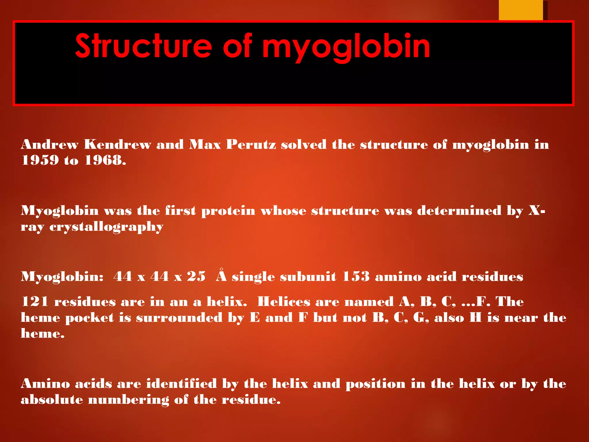
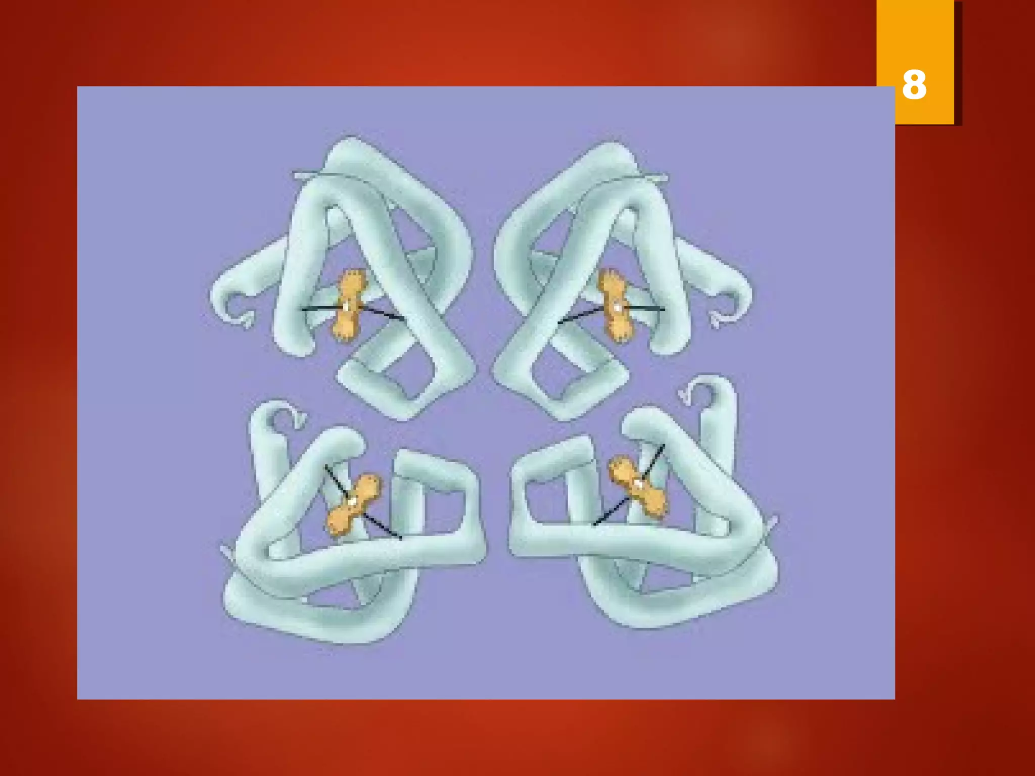
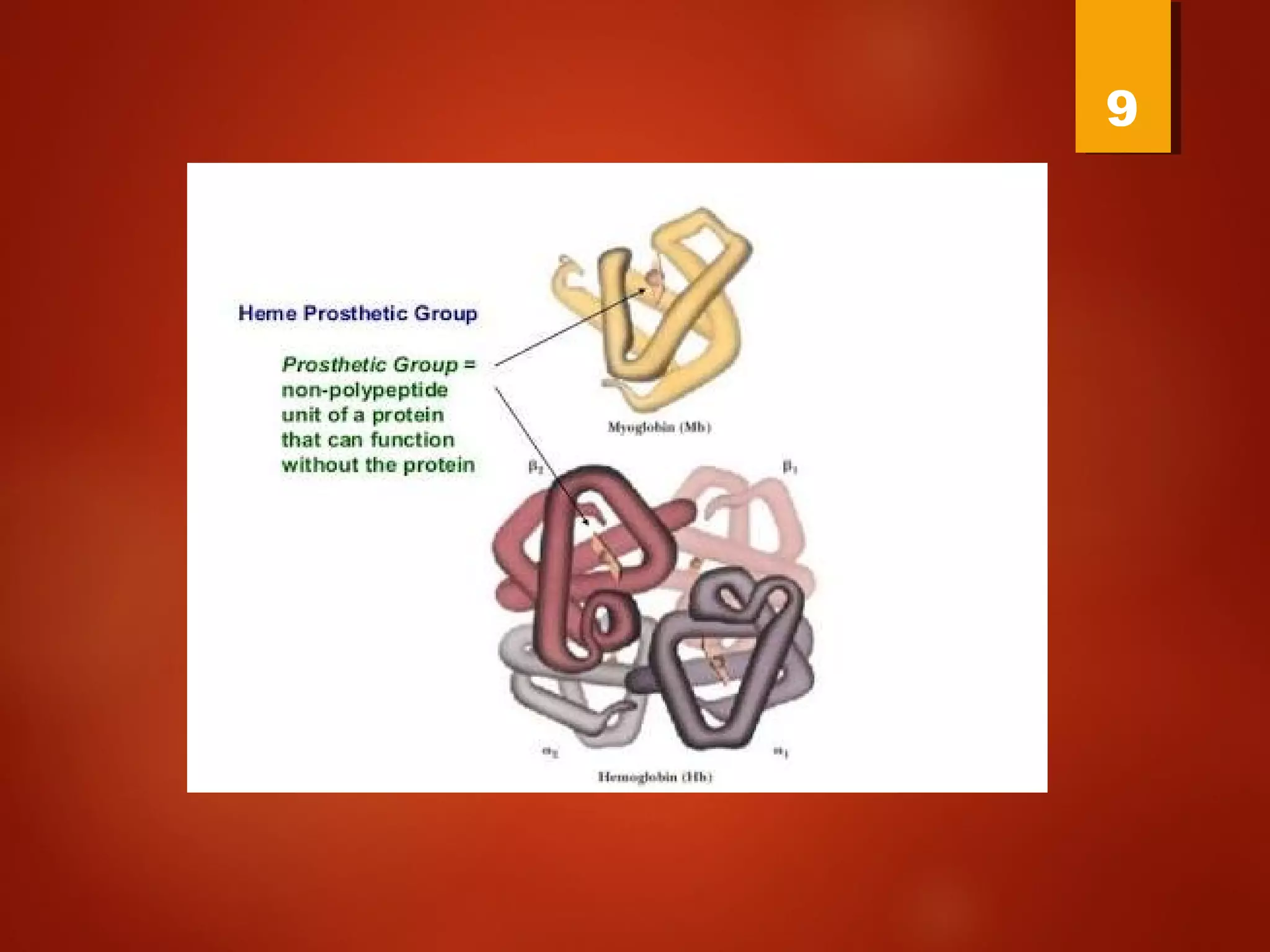
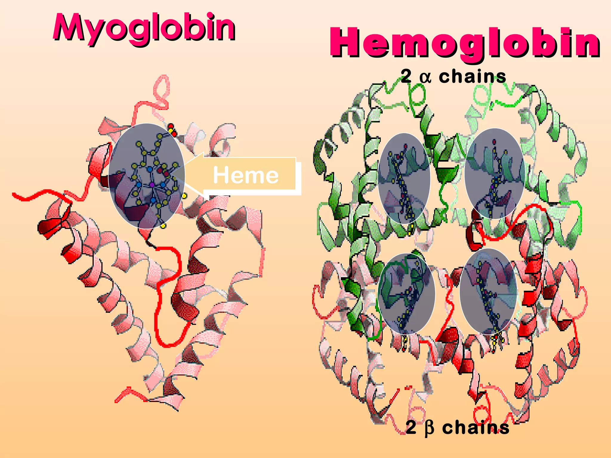
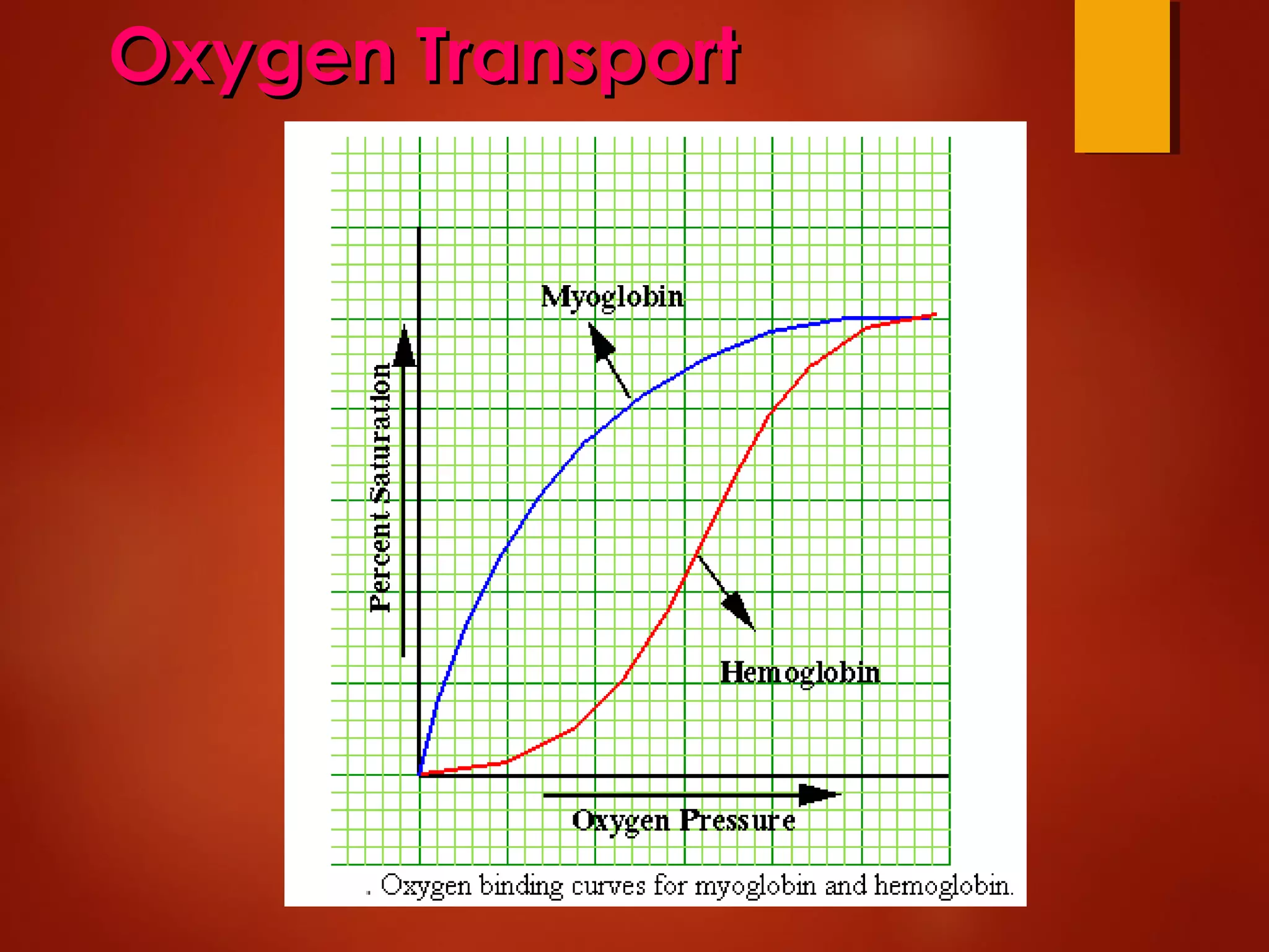
MYOGLOBIN
Dr.PRABHJOT KAUR BHINDER
DEPTT OF BIOCHEMISTRY
9/1/2017
1
Structure of myoglobin
Andrew Kendrew and Max Perutz solved the structure of myoglobin in
1959 to 1968.
Myoglobin was the first protein whose structure was determined by X-
ray crystallography
Myoglobin: 44 x 44 x 25 Å single subunit 153 amino acid residues
121 residues are in an a helix. Helices are named A, B, C, …F. The
heme pocket is surrounded by E and F but not B, C, G, also H is near the
heme.
Amino acids are identified by the helix and position in the helix or by the
absolute numbering of the residue.
4
X-RAY CRYSTALLOGRAPHY 5
Structure of HemoglobinStructure of Hemoglobin
7
8
9
MyoglobinMyoglobin
HemeHeme
2 α chains
2 β chains
HemoglobinHemoglobin
The Heme group
•Each subunit of hemoglobin or
myoglobin contains a heme.
•Binds one molecule of oxygen
•Protoporphyrin 9
The iron must be in the Fe(II) form or
reduced form. (ferrous oxidation) state.
The Conformation
Change The secret of Mb
 Oxygen binding changes the Mb
conformation
 Without oxygen bound, Fe is out of heme
plane
 Oxygen binding pulls the Fe into the heme
plane
 Fe pulls its His F8 ligand along with it
 The F helix moves when oxygen binds
 Total movement of Fe is 0.029 nm - 0.29 A
Function of the globin
Protoporphyrin binds oxygen to the
sixth ligand of Fe(II) out of the
plane of the heme. The fifth ligand
is a Histidine, F8 on the side across
the heme plane.
His F8 binds to the proximal side
and the oxygen binds to the distal
side.
The heme alone interacts with
oxygen such that the Fe(II) becomes
oxidized to Fe(III) and no longer
15
MyoglobinMyoglobin
oxygen storage protein of skeletal muscles.
As with the cytochrome example, both proteins use
heme groups. It acts as the binding site for molecular
oxygen.
Myoglobin facilitates rapidly
respiring muscle tissue
The rate of O2 diffusion from capillaries
to tissue is slow because of the
solubility of oxygen.
Myoglobin increases the solubility of
oxygen.
Myoglobin facilitates oxygen diffusion.
Oxygen storage is also a function
because Myoglobin concentrations are
10-fold greater in whales and seals than
in land mammals
 Myoglobin follows the michaelis- menten graph
 It follow michaelis-menton because it is a
simple chemical equilibrium
19
Oxygen TransportOxygen Transport
22
23
Myoglobin Structure
Mb is a monomeric heme protein
 Mb polypeptide "cradles" the heme
group
 Fe in Mb is Fe2+
- ferrous iron - the form
that binds oxygen
 Oxidation of Fe yields 3+ charge - ferric
iron -metmyoglobin does not bind
oxygen
 Oxygen binds as the sixth ligand to Fe
 Myoglobin an iron containing protein in muscles
,receive oxygen from rbc and transports it to the
mitochondria of muscle cells,where oxygen is used in
cellular respiration to produce energy
 Oxymyoglobin –oxygen supply and act as scavenger of
NO[MYOCYTES]
 Oxymyoglobin +NO= Harmless nitrates with ferric
myoglobin ,which is recycled by metmyoglobin
reductase
25
O2 binding to myoglobin
22 MbOOMb ↔+
][MbO
][Mb][O
Kd
2
2
=
][OKd
][O
][MbO[Mb]
][MbO
Y
2
2
2
2
O2
+
=
+
=
Written backwards
we can get the
dissociation
constant
Fractional Saturation solve for [MbO2]
and plug in
How do you measure the
concentration of oxygen?
Use the partial pressure of O2 or O2
tension = pO2
2d
2
O
pOK
pO
Y 2
+
=
P50 = the partial
oxygen pressure
when YO2 = 0.50
250
2
O
pOP
pO
Y 2
+
=
P50 value for myoglobin is 2.8 torr
or
1 torr = 1 mm Hg = 0.133 kPa
760 torr = 1 atm of pressure
Mb gives up little O2 over normal
physiological range of oxygen concentrations
in the tissue
i.e. 100 torr in arterial blood
30 torr in venous blood
YO2 = 0.97 to YO2 = 0.91
Contrast Mb O2 binding to
Hemoglobin
YO2 = 0.95 at 100 torr
but
0.55 at 30 torr
a ∆YO2 of 0.40
Hb gives up O2 easier than Mb and the
binding is Cooperative!!
Diseases
 Acute renal failure
 Heart attack
 Myoglobinuria [rhabdomyolysis]
33
34
Causes of MYOGLOBINURIA
 Mc Ardles disease
 Phosphofructokinase deficiency
 Carnitine palmitoyltransferase 2
 Malignant hyperthermia
 Polymyositis
 Lactate dehydrogenase deficiency
 Thermal or electric burns
35
Diagnosis
After centrifuging ,urine of myoglobinuria is
clear
Serum of myoglobinuria changes from pink
to red
36
Treatment
 Hospitalization and IV hydration
 Mannitol
 Sodium bicarbonates
37
38

myoglobin

  • 1.
    MYOGLOBIN Dr.PRABHJOT KAUR BHINDER DEPTTOF BIOCHEMISTRY 9/1/2017 1
  • 2.
    Structure of myoglobin AndrewKendrew and Max Perutz solved the structure of myoglobin in 1959 to 1968. Myoglobin was the first protein whose structure was determined by X- ray crystallography Myoglobin: 44 x 44 x 25 Å single subunit 153 amino acid residues 121 residues are in an a helix. Helices are named A, B, C, …F. The heme pocket is surrounded by E and F but not B, C, G, also H is near the heme. Amino acids are identified by the helix and position in the helix or by the absolute numbering of the residue.
  • 4.
  • 5.
  • 6.
  • 7.
  • 8.
  • 9.
  • 10.
    MyoglobinMyoglobin HemeHeme 2 α chains 2β chains HemoglobinHemoglobin
  • 11.
    The Heme group •Eachsubunit of hemoglobin or myoglobin contains a heme. •Binds one molecule of oxygen •Protoporphyrin 9 The iron must be in the Fe(II) form or reduced form. (ferrous oxidation) state.
  • 12.
    The Conformation Change Thesecret of Mb  Oxygen binding changes the Mb conformation  Without oxygen bound, Fe is out of heme plane  Oxygen binding pulls the Fe into the heme plane  Fe pulls its His F8 ligand along with it  The F helix moves when oxygen binds  Total movement of Fe is 0.029 nm - 0.29 A
  • 14.
    Function of theglobin Protoporphyrin binds oxygen to the sixth ligand of Fe(II) out of the plane of the heme. The fifth ligand is a Histidine, F8 on the side across the heme plane. His F8 binds to the proximal side and the oxygen binds to the distal side. The heme alone interacts with oxygen such that the Fe(II) becomes oxidized to Fe(III) and no longer
  • 15.
  • 16.
    MyoglobinMyoglobin oxygen storage proteinof skeletal muscles. As with the cytochrome example, both proteins use heme groups. It acts as the binding site for molecular oxygen.
  • 18.
    Myoglobin facilitates rapidly respiringmuscle tissue The rate of O2 diffusion from capillaries to tissue is slow because of the solubility of oxygen. Myoglobin increases the solubility of oxygen. Myoglobin facilitates oxygen diffusion. Oxygen storage is also a function because Myoglobin concentrations are 10-fold greater in whales and seals than in land mammals
  • 19.
     Myoglobin followsthe michaelis- menten graph  It follow michaelis-menton because it is a simple chemical equilibrium 19
  • 20.
  • 22.
  • 23.
  • 24.
    Myoglobin Structure Mb isa monomeric heme protein  Mb polypeptide "cradles" the heme group  Fe in Mb is Fe2+ - ferrous iron - the form that binds oxygen  Oxidation of Fe yields 3+ charge - ferric iron -metmyoglobin does not bind oxygen  Oxygen binds as the sixth ligand to Fe
  • 25.
     Myoglobin aniron containing protein in muscles ,receive oxygen from rbc and transports it to the mitochondria of muscle cells,where oxygen is used in cellular respiration to produce energy  Oxymyoglobin –oxygen supply and act as scavenger of NO[MYOCYTES]  Oxymyoglobin +NO= Harmless nitrates with ferric myoglobin ,which is recycled by metmyoglobin reductase 25
  • 28.
    O2 binding tomyoglobin 22 MbOOMb ↔+ ][MbO ][Mb][O Kd 2 2 = ][OKd ][O ][MbO[Mb] ][MbO Y 2 2 2 2 O2 + = + = Written backwards we can get the dissociation constant Fractional Saturation solve for [MbO2] and plug in
  • 29.
    How do youmeasure the concentration of oxygen? Use the partial pressure of O2 or O2 tension = pO2 2d 2 O pOK pO Y 2 + = P50 = the partial oxygen pressure when YO2 = 0.50 250 2 O pOP pO Y 2 + =
  • 30.
    P50 value formyoglobin is 2.8 torr or 1 torr = 1 mm Hg = 0.133 kPa 760 torr = 1 atm of pressure Mb gives up little O2 over normal physiological range of oxygen concentrations in the tissue i.e. 100 torr in arterial blood 30 torr in venous blood YO2 = 0.97 to YO2 = 0.91
  • 32.
    Contrast Mb O2binding to Hemoglobin YO2 = 0.95 at 100 torr but 0.55 at 30 torr a ∆YO2 of 0.40 Hb gives up O2 easier than Mb and the binding is Cooperative!!
  • 33.
    Diseases  Acute renalfailure  Heart attack  Myoglobinuria [rhabdomyolysis] 33
  • 34.
  • 35.
    Causes of MYOGLOBINURIA Mc Ardles disease  Phosphofructokinase deficiency  Carnitine palmitoyltransferase 2  Malignant hyperthermia  Polymyositis  Lactate dehydrogenase deficiency  Thermal or electric burns 35
  • 36.
    Diagnosis After centrifuging ,urineof myoglobinuria is clear Serum of myoglobinuria changes from pink to red 36
  • 37.
    Treatment  Hospitalization andIV hydration  Mannitol  Sodium bicarbonates 37
  • 38.