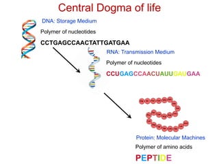
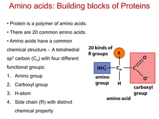
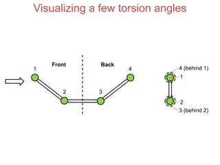
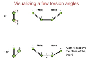
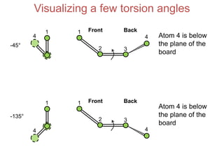
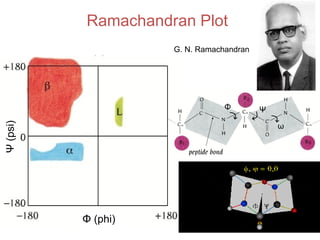
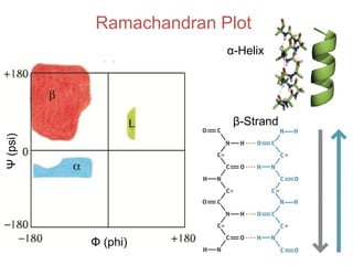
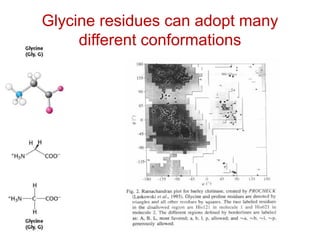
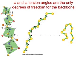
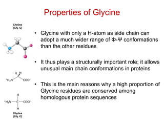
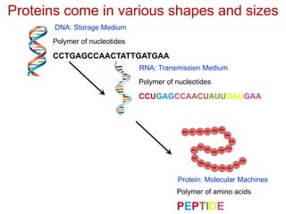
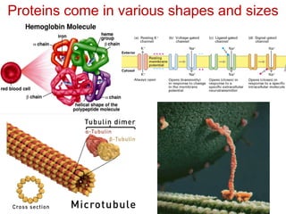
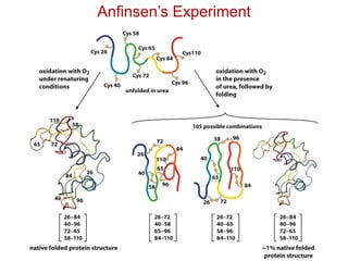
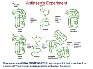
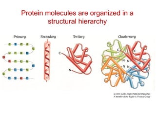
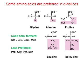
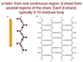
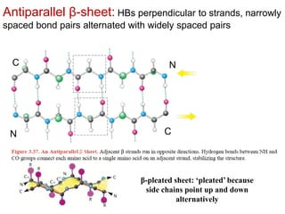
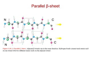
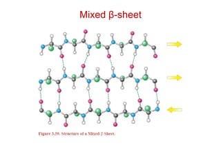
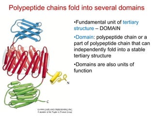
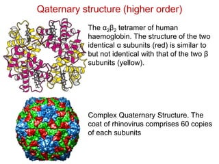
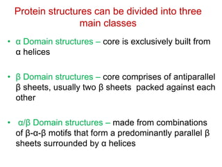
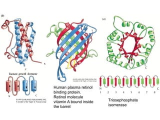
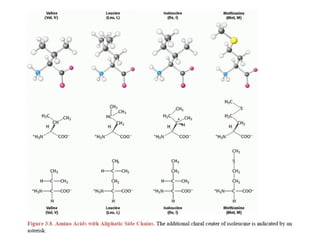
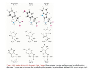
This document provides an overview of protein structure and function. It discusses the central dogma of life, the 20 common amino acids that make up proteins, and how they fold into defined structures like alpha helices and beta sheets. Key concepts covered include the hydrophobic effect that drives protein folding, domains as fundamental units of structure, and the three main classes of protein structures - alpha, beta, and alpha/beta domains. Real-world protein examples are also briefly mentioned.
Science of
Living System
SoumyaDe
School of Bio Science
Email: somde@iitkgp.ac.in
Tel: 03222-260514
BS20001
http://www0.cs.ucl.ac.uk/staff/d.jones/t42morph.html
2.
Books Followed:
• HowProteins Work (Mike Williamson)
• Introduction to protein structure (Carl
Branden & John Tooze)
• Biochemistry (Lubert Stryer)
Protein Structure, Function, Kinetics
and Energetics
3.
Central Dogma oflife
Polymer of nucleotides
DNA: Storage Medium
RNA: Transmission Medium
Polymer of nucleotides
CCUGAGCCAACUAUUGAUGAA
CCTGAGCCAACTATTGATGAA
PEPTIDE
Polymer of amino acids
Protein: Molecular Machines
4.
Amino acids: Buildingblocks of Proteins
• Protein is a polymer of amino acids.
• There are 20 common amino acids.
• Amino acids have a common
chemical structure - A tetrahedral
sp3 carbon (Cα) with four different
functional groups:
1. Amino group
2. Carboxyl group
3. H-atom
4. Side chain (R) with distinct
chemical property
5.
All amino acidsin protein have the “L-form”
H-atom is coming out of the whiteboard. Looking down the H-Cα
bond from the H-atom, the L-form amino acid has CO, R and N
going in a clockwise direction. The L-form reads “CORN” in
clockwise direction.
Visualizing a fewtorsion angles
Front Back
1
2 3
4
1
4 (behind 1)
2
3 (behind 2)
19.
Visualizing a fewtorsion angles
Front Back1
2 3
4
1
4
2
3
0°
+45°
1
4
Front Back1
2 3
4 Atom 4 is above
the plane of the
board
20.
4
Visualizing a fewtorsion angles
-45°
1
4
Front Back1
2 3
4 Atom 4 is below
the plane of the
board
-135°
1 Front Back1
2 3
Atom 4 is below
the plane of the
board
4
φ and ψtorsion angles are the only
degrees of freedom for the backbone
1
2
3
25.
Properties of Glycine
•Glycine with only a H-atom as side chain can
adopt a much wider range of Φ-Ψ conformations
than the other residues
• It thus plays a structurally important role; it allows
unusual main chain conformations in proteins
• This is the main reasons why a high proportion of
Glycine residues are conserved among
homologous protein sequences
26.
Proteins come invarious shapes and sizes
Polymer of nucleotides
DNA: Storage Medium
RNA: Transmission Medium
Polymer of nucleotides
CCUGAGCCAACUAUUGAUGAA
CCTGAGCCAACTATTGATGAA
PEPTIDE
Polymer of amino acids
Protein: Molecular Machines
The protein foldingproblem
- Consider a small protein with 100 residues.
- Cyrus Levinthal calculated that, if each residue can assume
three different conformations, the total number of structures
would be 3100, which is equal to 5 × 1047. If it takes 10-13 s to
convert one structure into another, the total search time would
be 5 × 1047 × 10-13 s, which is equal to 5 × 1034 s, or 1027 years
i.e. longer than the age of the universe!
- Clearly, it would take much too long for even a small protein to
fold properly by randomly trying out all possible conformations.
- The enormous difference between calculated and actual folding
times is called Levinthal's paradox.
29.
The 3D structureof a protein is encoded in its
primary sequence: Anfinsen’s Experiment
Thermodynamic hypothesis of
Protein Folding: The interactions
between the atoms in a protein control
the folding of the protein molecule into
a well-defined three-dimensional
structure.
In other words, the protein sequence
contains enough information required
for the proper folding of the protein
into its functional three-dimensional
structure.
Anfinsen’s Experiment
If weunderstand HOW PROTEINS FOLD, we can predict their structure from
sequence! Then we can design proteins with novel functions.
32.
Most important feature:The interior of proteins is hydrophobic!
The main driving force for folding water soluble globular protein
molecules is to pack hydrophobic side chains into the interior of
the molecule, thus creating a HYDROPHOBIC CORE and a
HYDROPHILLIC SURFACE.
Problem: How to create such a hydrophobic core from a linear
protein chain ???
- Hydrophobic effect
- Conformational entropy
- Electrostatics
- Hydrogen bonding
- van der Waals interaction
Forces that stabilize a protein structure
• Alpha helices
•Beta Sheets
• Characterized by main chain NH and CO
groups participating in H-bonds.
• Formed when a number of consecutive
residues have the similar phi and psi angles.
40.
Alpha Helices
Every 3.6residues
make one turn.
The distance (pitch of
helix) between two
turns is 5.4 Å.
The C=O of residue ‘n’
is hydrogen bonded to
N-H of residue ‘n+4’.
Alpha helices are
formed when a stretch
of consecutive
residues have the phi-
psi torsion angle pair
approx -60° & -50°.
This is in the allowed
region of
Ramchandran plot.
41.
The alpha-helix hasa dipole moment
The dipole of a peptide unit
Numbers in boxes give the
approximate fractional charges
of the atoms of the peptide unit
The dipoles of
peptide units are
aligned along
the α helical axis
+
-
42.
Good helix formers:
Ala, Glu, Leu , Met
Less Preferred:
Pro, Gly, Tyr, Ser
Some amino acids are preferred in α-helices
Helical Wheel: Each
residuecan be
plotted every
360/3.6=100° around
a circle or spiral
Hydrophobic
Hydrophilic
Charged
Totally buried Partially buried
Exposed
45.
Alpha helix canbe – Right-handed or Left
handed.
BUT, left handed helix is not possible for L-
amino acids due to close approach of the
side chains and CO group.
Right handed – most commonly observed
in proteins.
46.
α-helix: from onecontinuous region; β-sheet from
several regions of the chain; Each β-strand,
typically 5-10 residues long
47.
N
N
C
C
Antiparallel β-sheet: HBsperpendicular to strands, narrowly
spaced bond pairs alternated with widely spaced pairs
β-pleated sheet: ‘pleated’ because
side chains point up and down
alternatively
Polypeptide chains foldinto several domains
•Fundamental unit of tertiary
structure – DOMAIN
•Domain: polypeptide chain or a
part of polypeptide chain that can
independently fold into a stable
tertiary structure
•Domains are also units of
function
53.
Quaternary structure
Proteins containingmore than one polypeptide chain exhibit a fourth level of
structural organization. Each polypeptide chain in such a protein is called a
subunit. Quaternary structure refers to the spatial arrangement of subunits
and the nature of their interactions.
The simplest sort of quaternary structure is a dimer, consisting of two
identical subunits.
54.
Qaternary structure (higherorder)
Complex Quaternary Structure. The
coat of rhinovirus comprises 60 copies
of each subunits
The α2β2 tetramer of human
haemoglobin. The structure of the two
identical α subunits (red) is similar to
but not identical with that of the two β
subunits (yellow).
55.
Protein structures canbe divided into three
main classes
• α Domain structures – core is exclusively built from
α helices
• β Domain structures – core comprises of antiparallel
β sheets, usually two β sheets packed against each
other
• α/β Domain structures – made from combinations
of β-α-β motifs that form a predominantly parallel β
sheets surrounded by α helices
56.
Human plasma retinol
bindingprotein.
Retinol molecule
vitamin A bound inside
the barrel
Triosephosphate
isomerase