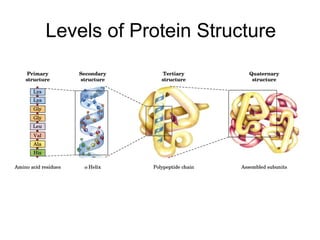
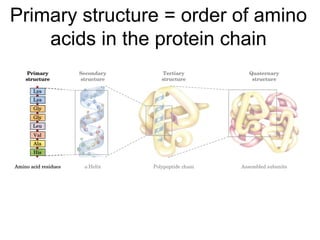
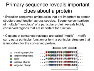
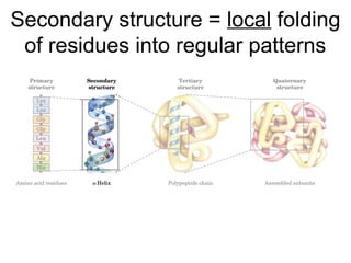
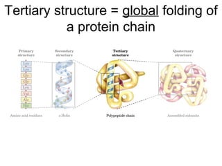
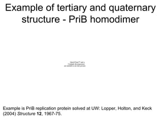
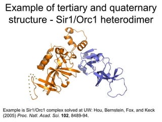
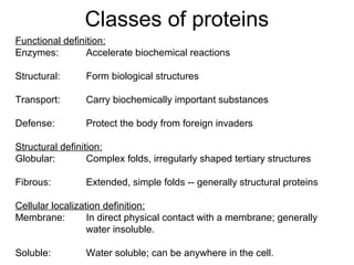
Lecture 1: Fundamentals of
Protein Structure
Wood, brick, nails, glass Materials Amino acids, cofactors
Temperature, earthquakes Environmental Factors Temperature, solubility
How many people? Population Factors # partner proteins, # reactants
How many doors and windows? Portals Passages for substrates and reactants
Spanish, Victorian, Motifs/Styles Conserved domains or protein folds
1950's blocky science building
Julia Morgan Architect Evolution
Traditional Architecture Molecular ArchitectureForm
fits
function
Levels of Protein Structure
Primary structure = order of amino
acids in the protein chain
Anatomy of an amino acid
Non-polar amino acids
Polar, non-charged amino acids
Negatively-charged amino acids
Positively-charged amino acids
Charged and polar R-groups tend
to map to protein surfaces
Non-polar R-groups tend to be
buried in the cores of proteins
Myoglobin
Blue = non-polar
R-group
Red = Heme
Some R-groups can be ionized
The Henderson-
Hasselbalch
equation allows
calculation of the
ratio of a weak acid
and its conjugate
base at any pH
General protein pK’ values
Approximate pK'
Group In a “Typical” Protein
α-carboxyl (free) 3 (C-terminal only)
β-carboxyl (Asp) 4
γ-carboxyl (Glu) 4
imidazole (His) 6
sulfhydryl (Cys) 8
1˚α-amino (free) 8 (N-terminal only)
ε-amino (Lys) 10
hydroxyl (Tyr) 10
2˚α-amino (Pro)(free) 9 (N-terminal only)
guanido (Arg) 12
Some R-groups can modified
Amino Acids Are Joined By
Peptide Bonds In Peptides
- α-carboxyl of one amino acid is joined to
α-amino of a second amino acid (with
removal of water)
- only α-carboxyl and α-amino groups are
used, not R-group carboxyl or amino
groups
Chemistry of peptide bond formation
The peptide bond is planar
This resonance
restricts the number
of conformations in
proteins -- main
chain rotations are
restricted to φ and ψ.
Primary sequence reveals important
clues about a protein
DnaG E. coli ...EPNRLLVVEGYMDVVAL...
DnaG S. typ ...EPQRLLVVEGYMDVVAL...
DnaG B. subt ...KQERAVLFEGFADVYTA...
gp4 T3 ...GGKKIVVTEGEIDMLTV...
gp4 T7 ...GGKKIVVTEGEIDALTV...
: *: :: * * : :
small hydrophobic
large hydrophobic
polar
positive charge
negative charge
• Evolution conserves amino acids that are important to protein
structure and function across species. Sequence comparison
of multiple “homologs” of a particular protein reveals highly
conserved regions that are important for function.
• Clusters of conserved residues are called “motifs” -- motifs
carry out a particular function or form a particular structure that
is important for the conserved protein.
motif
Secondary structure = local folding
of residues into regular patterns
The α-helix
• In the α-helix, the carbonyl
oxygen of residue “i” forms a
hydrogen bond with the
amide of residue “i+4”.
• Although each hydrogen
bond is relatively weak in
isolation, the sum of the
hydrogen bonds in a helix
makes it quite stable.
• The propensity of a peptide
for forming an α-helix also
depends on its sequence.
The β-sheet
• In a β-sheet, carbonyl
oxygens and amides form
hydrogen bonds.
• These secondary
structures can be either
antiparallel (as shown) or
parallel and need not be
planar (as shown) but can be
twisted.
• The propensity of a peptide
for forming β-sheet also
depends on its sequence.
β turns
• β-turns allow the protein backbone to make abrupt turns.
• Again, the propensity of a peptide for forming β-turns depends
on its sequence.
Which residues are common for α-
helix, β-sheet, and β-turn elements?
Ramachandran plot -- shows φ and ψ
angles for secondary structures
Tertiary structure = global folding of
a protein chain
Tertiary structures are quite varied
Quaternary structure = Higher-order
assembly of proteins
Example of tertiary and quaternary
structure - PriB homodimer
QuickTime™ and a
Cinepak decompressor
are needed to see this picture.
Example is PriB replication protein solved at UW: Lopper, Holton, and Keck
(2004) Structure 12, 1967-75.
Example of tertiary and quaternary
structure - Sir1/Orc1 heterodimer
Example is Sir1/Orc1 complex solved at UW: Hou, Bernstein, Fox, and Keck
(2005) Proc. Natl. Acad. Sci. 102, 8489-94.
Examples of other quaternary
structures
Tetramer Hexamer Filament
SSB DNA helicase Recombinase
Allows coordinated Allows coordinated DNA binding Allows complete
DNA binding and ATP hydrolysis coverage of an
extended molecule
Classes of proteins
Functional definition:
Enzymes: Accelerate biochemical reactions
Structural: Form biological structures
Transport: Carry biochemically important substances
Defense: Protect the body from foreign invaders
Structural definition:
Globular: Complex folds, irregularly shaped tertiary structures
Fibrous: Extended, simple folds -- generally structural proteins
Cellular localization definition:
Membrane: In direct physical contact with a membrane; generally
water insoluble.
Soluble: Water soluble; can be anywhere in the cell.

Protein structure

  • 1.
    Lecture 1: Fundamentalsof Protein Structure
  • 2.
    Wood, brick, nails,glass Materials Amino acids, cofactors Temperature, earthquakes Environmental Factors Temperature, solubility How many people? Population Factors # partner proteins, # reactants How many doors and windows? Portals Passages for substrates and reactants Spanish, Victorian, Motifs/Styles Conserved domains or protein folds 1950's blocky science building Julia Morgan Architect Evolution Traditional Architecture Molecular ArchitectureForm fits function
  • 3.
  • 4.
    Primary structure =order of amino acids in the protein chain
  • 5.
    Anatomy of anamino acid
  • 6.
  • 7.
  • 8.
  • 9.
  • 10.
    Charged and polarR-groups tend to map to protein surfaces
  • 11.
    Non-polar R-groups tendto be buried in the cores of proteins Myoglobin Blue = non-polar R-group Red = Heme
  • 12.
    Some R-groups canbe ionized The Henderson- Hasselbalch equation allows calculation of the ratio of a weak acid and its conjugate base at any pH
  • 13.
    General protein pK’values Approximate pK' Group In a “Typical” Protein α-carboxyl (free) 3 (C-terminal only) β-carboxyl (Asp) 4 γ-carboxyl (Glu) 4 imidazole (His) 6 sulfhydryl (Cys) 8 1˚α-amino (free) 8 (N-terminal only) ε-amino (Lys) 10 hydroxyl (Tyr) 10 2˚α-amino (Pro)(free) 9 (N-terminal only) guanido (Arg) 12
  • 14.
  • 15.
    Amino Acids AreJoined By Peptide Bonds In Peptides - α-carboxyl of one amino acid is joined to α-amino of a second amino acid (with removal of water) - only α-carboxyl and α-amino groups are used, not R-group carboxyl or amino groups
  • 16.
    Chemistry of peptidebond formation
  • 17.
    The peptide bondis planar This resonance restricts the number of conformations in proteins -- main chain rotations are restricted to φ and ψ.
  • 18.
    Primary sequence revealsimportant clues about a protein DnaG E. coli ...EPNRLLVVEGYMDVVAL... DnaG S. typ ...EPQRLLVVEGYMDVVAL... DnaG B. subt ...KQERAVLFEGFADVYTA... gp4 T3 ...GGKKIVVTEGEIDMLTV... gp4 T7 ...GGKKIVVTEGEIDALTV... : *: :: * * : : small hydrophobic large hydrophobic polar positive charge negative charge • Evolution conserves amino acids that are important to protein structure and function across species. Sequence comparison of multiple “homologs” of a particular protein reveals highly conserved regions that are important for function. • Clusters of conserved residues are called “motifs” -- motifs carry out a particular function or form a particular structure that is important for the conserved protein. motif
  • 19.
    Secondary structure =local folding of residues into regular patterns
  • 20.
    The α-helix • Inthe α-helix, the carbonyl oxygen of residue “i” forms a hydrogen bond with the amide of residue “i+4”. • Although each hydrogen bond is relatively weak in isolation, the sum of the hydrogen bonds in a helix makes it quite stable. • The propensity of a peptide for forming an α-helix also depends on its sequence.
  • 21.
    The β-sheet • Ina β-sheet, carbonyl oxygens and amides form hydrogen bonds. • These secondary structures can be either antiparallel (as shown) or parallel and need not be planar (as shown) but can be twisted. • The propensity of a peptide for forming β-sheet also depends on its sequence.
  • 22.
    β turns • β-turnsallow the protein backbone to make abrupt turns. • Again, the propensity of a peptide for forming β-turns depends on its sequence.
  • 23.
    Which residues arecommon for α- helix, β-sheet, and β-turn elements?
  • 24.
    Ramachandran plot --shows φ and ψ angles for secondary structures
  • 25.
    Tertiary structure =global folding of a protein chain
  • 26.
  • 27.
    Quaternary structure =Higher-order assembly of proteins
  • 28.
    Example of tertiaryand quaternary structure - PriB homodimer QuickTime™ and a Cinepak decompressor are needed to see this picture. Example is PriB replication protein solved at UW: Lopper, Holton, and Keck (2004) Structure 12, 1967-75.
  • 29.
    Example of tertiaryand quaternary structure - Sir1/Orc1 heterodimer Example is Sir1/Orc1 complex solved at UW: Hou, Bernstein, Fox, and Keck (2005) Proc. Natl. Acad. Sci. 102, 8489-94.
  • 30.
    Examples of otherquaternary structures Tetramer Hexamer Filament SSB DNA helicase Recombinase Allows coordinated Allows coordinated DNA binding Allows complete DNA binding and ATP hydrolysis coverage of an extended molecule
  • 31.
    Classes of proteins Functionaldefinition: Enzymes: Accelerate biochemical reactions Structural: Form biological structures Transport: Carry biochemically important substances Defense: Protect the body from foreign invaders Structural definition: Globular: Complex folds, irregularly shaped tertiary structures Fibrous: Extended, simple folds -- generally structural proteins Cellular localization definition: Membrane: In direct physical contact with a membrane; generally water insoluble. Soluble: Water soluble; can be anywhere in the cell.