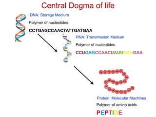
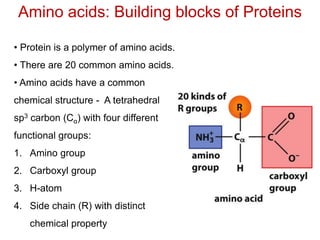
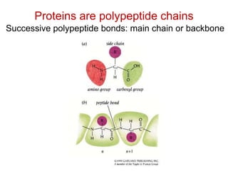
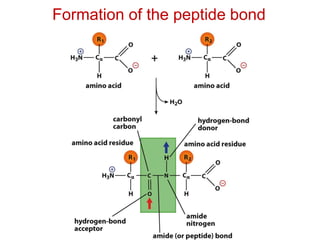
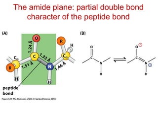
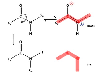
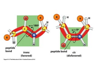
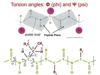
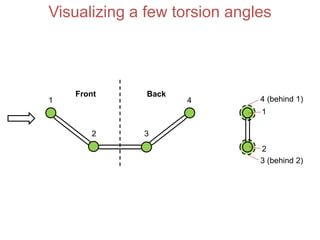
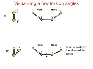
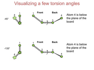
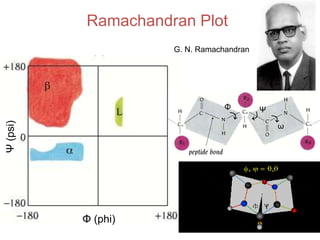
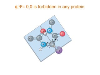
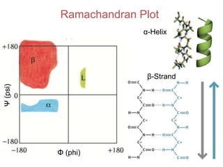
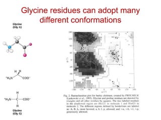
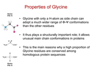
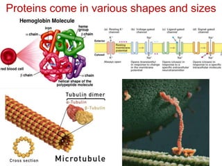
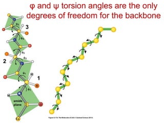
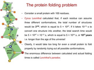
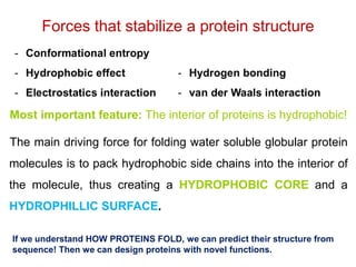
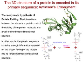
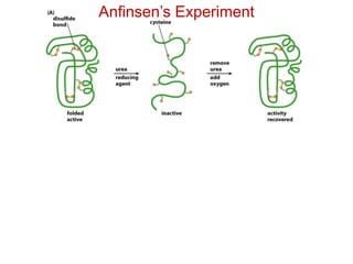
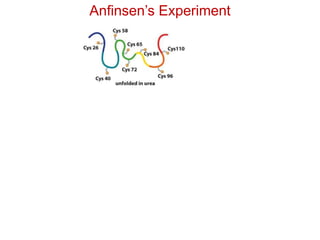
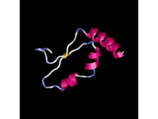
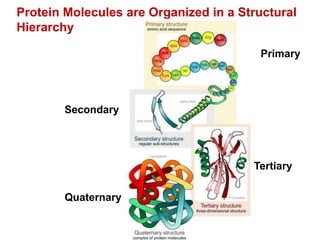
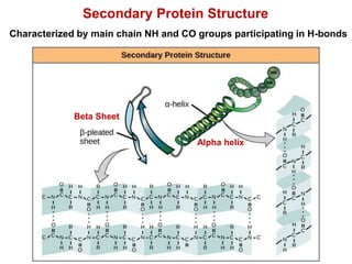
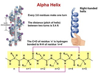
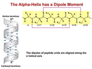
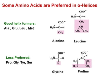
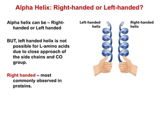
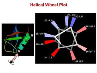
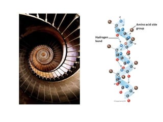
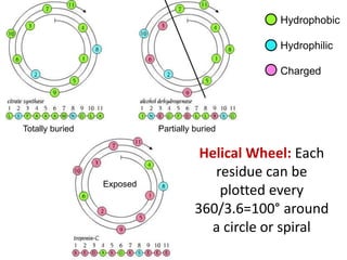
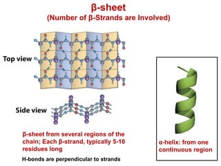
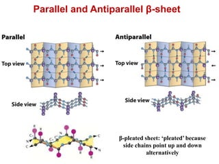
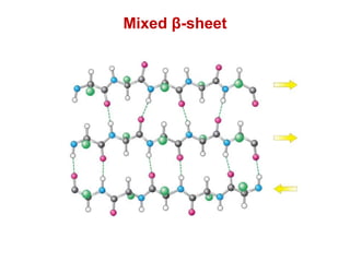
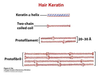
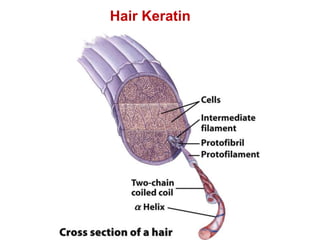
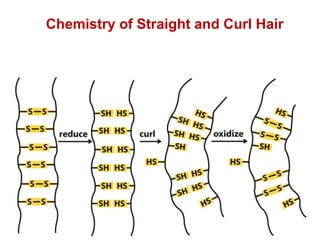
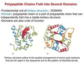
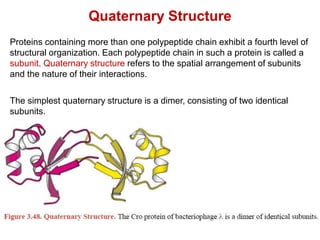
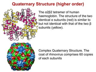
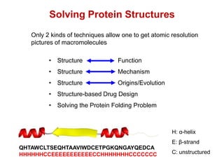
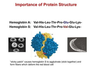
Science of
Living System
Arindam Mondal
School of Bio Science
Email:
arindam.mondal@iitkgp.ac.in
Tel: 03222-260516
BS20001
Books Followed:
• How Proteins Work (Mike Williamson)
• Introduction to protein structure (Carl
Branden & John Tooze)
• Biochemistry (Lubert Stryer)
Protein Structure, Function, Kinetics
and Energetics
Central Dogma of life
Polymer of nucleotides
DNA: Storage Medium
RNA: Transmission Medium
Polymer of nucleotides
CCUGAGCCAACUAUUGAUGAA
CCTGAGCCAACTATTGATGAA
PEPTIDE
Polymer of amino acids
Protein: Molecular Machines
Amino acids: Building blocks of Proteins
• Protein is a polymer of amino acids.
• There are 20 common amino acids.
• Amino acids have a common
chemical structure - A tetrahedral
sp3 carbon (Cα) with four different
functional groups:
1. Amino group
2. Carboxyl group
3. H-atom
4. Side chain (R) with distinct
chemical property
All amino acids in protein have the “L-form”
H-atom is coming out of the whiteboard. Looking down the H-Cα
bond from the H-atom, the L-form amino acid has CO, R and N
going in a clockwise direction. The L-form reads “CORN” in
clockwise direction.
Only imino acid
His can bind or release
protons near physiological pH
pKa ~ 6.4
Proteins are polypeptide chains
Successive polypeptide bonds: main chain or backbone
Formation of the peptide bond
The amide plane: partial double bond
character of the peptide bond
Cα
Cα
Cα
Cα
H
Cα
Cα
Cα
Cα
H
TRANS
CIS
Torsion angles: Φ (phi) and Ψ (psi)
Peptide Plane
Visualizing a few torsion angles
Front Back
1
2 3
4
1
4 (behind 1)
2
3 (behind 2)
Visualizing a few torsion angles
Front Back1
2 3
4
1
4
2
3
0°
+45°
1
4
Front Back1
2 3
4 Atom 4 is above
the plane of the
board
4
Visualizing a few torsion angles
-45°
1
4
Front Back1
2 3
4 Atom 4 is below
the plane of the
board
-135°
1 Front Back1
2 3
Atom 4 is below
the plane of the
board
4
Ramachandran Plot
G. N. Ramachandran
Ψ(psi)
Φ (phi)
Φ Ψ
ω
ɸ,Ψ= 0,0 is forbidden in any protein
Ramachandran Plot
Ψ(psi)
Φ (phi)
α-Helix
β-Strand
Glycine residues can adopt many
different conformations
Properties of Glycine
• Glycine with only a H-atom as side chain can
adopt a much wider range of Φ-Ψ conformations
than the other residues
• It thus plays a structurally important role; it allows
unusual main chain conformations in proteins
• This is the main reasons why a high proportion of
Glycine residues are conserved among
homologous protein sequences
Proteins come in various shapes and sizes
φ and ψ torsion angles are the only
degrees of freedom for the backbone
1
2
3
The protein folding problem
- Consider a small protein with 100 residues.
- Cyrus Levinthal calculated that, if each residue can assume
three different conformations, the total number of structures
would be 3100, which is equal to 5 × 1047. If it takes 10-13 s to
convert one structure into another, the total search time would
be 5 × 1047 × 10-13 s, which is equal to 5 × 1034 s, or 1027 years
i.e. longer than the age of the universe!
- Clearly, it would take too long for even a small protein to fold
properly by randomly trying out all possible conformations.
- The enormous difference between calculated and actual folding
times is called Levinthal's paradox.
Most important feature: The interior of proteins is hydrophobic!
The main driving force for folding water soluble globular protein
molecules is to pack hydrophobic side chains into the interior of
the molecule, thus creating a HYDROPHOBIC CORE and a
HYDROPHILLIC SURFACE.
Forces that stabilize a protein structure
If we understand HOW PROTEINS FOLD, we can predict their structure from
sequence! Then we can design proteins with novel functions.
The 3D structure of a protein is encoded in its
primary sequence: Anfinsen’s Experiment
Thermodynamic hypothesis of
Protein Folding: The interactions
between the atoms in a protein control
the folding of the protein molecule into
a well-defined three-dimensional
structure.
In other words, the protein sequence
contains enough information required
for the proper folding of the protein
into its functional three-dimensional
structure.
Anfinsen’s Experiment
The protein folding game - Foldit
https://fold.it/portal/
Hierarchy of Protein
Structure
Protein Molecules are Organized in a Structural
Hierarchy
Secondary
Tertiary
Primary
Quaternary
Secondary Protein Structure
Beta Sheet
Alpha helix
Characterized by main chain NH and CO groups participating in H-bonds
Alpha Helix
Every 3.6 residues make one turn
The distance (pitch of helix)
between two turns is 5.4 Å
The C=O of residue ‘n’ is hydrogen
bonded to N-H of residue ‘n+4’
n n+1 n+2 n+3 n+4 n+5
The Alpha-Helix has a Dipole Moment
n n+1 n+2 n+3 n+4 n+5
The dipoles of peptide units are aligned along the
α helical axis
Less Preferred:
Pro, Gly, Tyr, Ser
Some Amino Acids are Preferred in α-Helices
Good helix formers:
Ala , Glu, Leu , Met
Alpha helix can be – Right-
handed or Left handed
BUT, left handed helix is not
possible for L-amino acids
due to close approach of
the side chains and CO
group.
Right handed – most
commonly observed in
proteins.
Alpha Helix: Right-handed or Left-handed?
Helical Wheel Plot
N
C
Helical Wheel: Each
residue can be
plotted every
360/3.6=100° around
a circle or spiral
Hydrophobic
Hydrophilic
Charged
Totally buried Partially buried
Exposed
β-sheet
(Number of β-Strands are Involved)
β-sheet from several regions of the
chain; Each β-strand, typically 5-10
residues long
α-helix: from one
continuous region
H-bonds are perpendicular to strands
β-pleated sheet: ‘pleated’ because
side chains point up and down
alternatively
Parallel and Antiparallel β-sheet
Mixed β-sheet
Hair Keratin
Hair Keratin
Chemistry of Straight and Curl Hair
Polypeptide Chains Fold into Several Domains
•Fundamental unit of tertiary structure – DOMAIN
•Domain: polypeptide chain or a part of polypeptide chain that can
independently fold into a stable tertiary structure
•Domains are also units of function
Tertiary structure refers to the spatial arrangement of amino acid residues
that are far apart in the sequence and to the pattern of disulfide bonds.
Quaternary Structure
Proteins containing more than one polypeptide chain exhibit a fourth level of
structural organization. Each polypeptide chain in such a protein is called a
subunit. Quaternary structure refers to the spatial arrangement of subunits
and the nature of their interactions.
The simplest quaternary structure is a dimer, consisting of two identical
subunits.
Quaternary Structure (higher order)
Complex Quaternary Structure. The
coat of rhinovirus comprises 60 copies
of each subunits
The α2β2 tetramer of human
haemoglobin. The structure of the two
identical α subunits (red) is similar to
but not identical with that of the two β
subunits (yellow).
• Structure Function
• Structure Mechanism
• Structure Origins/Evolution
• Structure-based Drug Design
• Solving the Protein Folding Problem
Solving Protein Structures
Only 2 kinds of techniques allow one to get atomic resolution
pictures of macromolecules
QHTAWCLTSEQHTAAVIWDCETPGKQNGAYQEDCA
HHHHHHCCEEEEEEEEEEECCHHHHHHHCCCCCCC
H: α-helix
E: β-strand
C: unstructured
Hemoglobin A: Val-His-Leu-Thr-Pro-Glu-Glu-Lys-
Hemoglobin S: Val-His-Leu-Thr-Pro-Val-Glu-Lys-
“sticky patch” causes hemoglobin S to agglutinate (stick together) and
form fibers which deform the red blood cell
Importance of Protein Structure

Lecture3 intro to_proteins (1)

  • 1.
    Science of Living System ArindamMondal School of Bio Science Email: arindam.mondal@iitkgp.ac.in Tel: 03222-260516 BS20001
  • 2.
    Books Followed: • HowProteins Work (Mike Williamson) • Introduction to protein structure (Carl Branden & John Tooze) • Biochemistry (Lubert Stryer) Protein Structure, Function, Kinetics and Energetics
  • 3.
    Central Dogma oflife Polymer of nucleotides DNA: Storage Medium RNA: Transmission Medium Polymer of nucleotides CCUGAGCCAACUAUUGAUGAA CCTGAGCCAACTATTGATGAA PEPTIDE Polymer of amino acids Protein: Molecular Machines
  • 4.
    Amino acids: Buildingblocks of Proteins • Protein is a polymer of amino acids. • There are 20 common amino acids. • Amino acids have a common chemical structure - A tetrahedral sp3 carbon (Cα) with four different functional groups: 1. Amino group 2. Carboxyl group 3. H-atom 4. Side chain (R) with distinct chemical property
  • 5.
    All amino acidsin protein have the “L-form” H-atom is coming out of the whiteboard. Looking down the H-Cα bond from the H-atom, the L-form amino acid has CO, R and N going in a clockwise direction. The L-form reads “CORN” in clockwise direction.
  • 8.
  • 11.
    His can bindor release protons near physiological pH pKa ~ 6.4
  • 12.
    Proteins are polypeptidechains Successive polypeptide bonds: main chain or backbone
  • 13.
    Formation of thepeptide bond
  • 14.
    The amide plane:partial double bond character of the peptide bond
  • 15.
  • 17.
    Torsion angles: Φ(phi) and Ψ (psi) Peptide Plane
  • 18.
    Visualizing a fewtorsion angles Front Back 1 2 3 4 1 4 (behind 1) 2 3 (behind 2)
  • 19.
    Visualizing a fewtorsion angles Front Back1 2 3 4 1 4 2 3 0° +45° 1 4 Front Back1 2 3 4 Atom 4 is above the plane of the board
  • 20.
    4 Visualizing a fewtorsion angles -45° 1 4 Front Back1 2 3 4 Atom 4 is below the plane of the board -135° 1 Front Back1 2 3 Atom 4 is below the plane of the board 4
  • 21.
    Ramachandran Plot G. N.Ramachandran Ψ(psi) Φ (phi) Φ Ψ ω
  • 22.
    ɸ,Ψ= 0,0 isforbidden in any protein
  • 23.
  • 24.
    Glycine residues canadopt many different conformations
  • 25.
    Properties of Glycine •Glycine with only a H-atom as side chain can adopt a much wider range of Φ-Ψ conformations than the other residues • It thus plays a structurally important role; it allows unusual main chain conformations in proteins • This is the main reasons why a high proportion of Glycine residues are conserved among homologous protein sequences
  • 26.
    Proteins come invarious shapes and sizes
  • 27.
    φ and ψtorsion angles are the only degrees of freedom for the backbone 1 2 3
  • 28.
    The protein foldingproblem - Consider a small protein with 100 residues. - Cyrus Levinthal calculated that, if each residue can assume three different conformations, the total number of structures would be 3100, which is equal to 5 × 1047. If it takes 10-13 s to convert one structure into another, the total search time would be 5 × 1047 × 10-13 s, which is equal to 5 × 1034 s, or 1027 years i.e. longer than the age of the universe! - Clearly, it would take too long for even a small protein to fold properly by randomly trying out all possible conformations. - The enormous difference between calculated and actual folding times is called Levinthal's paradox.
  • 29.
    Most important feature:The interior of proteins is hydrophobic! The main driving force for folding water soluble globular protein molecules is to pack hydrophobic side chains into the interior of the molecule, thus creating a HYDROPHOBIC CORE and a HYDROPHILLIC SURFACE. Forces that stabilize a protein structure If we understand HOW PROTEINS FOLD, we can predict their structure from sequence! Then we can design proteins with novel functions.
  • 30.
    The 3D structureof a protein is encoded in its primary sequence: Anfinsen’s Experiment Thermodynamic hypothesis of Protein Folding: The interactions between the atoms in a protein control the folding of the protein molecule into a well-defined three-dimensional structure. In other words, the protein sequence contains enough information required for the proper folding of the protein into its functional three-dimensional structure.
  • 31.
  • 32.
  • 34.
    The protein foldinggame - Foldit https://fold.it/portal/
  • 35.
  • 36.
    Protein Molecules areOrganized in a Structural Hierarchy Secondary Tertiary Primary Quaternary
  • 37.
    Secondary Protein Structure BetaSheet Alpha helix Characterized by main chain NH and CO groups participating in H-bonds
  • 38.
    Alpha Helix Every 3.6residues make one turn The distance (pitch of helix) between two turns is 5.4 Å The C=O of residue ‘n’ is hydrogen bonded to N-H of residue ‘n+4’ n n+1 n+2 n+3 n+4 n+5
  • 39.
    The Alpha-Helix hasa Dipole Moment n n+1 n+2 n+3 n+4 n+5 The dipoles of peptide units are aligned along the α helical axis
  • 40.
    Less Preferred: Pro, Gly,Tyr, Ser Some Amino Acids are Preferred in α-Helices Good helix formers: Ala , Glu, Leu , Met
  • 41.
    Alpha helix canbe – Right- handed or Left handed BUT, left handed helix is not possible for L-amino acids due to close approach of the side chains and CO group. Right handed – most commonly observed in proteins. Alpha Helix: Right-handed or Left-handed?
  • 42.
  • 44.
    Helical Wheel: Each residuecan be plotted every 360/3.6=100° around a circle or spiral Hydrophobic Hydrophilic Charged Totally buried Partially buried Exposed
  • 45.
    β-sheet (Number of β-Strandsare Involved) β-sheet from several regions of the chain; Each β-strand, typically 5-10 residues long α-helix: from one continuous region H-bonds are perpendicular to strands
  • 46.
    β-pleated sheet: ‘pleated’because side chains point up and down alternatively Parallel and Antiparallel β-sheet
  • 47.
  • 48.
  • 49.
  • 50.
    Chemistry of Straightand Curl Hair
  • 51.
    Polypeptide Chains Foldinto Several Domains •Fundamental unit of tertiary structure – DOMAIN •Domain: polypeptide chain or a part of polypeptide chain that can independently fold into a stable tertiary structure •Domains are also units of function Tertiary structure refers to the spatial arrangement of amino acid residues that are far apart in the sequence and to the pattern of disulfide bonds.
  • 52.
    Quaternary Structure Proteins containingmore than one polypeptide chain exhibit a fourth level of structural organization. Each polypeptide chain in such a protein is called a subunit. Quaternary structure refers to the spatial arrangement of subunits and the nature of their interactions. The simplest quaternary structure is a dimer, consisting of two identical subunits.
  • 53.
    Quaternary Structure (higherorder) Complex Quaternary Structure. The coat of rhinovirus comprises 60 copies of each subunits The α2β2 tetramer of human haemoglobin. The structure of the two identical α subunits (red) is similar to but not identical with that of the two β subunits (yellow).
  • 54.
    • Structure Function •Structure Mechanism • Structure Origins/Evolution • Structure-based Drug Design • Solving the Protein Folding Problem Solving Protein Structures Only 2 kinds of techniques allow one to get atomic resolution pictures of macromolecules QHTAWCLTSEQHTAAVIWDCETPGKQNGAYQEDCA HHHHHHCCEEEEEEEEEEECCHHHHHHHCCCCCCC H: α-helix E: β-strand C: unstructured
  • 55.
    Hemoglobin A: Val-His-Leu-Thr-Pro-Glu-Glu-Lys- HemoglobinS: Val-His-Leu-Thr-Pro-Val-Glu-Lys- “sticky patch” causes hemoglobin S to agglutinate (stick together) and form fibers which deform the red blood cell Importance of Protein Structure