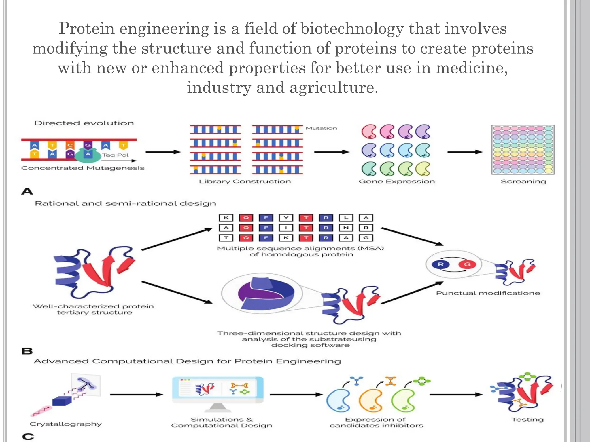
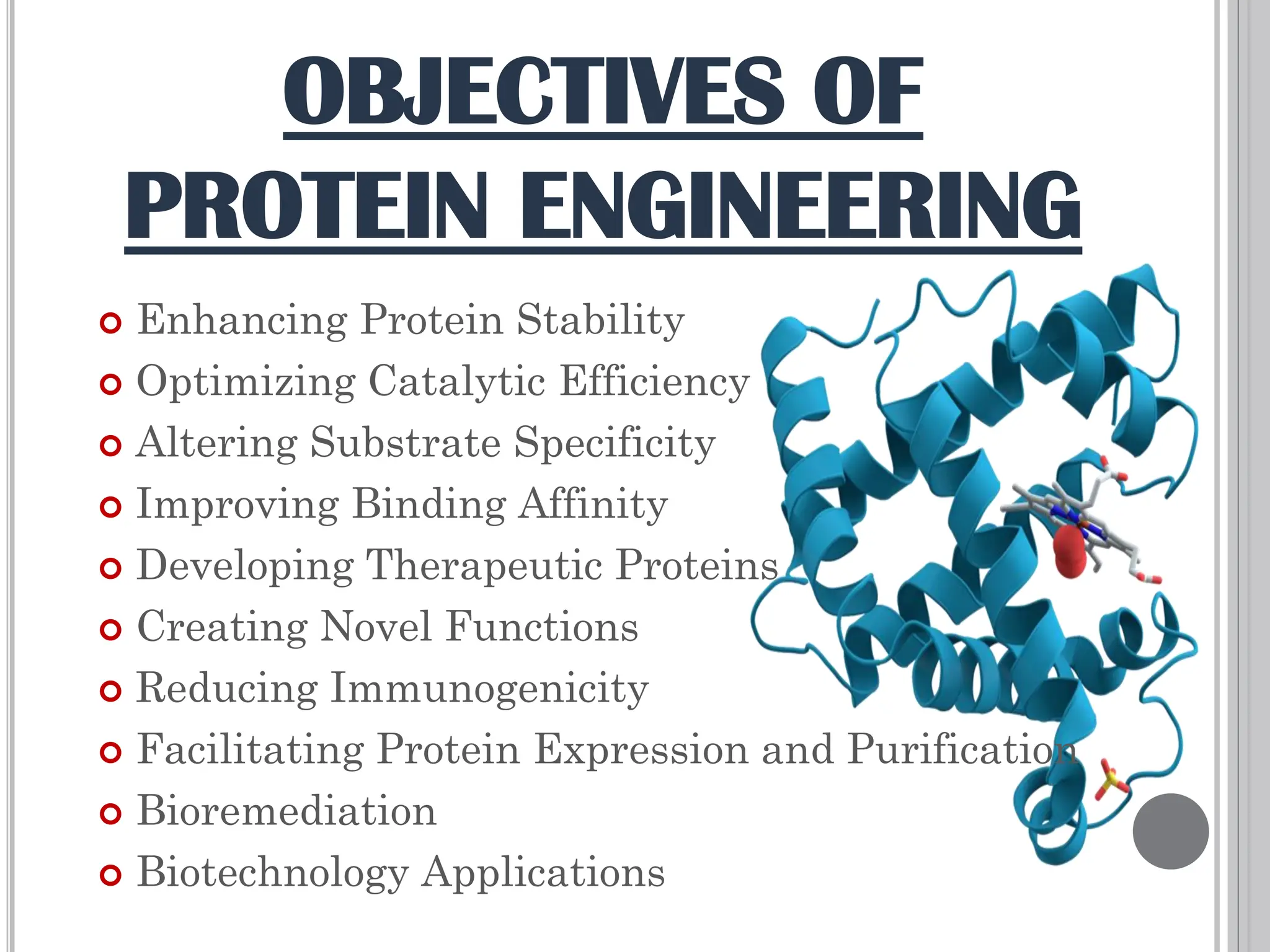
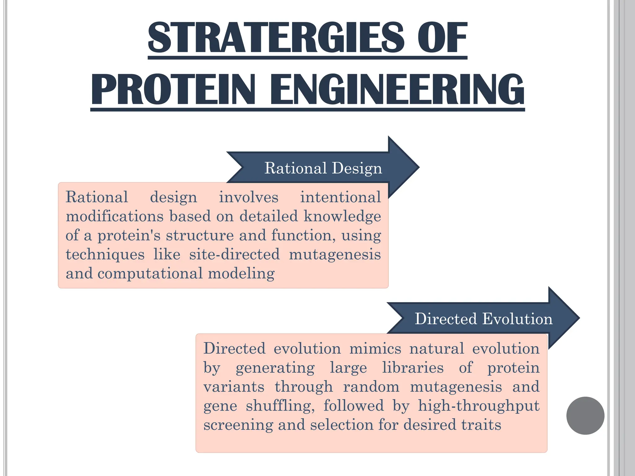
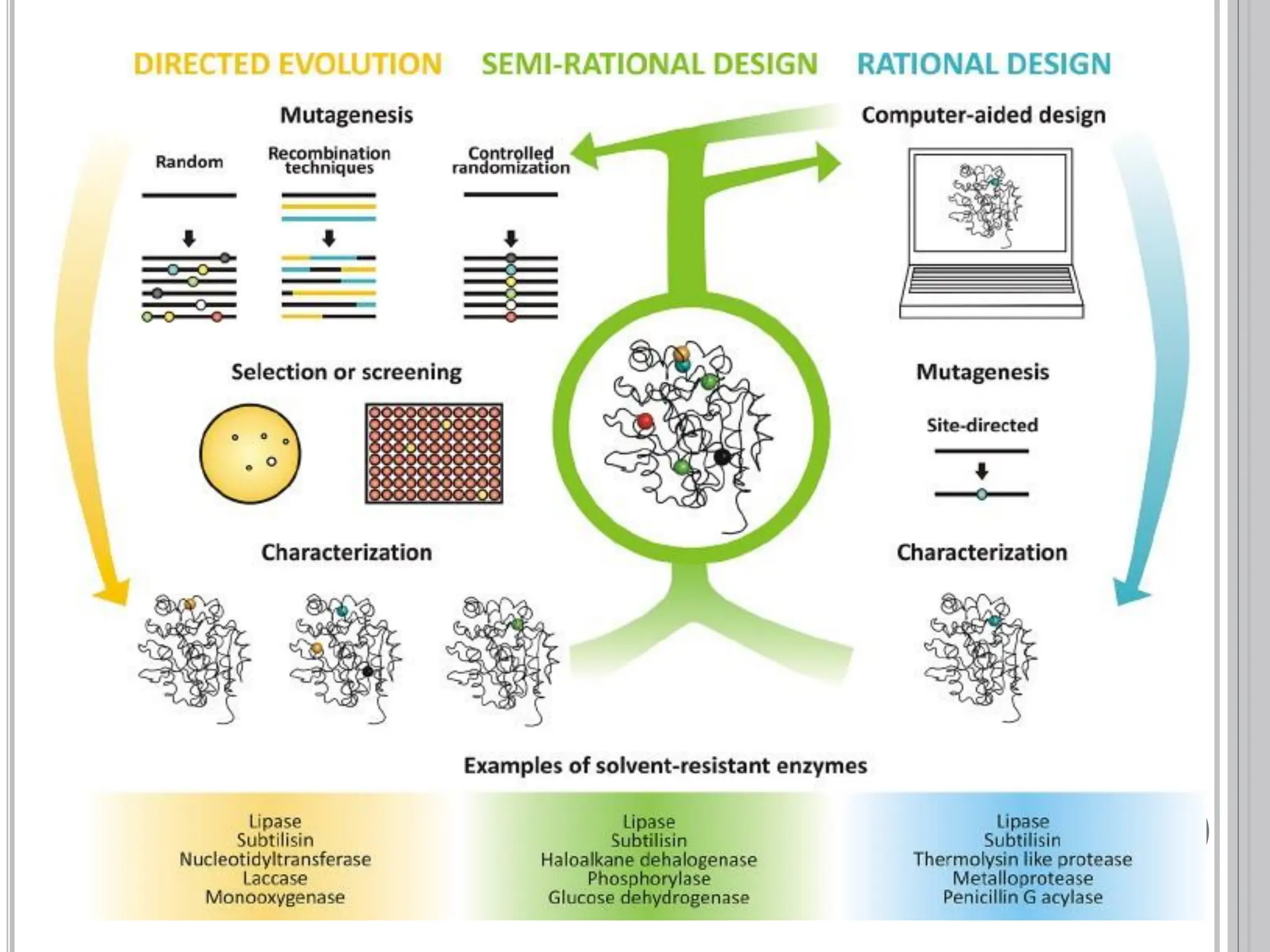
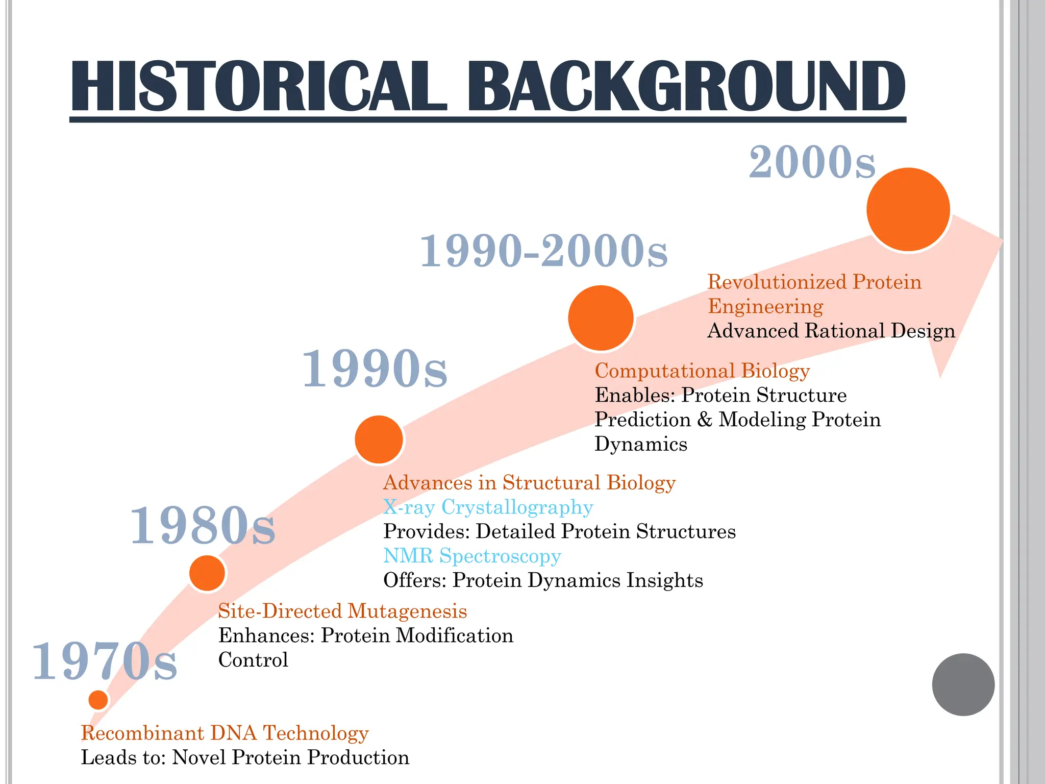
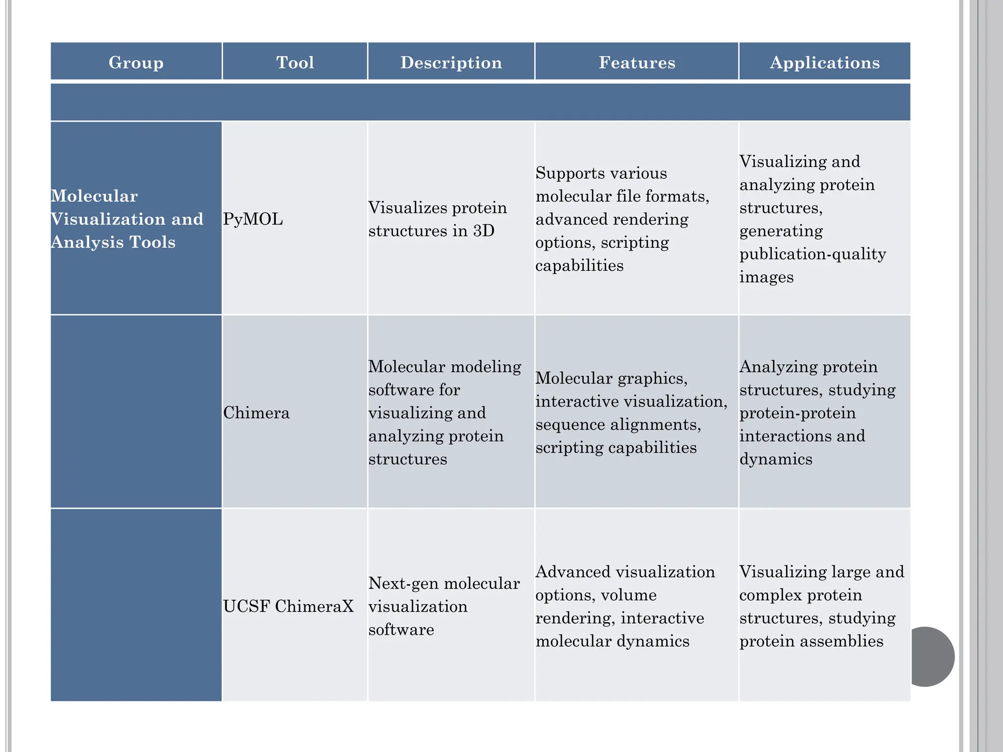
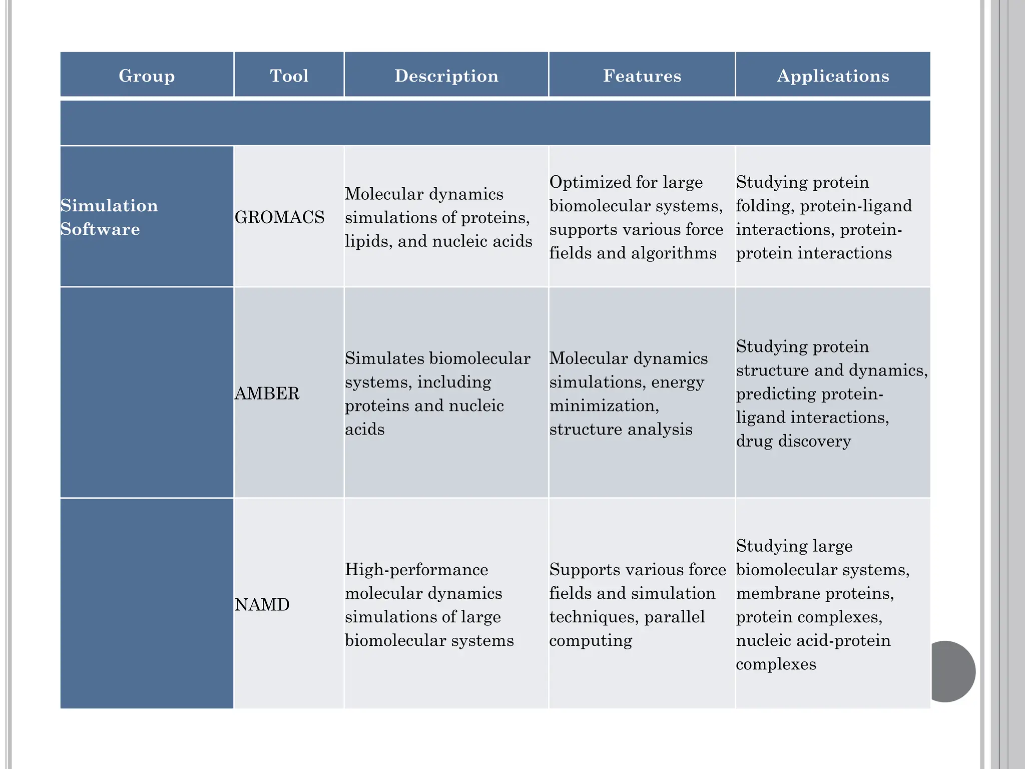
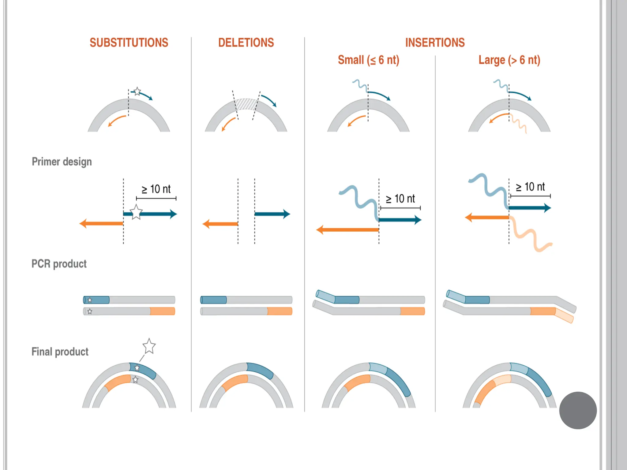
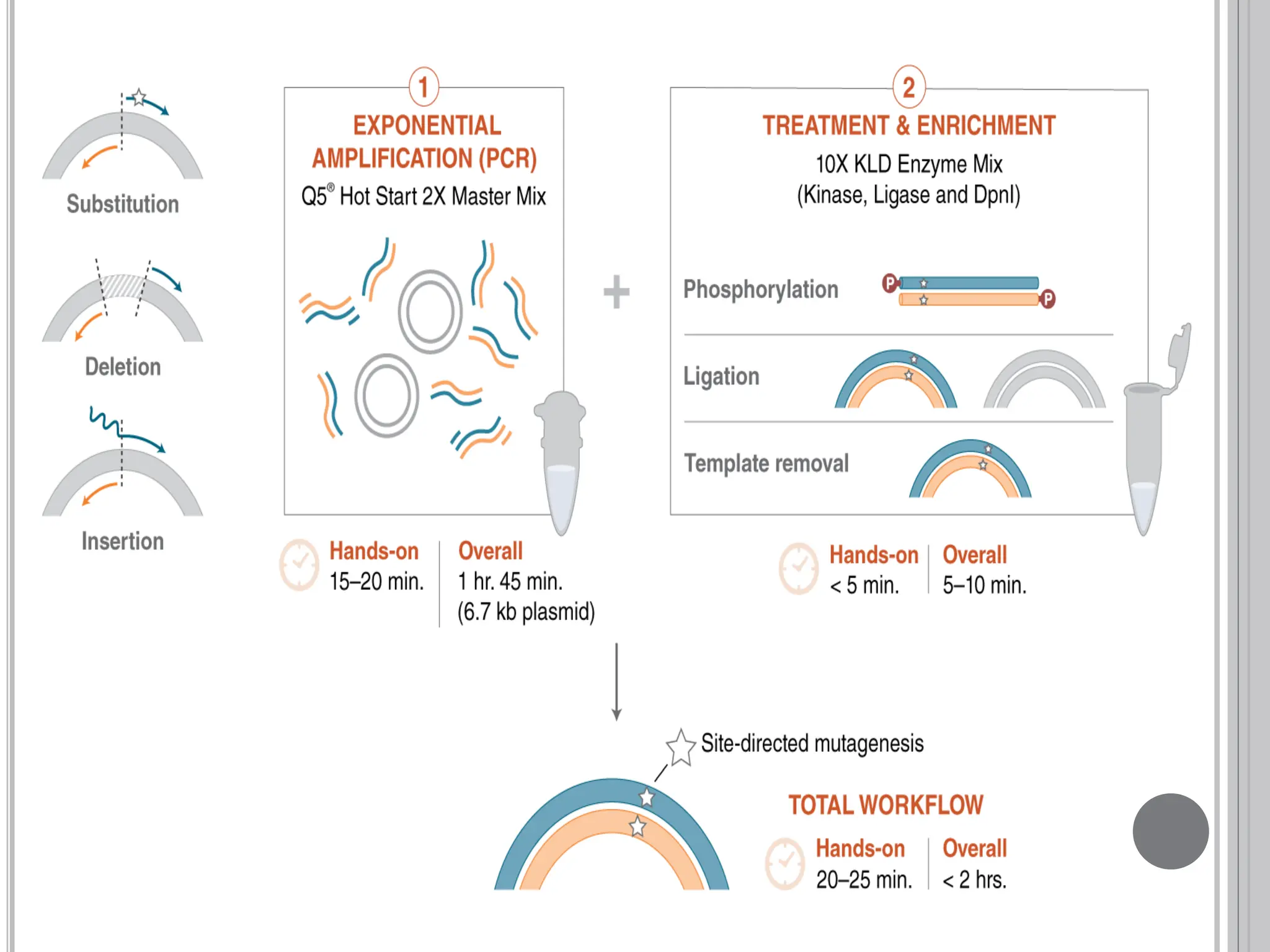
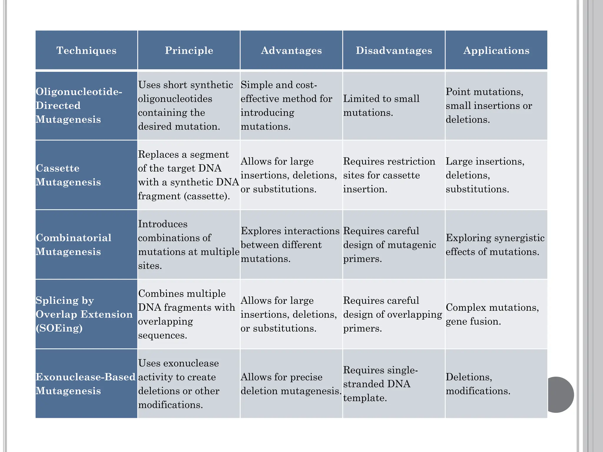
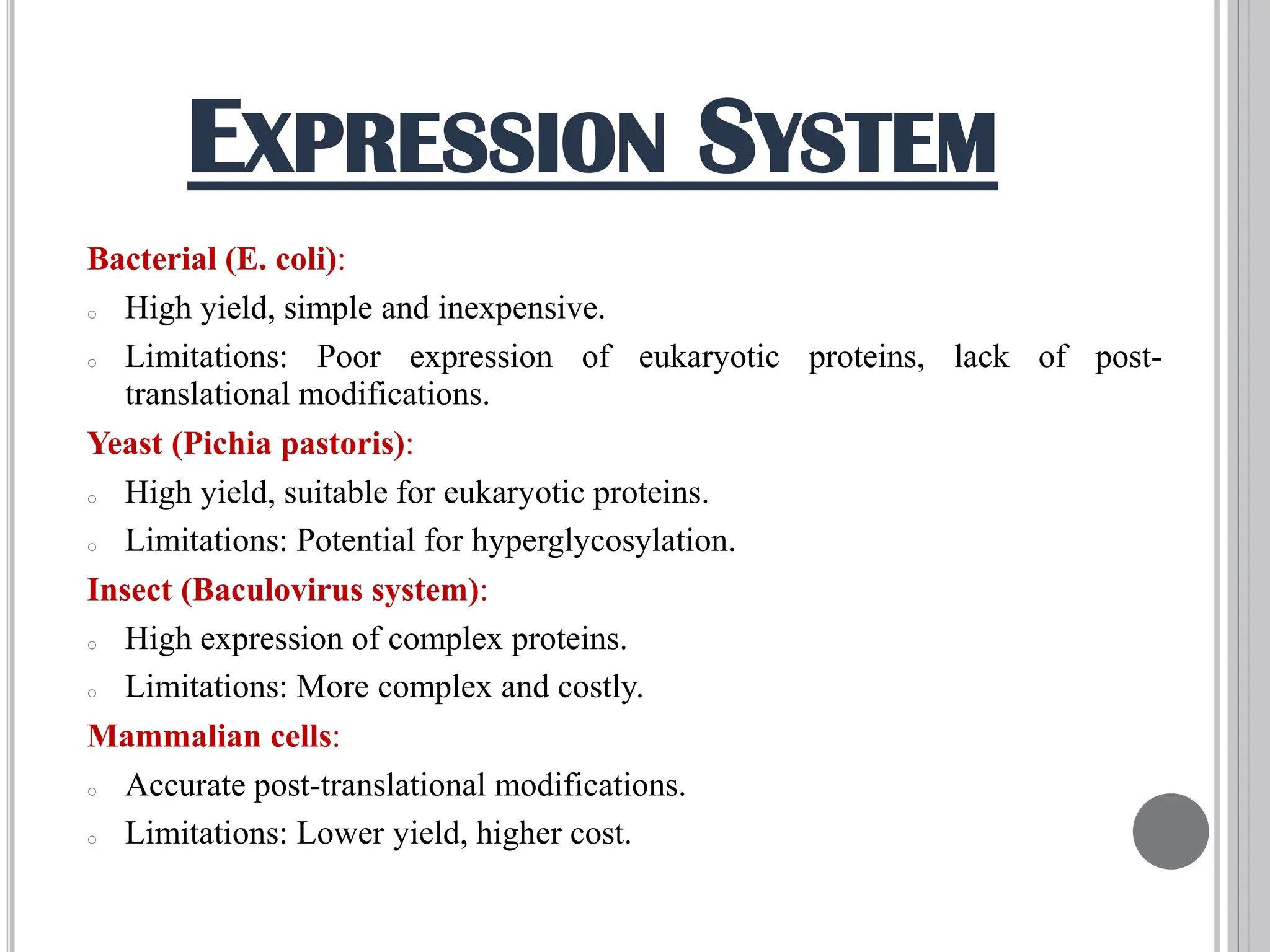
PROTEIN ENGINEERING
BY RATIONAL DESIGN
(TOOLS &TECHNIQUES)
PRESENTED BY
SUNMBAL AWAIS
23016160-003
PHD BIOCHEMISTRY
PROTEIN
ENGINEERING
Protein engineering is a field of biotechnology that involves
modifying the structure and function of proteins to create proteins
with new or enhanced properties for better use in medicine,
industry and agriculture.
OBJECTIVES OF
PROTEIN ENGINEERING
 Enhancing Protein Stability
 Optimizing Catalytic Efficiency
 Altering Substrate Specificity
 Improving Binding Affinity
 Developing Therapeutic Proteins
 Creating Novel Functions
 Reducing Immunogenicity
 Facilitating Protein Expression and Purification
 Bioremediation
 Biotechnology Applications
STEPS INVOLVED IN
PROTEIN ENGINEERING
STRATERGIES OF
PROTEIN ENGINEERING
Rational design involves intentional
modifications based on detailed knowledge
of a protein's structure and function, using
techniques like site-directed mutagenesis
and computational modeling
Directed evolution mimics natural evolution
by generating large libraries of protein
variants through random mutagenesis and
gene shuffling, followed by high-throughput
screening and selection for desired traits
Rational Design
Directed Evolution
PROTEIN
ENGINEERING
BY
RATIONAL DESIGN
RATIONAL DESIGN
Rational design
involves
intentional
modifications
based on
detailed
knowledge of a
protein's
structure and
function, using
techniques like
site-directed
mutagenesis
and
computational
modeling
HISTORICAL BACKGROUND
1970s
1980s
1990s
1990-2000s
Recombinant DNA Technology
Leads to: Novel Protein Production
Site-Directed Mutagenesis
Enhances: Protein Modification
Control
Advances in Structural Biology
X-ray Crystallography
Provides: Detailed Protein Structures
NMR Spectroscopy
Offers: Protein Dynamics Insights
Computational Biology
Enables: Protein Structure
Prediction & Modeling Protein
Dynamics
2000s
Revolutionized Protein
Engineering
Advanced Rational Design
 Understanding Structure-
Function Relationship
 Identifying Design Goals
 Designing Molecules / Materials
 Validation and Optimization
 Iterative Process
STEPS IN RATIONAL DESIGN
TOOLS & TECHNIQUES
IN
RATIONAL DESIGN
STEP 1
STRUCTURE
DETERMINATION
 Structure determination techniques play a crucial
role in rational design across various scientific
disciplines, including molecular biology,
biochemistry, pharmacology, and materials science.
 This information is fundamental for understanding
the structure-function relationships that govern
molecular behavior and is essential for the rational
design of new molecules and materials with specific
properties.
STRUCTURAL DETERMINATION
STRUCTURAL ANALYSIS TOOLS
X-RAY
CRYSTALLO
GRAPHY
NMR
SPECTRO
SCOPY
CRYO-
ELECTRO
MICROSCOPY
X-RAY CRYSTALLOGRAPHY
X-ray crystallography is a powerful technique used to
determine the three-dimensional structure of
biological macromolecules, such as proteins. This
technique plays a crucial role in protein engineering,
where the goal is to design and modify proteins for
specific functions or properties. By providing detailed
insights into the atomic-level structure of proteins, X-
ray crystallography enables researchers to
understand how protein structure relates to function
and to design proteins with enhanced or novel
properties.
PRINCIPLE OF X-RAY CRYSTALLOGRAPHY
X-ray crystallography relies on the principle that when
a crystal is exposed to X-rays, the X-rays are diffracted
by the atoms in the crystal lattice. The resulting
diffraction pattern contains information about the
spatial arrangement of atoms in the crystal. By
analyzing this diffraction pattern, the three
dimensional structure of the crystal can be
determined.
NUCLEAR MAGNETIC
RESONANCE SPECTROSCOPY
 NMR spectroscopy is a powerful technique used to
study the structure and dynamics of molecules in
solution form, including proteins, at the atomic
level.
 In protein engineering, NMR spectroscopy plays a
crucial role in elucidating the structure-function
relationships of proteins and designing proteins
with specific properties.
PRINCIPLE OF X-RAY CRYSTALLOGRAPHY
X-ray crystallography relies on the principle that when
a crystal is exposed to X-rays, the X-rays are diffracted
by the atoms in the crystal lattice. The resulting
diffraction pattern contains information about the
spatial arrangement of atoms in the crystal. By
analyzing this diffraction pattern, the three
dimensional structure of the crystal can be
determined.
CRYO-ELECTRON MICROSCOPY
 Cryo-Electron Microscopy (Cryo-EM) is a
revolutionary technique used to determine the
three dimensional structures of biological
macromolecules, such as proteins and nucleic acids,
at near atomic resolution.
 In protein engineering, Cryo-EM plays a crucial
role in elucidating the structures of proteins and
protein complexes, providing insights into their
functions and enabling the design of proteins with
specific properties.
PRINCIPLE OF CRYO-EM
Cryo-EM involves freezing samples in a thin layer of
ice to maintain their native structure and imaging
them using an electron microscope. By collecting
images from different orientations and combining them
computationally, a high-resolution 3D structure of the
sample can be reconstructed.
STEP 2
COMPUTER-AIDED
DESIGN (CAD)
Group Tool Description Features Applications
Molecular
Visualization and
Analysis Tools
PyMOL
Visualizes protein
structures in 3D
Supports various
molecular file formats,
advanced rendering
options, scripting
capabilities
Visualizing and
analyzing protein
structures,
generating
publication-quality
images
Chimera
Molecular modeling
software for
visualizing and
analyzing protein
structures
Molecular graphics,
interactive visualization,
sequence alignments,
scripting capabilities
Analyzing protein
structures, studying
protein-protein
interactions and
dynamics
UCSF ChimeraX
Next-gen molecular
visualization
software
Advanced visualization
options, volume
rendering, interactive
molecular dynamics
Visualizing large and
complex protein
structures, studying
protein assemblies
Group Tool Description Features Applications
Protein Structure
Prediction and
Modeling Tools
Rosetta
Predicts and designs
protein structures
using algorithms
De novo protein design,
protein-protein
docking, structure
refinement,
customizable
Designing novel protein
structures, predicting
protein-ligand
interactions
Modeller
Comparative protein
structure modeling
based on known
structures
Homology modeling,
loop modeling, side-
chain refinement,
visualization tools
Predicting protein
structures based on
homologous proteins
SWISS-MODEL
Web-based tool for
protein structure
modeling
User-friendly interface,
various modeling
methods, high-quality
models
Predicting protein
structures based on
known homologous
structures
Protein Stability
and Mutagenesis
Prediction Tools
FoldX
Predicts protein
stability changes
upon mutation
Empirical force fields,
predicts effects on
stability, protein-
protein and protein-
ligand interactions
Designing proteins
with specific properties,
studying genetic
disease mutations
Group Tool Description Features Applications
Simulation
Software
GROMACS
Molecular dynamics
simulations of proteins,
lipids, and nucleic acids
Optimized for large
biomolecular systems,
supports various force
fields and algorithms
Studying protein
folding, protein-ligand
interactions, protein-
protein interactions
AMBER
Simulates biomolecular
systems, including
proteins and nucleic
acids
Molecular dynamics
simulations, energy
minimization,
structure analysis
Studying protein
structure and dynamics,
predicting protein-
ligand interactions,
drug discovery
NAMD
High-performance
molecular dynamics
simulations of large
biomolecular systems
Supports various force
fields and simulation
techniques, parallel
computing
Studying large
biomolecular systems,
membrane proteins,
protein complexes,
nucleic acid-protein
complexes
Group Tool Description Features Applications
Bioinformatics
Tools
PyRosetta
Python-based interface
for Rosetta, for protein
structure prediction and
design
Access to Rosetta's
energy and scoring
functions, customizable
protocols and workflows
Developing custom
algorithms for protein
structure prediction,
protein-ligand
docking, protein
design
Biopython
Comprehensive tools for
biological computation
Sequence alignment,
motif analysis, protein
structure analysis,
supports various
bioinformatics formats
Sequence analysis,
structure prediction,
phylogenetic analysis,
bioinformatics
research
Systems Biology
Software
COPASI
Modeling and simulation
of biochemical networks
and metabolic pathways
Supports ODEs, SSAs,
Petri nets, parameter
estimation, sensitivity
analysis, optimization
Modeling and
simulating
biochemical networks,
studying dynamics of
complex biological
systems
Cell Designer
Modeling tool for
biological systems,
including signaling
pathways and networks
Graphical interface,
supports SBML and
SBGN, library of
biological components,
simulation and analysis
Modeling and
simulating biological
systems at cellular
and molecular levels,
studying signaling
pathways
STEP 3
SITE-DIRECTED
MUTAGENESIS
 Mutagenesis is a powerful DNA methodology that is used to alter
the nucleotide sequence of DNA to study its effect on gene or DNA
function.
 The mutagenesis can be conducted in vivo (in studies of model
organisms or cultured cells) or in vitro (typically using plasmid
constructs) and can be either generated at a specific site in a
predetermined way (site-directed mutagenesis) or randomly
incorporated in the DNA sequence of interest.
 Site-directed mutagenesis is a fundamental technique in
molecular biology that allows for precise and targeted modifications
in the specific nucleotide changes in a DNA sequence of a gene,
which in turn alters the amino acid sequence of the encoded protein.
This technique is indispensable in rational protein design, where the
goal is to create proteins with specific properties or functionalities
by understanding and manipulating their structure-function
relationships. This precision allows researchers to study the effects
of individual amino acids on protein structure and function.
MECHANISM
The basic procedure requires the synthesis of a short DNA
primer. This synthetic primer contains the desired mutation and
is complementary to the template DNA around the mutation
site so it can hybridize with the DNA in the gene of interest. The
mutation may be a single base change (a point mutation),
multiple base changes, deletion, or insertion. The single-strand
primer is then extended using a DNA polymerase, which copies
the rest of the gene. The gene thus copied contains the mutated
site, and is then introduced into a host cell in a vector and
cloned. Finally, mutants are selected by DNA sequencing to
check that they contain the desired mutation
Techniques Principle Advantages Disadvantages Applications
Primer Extension
Mutagenesis
Uses a synthetic
oligonucleotide
(primer) containing
the desired mutation
to amplify a specific
region of DNA.
Simple and widely
used method for
introducing point
mutations.
Limited to small
mutations.
Protein engineering,
gene function
analysis.
QuikChange PCR
Mutagenesis
Uses a single PCR
step with a mutagenic
primer to introduce
mutations.
Rapid and efficient
method for
introducing point
mutations.
Limited to small
mutations.
Rapid site-directed
mutagenesis.
Inverse PCR
Mutagenesis
Introduces mutations
into circular DNA,
such as plasmids.
Useful for mutating
circular DNA.
Limited to circular
DNA.
Plasmid mutagenesis.
Overlap Extension
PCR (OE-PCR)
Combines two DNA
fragments with
overlapping ends to
create mutations.
Allows for larger
insertions, deletions,
or substitutions.
Requires multiple
PCR steps.
Complex mutations,
gene fusion.
Mega-Primer PCR
Mutagenesis
Uses a large
mutagenic primer
(mega-primer) to
introduce mutations.
Allows for medium-
sized mutations.
Requires careful
design of mega-
primer.
Medium-sized
mutations.
Techniques Principle Advantages Disadvantages Applications
Oligonucleotide-
Directed
Mutagenesis
Uses short synthetic
oligonucleotides
containing the
desired mutation.
Simple and cost-
effective method for
introducing
mutations.
Limited to small
mutations.
Point mutations,
small insertions or
deletions.
Cassette
Mutagenesis
Replaces a segment
of the target DNA
with a synthetic DNA
fragment (cassette).
Allows for large
insertions, deletions,
or substitutions.
Requires restriction
sites for cassette
insertion.
Large insertions,
deletions,
substitutions.
Combinatorial
Mutagenesis
Introduces
combinations of
mutations at multiple
sites.
Explores interactions
between different
mutations.
Requires careful
design of mutagenic
primers.
Exploring synergistic
effects of mutations.
Splicing by
Overlap Extension
(SOEing)
Combines multiple
DNA fragments with
overlapping
sequences.
Allows for large
insertions, deletions,
or substitutions.
Requires careful
design of overlapping
primers.
Complex mutations,
gene fusion.
Exonuclease-Based
Mutagenesis
Uses exonuclease
activity to create
deletions or other
modifications.
Allows for precise
deletion mutagenesis.
Requires single-
stranded DNA
template.
Deletions,
modifications.
Techniques Principle Advantages Disadvantages Applications
Alanine-Scanning
Mutagenesis
Replaces amino acids
with alanine to study
the function of
specific residues.
Identifies residues
important for protein
function.
Limited to alanine
substitutions.
Studying protein
structure and
function.
Random
Mutagenesis
Introduces random
mutations
throughout a DNA
sequence.
Generates libraries of
variants for directed
evolution.
Creates a large
number of non-
functional mutants.
Directed evolution,
studying mutation
effects.
Deletion
Mutagenesis
Removes specific
regions of a DNA
sequence.
Creates truncated
proteins or studies
functional regions.
May disrupt essential
gene functions.
Studying functional
regions of genes.
Insertional
Mutagenesis
Introduces specific
sequences into a
DNA sequence.
Creates fusion
proteins or studies
the effects of
insertions.
May disrupt protein
function.
Creating fusion
proteins, studying
insertional effects.
STEP 4
EXPRESSION
AND
PURIFICATION
Bacterial (E. coli):
o High yield, simple and inexpensive.
o Limitations: Poor expression of eukaryotic proteins, lack of post-
translational modifications.
Yeast (Pichia pastoris):
o High yield, suitable for eukaryotic proteins.
o Limitations: Potential for hyperglycosylation.
Insect (Baculovirus system):
o High expression of complex proteins.
o Limitations: More complex and costly.
Mammalian cells:
o Accurate post-translational modifications.
o Limitations: Lower yield, higher cost.
EXPRESSION SYSTEM
PROTEIN PURIFICATION
TECHNIQUES
 Affinity Chromatography:
Uses a specific tag (e.g., His-tag) to purify the protein. Highly selective
and efficient.
 Ion Exchange Chromatography:
Separates proteins based on charge. Useful for further purification steps.
 Size Exclusion Chromatography:
Separates proteins based on size. Useful for removing aggregates and
buffer exchange.
STEP 5
FUNCTIONAL
ASSAYS
ENZYME ACTIVITY ASSAYS
Enzyme activity assays measure the catalytic efficiency of
engineered enzymes.
 Methods:
Spectrophotometric assays, fluorometric assays, and
colorimetric assays.
 Applications:
Determining kinetic parameters (Km, Vmax), assessing
substrate specificity, and evaluating the effect of mutations.
BINDING STUDIES
Binding studies evaluate the interaction between proteins
and ligands or other proteins.
 Surface Plasmon Resonance (SPR):
Measures binding kinetics and affinities in real-time.
 Isothermal Titration Calorimetry (ITC):
Measures binding affinity and thermodynamics.
Assessing the stability and folding of engineered proteins is
crucial for their functionality.
 Differential Scanning Calorimetry (DSC):
Measures thermal stability by monitoring heat changes during
protein unfolding.
 Circular Dichroism (CD):
Analyzes secondary structure content and folding properties.
STABILITY AND FOLDING
ASSAYS
REFERENCES
1. Wlodawer, A., & Dauter, Z. (2017). High-Resolution X-ray
Crystallography of Protein Structures. In A. Wlodawer, Z.
Dauter, & M. Jaskolski (Eds.), Protein Crystallography: Methods
and Protocols (pp. 229-235). Humana Press.
2. Drenth, J. (2007). Principles of Protein X-ray Crystallography.
Springer Science & Business Media.
3. Carrigan, P. E., Ballar, P., & Tuzmen, S. (2011). Site-directed
mutagenesis. Disease Gene Identification: Methods and Protocols,
107-124.
4. Carter, Paul. "Site-directed mutagenesis." Biochemical Journal
237.1 (1986): 1.
5. Kazlauskas, R. J., & Bornscheuer, U. T. (2009). Finding better
protein engineering strategies. Nature chemical biology, 5(8),
526-529.
THANK YOU

Protein Engineering by Rational Design (Tools & Techniques).pdf

  • 1.
    PROTEIN ENGINEERING BY RATIONALDESIGN (TOOLS &TECHNIQUES) PRESENTED BY SUNMBAL AWAIS 23016160-003 PHD BIOCHEMISTRY
  • 2.
  • 3.
    Protein engineering isa field of biotechnology that involves modifying the structure and function of proteins to create proteins with new or enhanced properties for better use in medicine, industry and agriculture.
  • 4.
    OBJECTIVES OF PROTEIN ENGINEERING Enhancing Protein Stability  Optimizing Catalytic Efficiency  Altering Substrate Specificity  Improving Binding Affinity  Developing Therapeutic Proteins  Creating Novel Functions  Reducing Immunogenicity  Facilitating Protein Expression and Purification  Bioremediation  Biotechnology Applications
  • 5.
  • 6.
    STRATERGIES OF PROTEIN ENGINEERING Rationaldesign involves intentional modifications based on detailed knowledge of a protein's structure and function, using techniques like site-directed mutagenesis and computational modeling Directed evolution mimics natural evolution by generating large libraries of protein variants through random mutagenesis and gene shuffling, followed by high-throughput screening and selection for desired traits Rational Design Directed Evolution
  • 8.
  • 9.
    RATIONAL DESIGN Rational design involves intentional modifications basedon detailed knowledge of a protein's structure and function, using techniques like site-directed mutagenesis and computational modeling
  • 10.
    HISTORICAL BACKGROUND 1970s 1980s 1990s 1990-2000s Recombinant DNATechnology Leads to: Novel Protein Production Site-Directed Mutagenesis Enhances: Protein Modification Control Advances in Structural Biology X-ray Crystallography Provides: Detailed Protein Structures NMR Spectroscopy Offers: Protein Dynamics Insights Computational Biology Enables: Protein Structure Prediction & Modeling Protein Dynamics 2000s Revolutionized Protein Engineering Advanced Rational Design
  • 11.
     Understanding Structure- FunctionRelationship  Identifying Design Goals  Designing Molecules / Materials  Validation and Optimization  Iterative Process STEPS IN RATIONAL DESIGN
  • 12.
  • 14.
  • 15.
     Structure determinationtechniques play a crucial role in rational design across various scientific disciplines, including molecular biology, biochemistry, pharmacology, and materials science.  This information is fundamental for understanding the structure-function relationships that govern molecular behavior and is essential for the rational design of new molecules and materials with specific properties. STRUCTURAL DETERMINATION
  • 16.
  • 17.
    X-RAY CRYSTALLOGRAPHY X-ray crystallographyis a powerful technique used to determine the three-dimensional structure of biological macromolecules, such as proteins. This technique plays a crucial role in protein engineering, where the goal is to design and modify proteins for specific functions or properties. By providing detailed insights into the atomic-level structure of proteins, X- ray crystallography enables researchers to understand how protein structure relates to function and to design proteins with enhanced or novel properties.
  • 18.
    PRINCIPLE OF X-RAYCRYSTALLOGRAPHY X-ray crystallography relies on the principle that when a crystal is exposed to X-rays, the X-rays are diffracted by the atoms in the crystal lattice. The resulting diffraction pattern contains information about the spatial arrangement of atoms in the crystal. By analyzing this diffraction pattern, the three dimensional structure of the crystal can be determined.
  • 19.
    NUCLEAR MAGNETIC RESONANCE SPECTROSCOPY NMR spectroscopy is a powerful technique used to study the structure and dynamics of molecules in solution form, including proteins, at the atomic level.  In protein engineering, NMR spectroscopy plays a crucial role in elucidating the structure-function relationships of proteins and designing proteins with specific properties.
  • 20.
    PRINCIPLE OF X-RAYCRYSTALLOGRAPHY X-ray crystallography relies on the principle that when a crystal is exposed to X-rays, the X-rays are diffracted by the atoms in the crystal lattice. The resulting diffraction pattern contains information about the spatial arrangement of atoms in the crystal. By analyzing this diffraction pattern, the three dimensional structure of the crystal can be determined.
  • 21.
    CRYO-ELECTRON MICROSCOPY  Cryo-ElectronMicroscopy (Cryo-EM) is a revolutionary technique used to determine the three dimensional structures of biological macromolecules, such as proteins and nucleic acids, at near atomic resolution.  In protein engineering, Cryo-EM plays a crucial role in elucidating the structures of proteins and protein complexes, providing insights into their functions and enabling the design of proteins with specific properties.
  • 22.
    PRINCIPLE OF CRYO-EM Cryo-EMinvolves freezing samples in a thin layer of ice to maintain their native structure and imaging them using an electron microscope. By collecting images from different orientations and combining them computationally, a high-resolution 3D structure of the sample can be reconstructed.
  • 23.
  • 24.
    Group Tool DescriptionFeatures Applications Molecular Visualization and Analysis Tools PyMOL Visualizes protein structures in 3D Supports various molecular file formats, advanced rendering options, scripting capabilities Visualizing and analyzing protein structures, generating publication-quality images Chimera Molecular modeling software for visualizing and analyzing protein structures Molecular graphics, interactive visualization, sequence alignments, scripting capabilities Analyzing protein structures, studying protein-protein interactions and dynamics UCSF ChimeraX Next-gen molecular visualization software Advanced visualization options, volume rendering, interactive molecular dynamics Visualizing large and complex protein structures, studying protein assemblies
  • 25.
    Group Tool DescriptionFeatures Applications Protein Structure Prediction and Modeling Tools Rosetta Predicts and designs protein structures using algorithms De novo protein design, protein-protein docking, structure refinement, customizable Designing novel protein structures, predicting protein-ligand interactions Modeller Comparative protein structure modeling based on known structures Homology modeling, loop modeling, side- chain refinement, visualization tools Predicting protein structures based on homologous proteins SWISS-MODEL Web-based tool for protein structure modeling User-friendly interface, various modeling methods, high-quality models Predicting protein structures based on known homologous structures Protein Stability and Mutagenesis Prediction Tools FoldX Predicts protein stability changes upon mutation Empirical force fields, predicts effects on stability, protein- protein and protein- ligand interactions Designing proteins with specific properties, studying genetic disease mutations
  • 26.
    Group Tool DescriptionFeatures Applications Simulation Software GROMACS Molecular dynamics simulations of proteins, lipids, and nucleic acids Optimized for large biomolecular systems, supports various force fields and algorithms Studying protein folding, protein-ligand interactions, protein- protein interactions AMBER Simulates biomolecular systems, including proteins and nucleic acids Molecular dynamics simulations, energy minimization, structure analysis Studying protein structure and dynamics, predicting protein- ligand interactions, drug discovery NAMD High-performance molecular dynamics simulations of large biomolecular systems Supports various force fields and simulation techniques, parallel computing Studying large biomolecular systems, membrane proteins, protein complexes, nucleic acid-protein complexes
  • 27.
    Group Tool DescriptionFeatures Applications Bioinformatics Tools PyRosetta Python-based interface for Rosetta, for protein structure prediction and design Access to Rosetta's energy and scoring functions, customizable protocols and workflows Developing custom algorithms for protein structure prediction, protein-ligand docking, protein design Biopython Comprehensive tools for biological computation Sequence alignment, motif analysis, protein structure analysis, supports various bioinformatics formats Sequence analysis, structure prediction, phylogenetic analysis, bioinformatics research Systems Biology Software COPASI Modeling and simulation of biochemical networks and metabolic pathways Supports ODEs, SSAs, Petri nets, parameter estimation, sensitivity analysis, optimization Modeling and simulating biochemical networks, studying dynamics of complex biological systems Cell Designer Modeling tool for biological systems, including signaling pathways and networks Graphical interface, supports SBML and SBGN, library of biological components, simulation and analysis Modeling and simulating biological systems at cellular and molecular levels, studying signaling pathways
  • 28.
  • 29.
     Mutagenesis isa powerful DNA methodology that is used to alter the nucleotide sequence of DNA to study its effect on gene or DNA function.  The mutagenesis can be conducted in vivo (in studies of model organisms or cultured cells) or in vitro (typically using plasmid constructs) and can be either generated at a specific site in a predetermined way (site-directed mutagenesis) or randomly incorporated in the DNA sequence of interest.  Site-directed mutagenesis is a fundamental technique in molecular biology that allows for precise and targeted modifications in the specific nucleotide changes in a DNA sequence of a gene, which in turn alters the amino acid sequence of the encoded protein. This technique is indispensable in rational protein design, where the goal is to create proteins with specific properties or functionalities by understanding and manipulating their structure-function relationships. This precision allows researchers to study the effects of individual amino acids on protein structure and function.
  • 31.
    MECHANISM The basic procedurerequires the synthesis of a short DNA primer. This synthetic primer contains the desired mutation and is complementary to the template DNA around the mutation site so it can hybridize with the DNA in the gene of interest. The mutation may be a single base change (a point mutation), multiple base changes, deletion, or insertion. The single-strand primer is then extended using a DNA polymerase, which copies the rest of the gene. The gene thus copied contains the mutated site, and is then introduced into a host cell in a vector and cloned. Finally, mutants are selected by DNA sequencing to check that they contain the desired mutation
  • 33.
    Techniques Principle AdvantagesDisadvantages Applications Primer Extension Mutagenesis Uses a synthetic oligonucleotide (primer) containing the desired mutation to amplify a specific region of DNA. Simple and widely used method for introducing point mutations. Limited to small mutations. Protein engineering, gene function analysis. QuikChange PCR Mutagenesis Uses a single PCR step with a mutagenic primer to introduce mutations. Rapid and efficient method for introducing point mutations. Limited to small mutations. Rapid site-directed mutagenesis. Inverse PCR Mutagenesis Introduces mutations into circular DNA, such as plasmids. Useful for mutating circular DNA. Limited to circular DNA. Plasmid mutagenesis. Overlap Extension PCR (OE-PCR) Combines two DNA fragments with overlapping ends to create mutations. Allows for larger insertions, deletions, or substitutions. Requires multiple PCR steps. Complex mutations, gene fusion. Mega-Primer PCR Mutagenesis Uses a large mutagenic primer (mega-primer) to introduce mutations. Allows for medium- sized mutations. Requires careful design of mega- primer. Medium-sized mutations.
  • 34.
    Techniques Principle AdvantagesDisadvantages Applications Oligonucleotide- Directed Mutagenesis Uses short synthetic oligonucleotides containing the desired mutation. Simple and cost- effective method for introducing mutations. Limited to small mutations. Point mutations, small insertions or deletions. Cassette Mutagenesis Replaces a segment of the target DNA with a synthetic DNA fragment (cassette). Allows for large insertions, deletions, or substitutions. Requires restriction sites for cassette insertion. Large insertions, deletions, substitutions. Combinatorial Mutagenesis Introduces combinations of mutations at multiple sites. Explores interactions between different mutations. Requires careful design of mutagenic primers. Exploring synergistic effects of mutations. Splicing by Overlap Extension (SOEing) Combines multiple DNA fragments with overlapping sequences. Allows for large insertions, deletions, or substitutions. Requires careful design of overlapping primers. Complex mutations, gene fusion. Exonuclease-Based Mutagenesis Uses exonuclease activity to create deletions or other modifications. Allows for precise deletion mutagenesis. Requires single- stranded DNA template. Deletions, modifications.
  • 35.
    Techniques Principle AdvantagesDisadvantages Applications Alanine-Scanning Mutagenesis Replaces amino acids with alanine to study the function of specific residues. Identifies residues important for protein function. Limited to alanine substitutions. Studying protein structure and function. Random Mutagenesis Introduces random mutations throughout a DNA sequence. Generates libraries of variants for directed evolution. Creates a large number of non- functional mutants. Directed evolution, studying mutation effects. Deletion Mutagenesis Removes specific regions of a DNA sequence. Creates truncated proteins or studies functional regions. May disrupt essential gene functions. Studying functional regions of genes. Insertional Mutagenesis Introduces specific sequences into a DNA sequence. Creates fusion proteins or studies the effects of insertions. May disrupt protein function. Creating fusion proteins, studying insertional effects.
  • 36.
  • 37.
    Bacterial (E. coli): oHigh yield, simple and inexpensive. o Limitations: Poor expression of eukaryotic proteins, lack of post- translational modifications. Yeast (Pichia pastoris): o High yield, suitable for eukaryotic proteins. o Limitations: Potential for hyperglycosylation. Insect (Baculovirus system): o High expression of complex proteins. o Limitations: More complex and costly. Mammalian cells: o Accurate post-translational modifications. o Limitations: Lower yield, higher cost. EXPRESSION SYSTEM
  • 38.
    PROTEIN PURIFICATION TECHNIQUES  AffinityChromatography: Uses a specific tag (e.g., His-tag) to purify the protein. Highly selective and efficient.  Ion Exchange Chromatography: Separates proteins based on charge. Useful for further purification steps.  Size Exclusion Chromatography: Separates proteins based on size. Useful for removing aggregates and buffer exchange.
  • 39.
  • 40.
    ENZYME ACTIVITY ASSAYS Enzymeactivity assays measure the catalytic efficiency of engineered enzymes.  Methods: Spectrophotometric assays, fluorometric assays, and colorimetric assays.  Applications: Determining kinetic parameters (Km, Vmax), assessing substrate specificity, and evaluating the effect of mutations.
  • 41.
    BINDING STUDIES Binding studiesevaluate the interaction between proteins and ligands or other proteins.  Surface Plasmon Resonance (SPR): Measures binding kinetics and affinities in real-time.  Isothermal Titration Calorimetry (ITC): Measures binding affinity and thermodynamics.
  • 42.
    Assessing the stabilityand folding of engineered proteins is crucial for their functionality.  Differential Scanning Calorimetry (DSC): Measures thermal stability by monitoring heat changes during protein unfolding.  Circular Dichroism (CD): Analyzes secondary structure content and folding properties. STABILITY AND FOLDING ASSAYS
  • 43.
    REFERENCES 1. Wlodawer, A.,& Dauter, Z. (2017). High-Resolution X-ray Crystallography of Protein Structures. In A. Wlodawer, Z. Dauter, & M. Jaskolski (Eds.), Protein Crystallography: Methods and Protocols (pp. 229-235). Humana Press. 2. Drenth, J. (2007). Principles of Protein X-ray Crystallography. Springer Science & Business Media. 3. Carrigan, P. E., Ballar, P., & Tuzmen, S. (2011). Site-directed mutagenesis. Disease Gene Identification: Methods and Protocols, 107-124. 4. Carter, Paul. "Site-directed mutagenesis." Biochemical Journal 237.1 (1986): 1. 5. Kazlauskas, R. J., & Bornscheuer, U. T. (2009). Finding better protein engineering strategies. Nature chemical biology, 5(8), 526-529.
  • 44.