More Related Content
Similar to THE THREE DIMENSIONAL STRUCTURE OF PROTEINS.pdf (20)
THE THREE DIMENSIONAL STRUCTURE OF PROTEINS.pdf
- 1. 4
4-1
© 2003 Thomson Learning, Inc.
All rights reserved
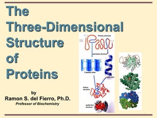
The
Three-Dimensional
Structure
of
Proteins
by
Ramon S. del Fierro, Ph.D.
Professor of Biochemistry
- 2. 4
4-1
© 2003 Thomson Learning, Inc.
All rights reserved
Topic Outline :
Classifications of proteins
Hierarchy of protein structure
Shapes/configuration of proteins
Factors which stabilize protein structure
Denaturation of proteins
Myoglobin
Hemoglobin
- 3. 4
4-1
© 2003 Thomson Learning, Inc.
All rights reserved
Classification of Proteins
Enzymes Essentially all physiological
reactions are catalyzed by
biological catalysts, e.g., amylase
Transport Many molecules and ions are
transported through plasma bound
to proteins, e.g., hemoglobin
Contractile Proteins such as actin and myosin
in muscle cells have the ability to
contract and expand. This gives
the property of motion
- 4. 4
4-1
© 2003 Thomson Learning, Inc.
All rights reserved
Classification of Proteins
Structural The protein collagen is the major
component of tendons, cartilage
and skin. This gives a high tensile
strength to tissues.
Defense In vertebrates, specific proteins
serve as antibodies in the immune
system. Antibodies recognize, complex
with and thus neutralize foreign proteins
in other organisms such as viruses or
bacteria. Toxins such as snake
venoms serve as protective devices for
the organism producing them.
- 5. 4
4-1
© 2003 Thomson Learning, Inc.
All rights reserved
Classification of Proteins
Regulatory Proteins are widely involved in the
regulation and control of metabolism,
enzymatic biosynthesis and nerve
transmission. Metabolism is mediated
by protein hormones such as insulin and
parathyroid hormone. Receptor sites at
nerve synapses are proteins.
Nutrient Some proteins serve as storage forms
of nutrients for a developing organism.
Examples of nutrient proteins are seed
proteins of grain plants; ovalbumin of
egg white ; and casein in milk.
- 6. 4
4-1
© 2003 Thomson Learning, Inc.
All rights reserved
Hierarchy of Protein Structure
β-Pleated Sheet
- 8. 4
4-1
© 2003 Thomson Learning, Inc.
All rights reserved
Protein Structure
• 1° structure: the sequence of amino acids in a
polypeptide chain, read from the N-terminal end
to the C-terminal end
• 2° structure: the ordered 3-dimensional
arrangements (conformations) in localized
regions of a polypeptide chain; refers only to
interactions of the peptide backbone
• e. g., -helix and -pleated sheet
- 9. 4
4-1
© 2003 Thomson Learning, Inc.
All rights reserved
3° and 4° Structure
• Tertiary (3°) structure: the arrangement in space
of all atoms in a polypeptide chain
• it is not always possible to draw a clear distinction
between 2° and 3° structure
• Quaternary (4°) structure: the association of
polypeptide chains into aggregations
Proteins are divided into two large classes
based on their three-dimensional structure
• fibrous proteins
• globular proteins
- 10. 4
4-1
© 2003 Thomson Learning, Inc.
All rights reserved
- is simply a long sequence of amino acid residues combined together
forming a polypeptide chain.
Primary Structure
COOH
Carboxyl
end
- 12. 4
4-1
© 2003 Thomson Learning, Inc.
All rights reserved
- develops when the primary structure of a polypeptide has group
projecting from the N-C-C backbone.
Secondary structure
H-Bonds
- 14. 4
4-1
© 2003 Thomson Learning, Inc.
All rights reserved
-Helix
• coil of the helix is right-handed
• there are 3.6 amino acids per
turn
• repeat distance is 5.4Å
• each peptide bond is s-trans and
planar
• C=O of each peptide bond is
hydrogen bonded to the N-H of
the fourth amino acid away
• C=O----H-N hydrogen bonds are parallel to helical axis
• all R groups point outward from helix
3.6 amino acids
H-Bonds
-R
-R
-R
R-
-R
R-
R-
- 19. 4
4-1
© 2003 Thomson Learning, Inc.
All rights reserved
-Helix
Several factors can disrupt an -helix
• Proline creates a bend because of :
(1) the restricted rotation due to its cyclic structure
(2) its -amino group has no N-H for hydrogen
bonding
• strong electrostatic repulsion caused by the proximity
of several side chains of like charge, e.g., Lys and Arg
or Glu and Asp
• steric crowding caused by the proximity of bulky side
chains, e.g., Val, Ile, Trp
- 20. 4
4-1
© 2003 Thomson Learning, Inc.
All rights reserved
-Helix
without Pro
-Helix
Pro
Pro
Peptide bond -N
- 21. 4
4-1
© 2003 Thomson Learning, Inc.
All rights reserved
If Bulky Groups are adjacent
- there will be steric hindrance
If Similarly charged amino acids
are adjacent
- there will be repulsive forces
- 25. 4
4-1
© 2003 Thomson Learning, Inc.
All rights reserved
-Pleated Sheet
• polypeptide chains lie adjacent to one another; may
be parallel or antiparallel
• R groups alternate, first above and then below plane
• each peptide bond is s-trans and planar
• C=O and N-H groups of each peptide bond are
perpendicular to axis of the sheet
• C=O---H-N hydrogen bonds are between adjacent
sheets and perpendicular to the direction of the sheet
R R
R
R
R R
R
R
R
R R
R
H-Bonds
- 29. 4
4-1
© 2003 Thomson Learning, Inc.
All rights reserved
Three-dimensional form
of the antiparallel
ß-pleated sheet arrangement
H-Bonds
- 31. 4
4-1
© 2003 Thomson Learning, Inc.
All rights reserved
- is when the molecule is further folded and held in a particular complex
shape forming precise and compact structure, unique to that protein.
The shape is maintained permanently by the intra- molecular bonds
Tertiary structure
NH2
COOH
- 32. 4
4-1
© 2003 Thomson Learning, Inc.
All rights reserved
3˚ Structure
• The 3-dimensional arrangement of atoms in the
molecule.
• In fibrous protein, backbone of protein does not fall
back on itself, it is important aspect of 3˚ not specified
by 2˚ structure.
• In globular protein, more information needed. 3D
structure allows for the determination of the way
helical and pleated-sheet sections fold back on each
other.
• Interactions between side chains also plays a role.
- 33. 4
4-1
© 2003 Thomson Learning, Inc.
All rights reserved
Fibrous Proteins
• Fibrous proteins: contain polypeptide chains
organized approximately parallel along a single
axis. They
• consist of long fibers or large sheets
• tend to be mechanically strong
• are insoluble in water and dilute salt solutions
• play important structural roles in nature
• Examples are
• keratin of hair and wool
• collagen of connective tissue of animals including
cartilage, bones, teeth, skin, and blood vessels
- 34. 4
4-1
© 2003 Thomson Learning, Inc.
All rights reserved
Collagen Triple Helix
• consists of three polypeptide chains wrapped around
each other in a ropelike twist to form a triple helix
called tropocollagen; MW approx. 300,000
• 30% of amino acids in each chain are Pro and Hyp
(hydroxyproline); hydroxylysine also occurs
• every third position is Gly and repeating sequences
are X-Pro-Gly and X-Hyp-Gly
• each polypeptide chain is a helix but not an -helix
• the three strands are held together by hydrogen
bonding involving hydroxyproline and hydroxylysine
• with age, collagen helices become cross linked by
covalent bonds formed between Lys and His residues
• deficiency of Hyp results in fragile collagen
- 36. 4
4-1
© 2003 Thomson Learning, Inc.
All rights reserved
Poly(Gly-Pro-Pro),
A collagen-like
right-handed
triple helix,
composed of
three left-handed
helical chains
Proline
H-Bonds
- 37. 4
4-1
© 2003 Thomson Learning, Inc.
All rights reserved
Globular Proteins
• Globular proteins: proteins which are folded to a
more or less spherical shape
• they tend to be soluble in water and salt solutions
• most of their polar side chains are on the outside and
interact with the aqueous environment by hydrogen
bonding and ion-dipole interactions
• most of their nonpolar side chains are buried inside
• nearly all have substantial sections of -helix or
-sheet
• Examples are
• myoglobin
• hemoglobin
- 38. 4
4-1
© 2003 Thomson Learning, Inc.
All rights reserved
Comparison of fibrous and globular proteins
- 39. 4
4-1
© 2003 Thomson Learning, Inc.
All rights reserved
Factors Directing Folding
• Noncovalent interactions, including
• hydrogen bonding between polar side chains,
e.g., Ser and Thr
• hydrophobic interaction between nonpolar side
chains, e.g., Val and Ile
• electrostatic attraction between side chains of
opposite charge, e.g., Lys and Glu
• electrostatic repulsion between side chains of like
charge, e.g., Lys and Arg, Glu and Asp
• Formation of disulfide (-S-S-) bonds between
side chains of cysteines
- 40. 4
4-1
© 2003 Thomson Learning, Inc.
All rights reserved
Forces that stabilize tertiary structure
H-Bonds
- 42. 4
4-1
© 2003 Thomson Learning, Inc.
All rights reserved
3° Structure
• x-ray crystallography
• uses a perfect crystal; that is, one in which all
individual protein molecules have the same 3D
structure and orientation
• exposure to a beam of x-rays gives a series diffraction
patterns
• information on molecular coordinates is extracted by a
mathematical analysis called a Fourier series
• 2-D Nuclear magnetic resonance
• can be done on protein samples in aqueous solution
- 43. 4
4-1
© 2003 Thomson Learning, Inc.
All rights reserved
Tertiary structure of
-lactalbumin
-helix
Pleated Sheet
- 45. 4
4-1
© 2003 Thomson Learning, Inc.
All rights reserved
Myoglobin
• a single polypeptide chain of 153 amino acids
• a single heme group in a hydrophobic pocket
• 8 regions of -helix; no regions of -sheet
• most polar side chains are on the surface
• nonpolar side chains are folded to the interior
• two His side chains are in the interior, involved with
interaction with the heme group
• Fe(II) of heme has 6 coordinates sites; 4 interact with
N atoms of heme, 1 with N of a His side chain, and 1
with either an O2 molecule or an N of the second His
side chain
- 47. 4
4-1
© 2003 Thomson Learning, Inc.
All rights reserved
Heme structure
Protoporphyrin IX
methylene
=CH-
- 48. 4
4-1
© 2003 Thomson Learning, Inc.
All rights reserved
Oxygen-binding
site for myoglobin
The porphyrin ring occupies
four of the six coordination
sites of the Fe(II).
Histidine F8 occupies
the fifth coordination
site of the iron
- 49. 4
4-1
© 2003 Thomson Learning, Inc.
All rights reserved
Oxygen and carbon monoxide binding to the heme group of myoglobin
- 50. 4
4-1
© 2003 Thomson Learning, Inc.
All rights reserved
Hierarchy of Protein Structure
β-Pleated Sheet
- 51. 4
4-1
© 2003 Thomson Learning, Inc.
All rights reserved
Quaternary Structure
• Quaternary (4°) structure: the association of
polypepetide monomers into multisubunit
proteins
• examples
Globular Protein Subunits
Alcohol dehydrogenase 2
Triosephosphate isomerase 2
Aldolase 3
Lactate dehydrogenase 4
Hemoglobin 2 + 2
Pyruvate kinase 4
Insulin 6
- 52. 4
4-1
© 2003 Thomson Learning, Inc.
All rights reserved
Quaternary Structure
• Quaternary (4°) structure: the association of
polypepetide monomers into multisubunit
proteins
• dimers
• trimers
• tetramers
• Noncovalent interactions
• electrostatics, hydrogen bonds, hydrophobic
- 53. 4
4-1
© 2003 Thomson Learning, Inc.
All rights reserved
Quaternary structure
- arise when a number of tertiary polypeptides joined together forming a
complex, biologically active molecule
- 54. 4
4-1
© 2003 Thomson Learning, Inc.
All rights reserved Hemoglobin
Two of - and two
of -chains
- 55. 4
4-1
© 2003 Thomson Learning, Inc.
All rights reserved
Oxygen Binding of Hb
• a tetramer of two -chains (146 amino acids each) and
two -chains (153 amino acids each); 22
• each chain has 1 heme group; hemoglobin can bind up
to 4 molecules of O2
• binding is cooperative; when one O2 is bound, it
becomes easier for the next O2 to bind
• the function of hemoglobin is to transport oxygen
• the structure of oxygenated Hb is different from that of
unoxygenated Hb
• H+, CO2, Cl-, and 2,3-bisphosphoglycerate (BPG) affect
the ability of Hb to bind and transport oxygen
- 58. 4
4-1
© 2003 Thomson Learning, Inc.
All rights reserved
Oxygen Binding of Hb
• The effect of pH on the oxygen-binding ability of
Hb is called the Bohr effect
as pH decreases (more acidic), oxygen is released
• CO2 promotes release of O2 from HbO2
HbO2 HbH+
+ H+
O2
+
CO2 + H2 O
carbonic
anhydrase
H2 CO3
H2 CO3 H+
+ HCO3
-
- 59. 4
4-1
© 2003 Thomson Learning, Inc.
All rights reserved
Oxygen Binding of Hb
Release of oxygen influenced by carbon dioxide
- 60. 4
4-1
© 2003 Thomson Learning, Inc.
All rights reserved
Oxygen Binding of Hb
Summary of the Bohr effect
Lungs Actively Metabolizing Muscle
Higher pH than actively
metabolizing tissue
Hemoglobin binds O 2
Hemoglobin releases H
+
Lower pH due to production of H
+
Hemoglobin releases O2
Hemoglobin binds H
+
- 61. 4
4-1
© 2003 Thomson Learning, Inc.
All rights reserved Oxygen saturation curves for myoglobin and Hb
at five different pH values
As pH is increased,
percent saturation is
increased
As pH is dereased,
percent saturation is
dereased
As pH is decreased,
oxygen saturation curve
shifts to the right
Hemoglobin
- 62. 4
4-1
© 2003 Thomson Learning, Inc.
All rights reserved
Hemoglobin (Hb)
• Hemoglobin in blood is bound to BPG
• interaction is electrostatic, between negative
charges on BPG and positive side chains
(e.g., Lys, Arg) of hemoglobin
• BPG promotes O2 dissociation
• Hb stripped of BPG remains saturated with O2
C
C
O-
O
CH2 OPO3
2 -
OPO3
2 -
H
2,3-Bisphosphoglycerate
(BPG)
- 65. 4
4-1
© 2003 Thomson Learning, Inc.
All rights reserved
Fetal Hemoglobin, Hb F
• has a higher affinity for O2 than maternal Hb A
• structure is 2g2
• binds less strongly to BPG that does Hb A
Oxygen binding capacity of Hb F
- 67. 4
4-1
© 2003 Thomson Learning, Inc.
All rights reserved
Abnormal Human Hb
• Hb S: substitution of Val for Glu at 26
• Hb E: Glu B8(26) -> Lys; change is on the surface and
has little effect on Hb stability or function
• Hb Savannah: Gly B6(24) -> Val; not enough room for
Val between B-helix and E-helix which disrupts entire
structure
• Hb Bibba: Leu H19(136) -> Pro; proline disrupts the
H-helix
• Hb M Iwate: His F8(87) -> Tyr; blood contains
methemoglobin and blood is chocolate brown
• Hb Milwaukee: Val E11(67) -> Glu; glutamate side
chain forms an ion pair with heme iron which
stabilizes Fe(III) and prevents O2 binding
- 68. 4
4-1
© 2003 Thomson Learning, Inc.
All rights reserved
Defects from Hemoglobin Mutations
1. Weakened heme binding.
2. Disruption of secondary structure.
3. Disruption of quaternary structure.
4. Defective oxygen transfer.
5. Altered affinity for oxygen.
6. Oxidation of Fe(II) to Fe(III).
7. Aggregation in the T state (Hemoglobin S). Sickle cell
anemia results from aggregation of Hb into insoluble
fibers causing mishapen blood cells that cannot pass
through capillaries and block blood flow to tissues.
- 70. 4
4-1
© 2003 Thomson Learning, Inc.
All rights reserved
• The oxygenated molecules are soluble, but upon de-
oxygenation, the conformation of HbS differs
considerably from HbA, and it aggregates into insoluble
fibers.
• These fibers deform the RBCs into spiny or sickle-
shaped cells.
A genetic disease resulting from a
mutation that converts Glu6 (acidic)
in the -chains to Val (nonpolar).
This substitution creates
hydrophobic “sticky” patches on the
normally charged surface of the -
chains.
Sickle-cell anemia
- 72. 4
4-1
© 2003 Thomson Learning, Inc.
All rights reserved
The gene defect is a known mutation of a single nucleotide (A to
T) of the β-globin gene, which results in glutamate being substituted by
valine at position 6. Hemoglobin S with this mutation are referred to as
HbS, as opposed to the normal adult HbA.
Transversion type of mutation
- 74. 4
4-1
© 2003 Thomson Learning, Inc.
All rights reserved
Carbon Monoxide Poisoning
• Heme Fe(II) binds many other small molecules with
structures similar to O2 including: CO, NO, H2S
• O2 is actually a fairly weak binder relative to these
other molecules, particularly CO.
• When exposed to CO, even at low concentrations, O2
transport proteins will be filled with CO limiting their
vital O2 capacity.
- 76. 4
4-1
© 2003 Thomson Learning, Inc.
All rights reserved
Denaturation
• Denaturation: the loss of the structural order (2°,
3°, 4°, or a combination of these) that gives a
protein its biological activity; that is, the loss of
biological activity
• Denaturation can be brought about by
• heat
• large changes in pH, which alter charges on side
chains, e.g., -COO- to -COOH or -NH
+ to -NH
• detergents such as sodium dodecyl sulfate (SDS)
which disrupt hydrophobic interactions
• urea or guanidine, which disrupt hydrogen bonding
• mercaptoethanol, which reduces disulfide bonds
- 78. 4
4-1
© 2003 Thomson Learning, Inc.
All rights reserved
Denaturation and
refolding in
ribonuclease
Several ways to denature
proteins
• Heat
• pH
• Detergents
• Urea
• Guanadine hydrochloride
- 79. 4
4-1
© 2003 Thomson Learning, Inc.
All rights reserved
SUMMARY
• Proteins may be classified on the basis of the
solubility, shape, or function or of the presence
of a prosthetic group such as heme.
• Proteins perform complex physical and catalytic
functions by positioning specific chemical
groups in a precise three-dimensional
arrangement that is both functionally efficient
and physically strong.
• The hierarchy of proteins depend on the forces
which stabilize them
- 80. 4
4-1
© 2003 Thomson Learning, Inc.
All rights reserved
• Tertiary structure concerns the relationships
between secondary structural domains.
• Quaternary structure of proteins with two or
more polypeptides (oligomeric proteins) is a
feature based on the spatial relationships
between various polypeptides
• Primary structures are stabilized by covalent
peptide bonds
• Higher order structures are stabilized by weak
forces: multiple H bonds, salt (electrostatic)
bonds and association of hydrophobic R groups
- 81. 4
4-1
© 2003 Thomson Learning, Inc.
All rights reserved
• Myoglobin is monomeric; hemoglobin is a
tetramer of two subunit types. Despite having
different primary structures, myoglobin and the
subunits of hemoglobin have nearly identical
secondary and tertiary structures.
• The O2-binding curve for myoglobin is
hyperbolic, but for hemoglobin is sigmoidal, a
consequence of cooperative interactions in the
tetramer.
- 83. 4
4-1
© 2003 Thomson Learning, Inc.
All rights reserved
… hope you have learned something!
RAMON S. DEL FIERRO, Ph.D. (Tokyo)
All the Best!