
Recommended
More Related Content
What's hot
What's hot (20)
Similar to Chymotrypsin Serine Protease Mechanism
Similar to Chymotrypsin Serine Protease Mechanism (20)
Recently uploaded
Recently uploaded (20)
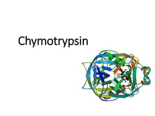
Chymotrypsin Serine Protease Mechanism
- 1. Chymotrypsin
- 2. The presentation includes: • Introduction to SERINE PROTEASES • Chymotrypsin: Overview • History • Properties • Occurrence • Functions • Side Effects • Structure and Active site • Discovery of active site residues • Activation from precursors • Kinetics • Mechanism of Action • Catalytic triad • Acylation • Deacylation • pH curve • Inhibition • Diseases & Disorders
- 3. Introduction to SERINE PROTEASES 67% 33% Proteases Serine Proteases Others Enzyme Cleavage site (C-terminal) Chymotrypsin aromatic residues- tyrosine, tryptophan, phenylalanine Trypsin basic residues - lysine & arginine Elastase smaller neutral residues- valine, glycine, alanine • Enzymes that cleave peptide bonds in proteins; serine serves as nucleophilic amino acid at enzyme’s active site. • Account for 1/3rd of all proteolytic enzymes • Some common examples :
- 5. Serine Protease Family • Serine Proteases I. Chymotrypsin II. Trypsin III. Elastase • Similarity I. Similar 3D structure II. Catalytic triad III. Oxyanion hole IV. Covalent acyl-enzyme intermediate V. Secreted by pancreases as inactive precursors Superposition of Trypsin (yellow), Elastase (green), and Chymotrypsin (blue) Backbones Image reference: http://chemistry.umeche.maine.edu/CHY252/Peptidase3.html
- 6. Specificity Difference of Chymotrypsin, Trypsin, and Elastase • Substrate specificity • Chymotrypsin: aromatic or bulky nonpolar side chain • Trypsin: basic residues; Lys or Arg • Elastase: smaller & uncharged side chains
- 7. • Small structural difference in the binding site explains the substrate specificity nonpolar pocket Asp (negatively charged) vs. Ser in Chymotrypsin no pocket present as two Gly in chymotrypsin are replaced by Val and Thr
- 8. Chymotrypsin • Bovine pancreatic chymotrypsin (Mr 25,191), a protease that catalyzes hydrolytic cleavage of peptide bonds. • Specific for peptide bonds adjacent to aromatic amino acid residues (Trp, Phe, Tyr).
- 9. History Early 1900s • Vernon proposed that pancreatic preparations could give rise to an intrinsic activator of its own enzyme 1902 • Vernon’s milk-clotting experiments determined there were at least two enzymes present and that one was more stable than the other 1938 • Kunitz isolated different active forms of chymotrypsin - alpha, beta, gamma 1942 • Fruton and Bergmann further studied the specificity of chymotrypsin, reporting on several new substrates 1947 • Jacobsen soon identified additional forms of chymotrypsin, designating them as delta and pi 1948 • Schwert further characterized the molecular weights of chymotrypsin and chymotrypsinogen 1954 • 1st evidence for 3-step mechanism of chymotrypsin hydrolyzing amide and ester substrates was reported on by Hartley and Kilby
- 10. Properties of Chymotrypsin: • Molecular Weight : 25.6 kDa • Optimal pH : 7.8-8.0 • Optimal Temperature : 40-50 °C • Isoelectric Point : 8.52 (Chymotrypsinogen, Theoretical) 8.33 (Chymotrypsin, Theoretical) • Extinction Coefficient : 51,840 cm-1 M-1 (Theoretical)
- 11. Occurrence of Chymotrypsin: • Digestive enzyme component of pancreatic juice • Pancreatic Acinar cells filled with granules, contain the digestive enzymes(secreted in an inactive form). • When released into the duodenum, they are activated by the enzyme enter peptidase present in the lining of the duodenum, performs proteolysis. Image Reference: http://pathology.jhu.edu/pancreas/basicoverview3.php?area= ba
- 12. Functions • Aid in digestion. • Treat inflammation and reduce swelling (i.e., soft tissue injuries, acute traumatic injuries, sprains, infections, edema of the eyelids and genitalia, muscle cramps, and sports injuries). • Liquefy mucus secretions. • Kill enterozoic worms and other parasites in the digestive tract. • Alleviate effects of chemotherapy. • Act as wound cleaner.
- 13. Chymotrypsin as medicine It is taken orally, inhaled, injected or applied to skin. Used to treat: • Ulcers • shingles and acne. • surgical or traumatic injuries • necrotic tissue • help loosen phlegm in asthma, bronchitis, lung diseases, and sinus infections. • Wound . • Fracture and burn treatments • Arthritis and such other autoimmune diseases as lupus, scleroderma, and multiple sclerosis. • Pelvic inflammatory diseases shingles
- 14. Side-effects: • Temporary side effects: changes in the color, consistency, and odor of the stool. • gastrointestinal disturbances, such as, a feeling of fullness, • Diarrhea • Constipation • Nausea • With high doses, minor allergic reactions like reddening of the skin may occur • Corneal edema Redness on face
- 15. Structure and active site: • 247-amino acid residues • 3 peptide chains • 4 disulfide bridges • 14% α-helix and 45% β- conformation present in peptides. • folded into two domains, beta strands arranged as antiparallel sheets which form a circular structure known as a beta barrel. • Catalytic triad - His57 and Ser195 located at substrate binding site along with Asp102.
- 16. Identification of active site residues: • When Dixon & Neurath treated Chymotrypsin with diisopropylphosphofluoridate (DIPF), it became inactivate. Since only serine-195 was modified by diisopropylphosphofluoridate, it indicates that Serine-195 plays the crucial role in the mechanism as a nucleophile. • Similarly, Histidine-57 was identified using chemical affinity labelling.TPCK (N-tosyl l-phenylalanyl chloromethyl ketone), specifically reacted with histidine 57, in chymotrypsin. • Asp-102 was identified using X-ray Crystallography.
- 17. Reaction type • Chymotrypsin operates through a ping-pong mechanism called covalent hydrolysis. • Enzyme first forms covalent bond with target substrate, displacing more stable moiety into solution. • Enzyme-substrate complex is called enzyme intermediate. • Intermediate reacts with water, which displaces the remaining part of the initial substrate and reforms the initial enzyme.
- 18. Kinetics • Experiments were conducted in 1953 by B.S. Hartley and B.A. Kilby to investigate the kinetics of chymotrypsin-catalyzed hydrolysis. Instead of using a poly-peptide chain as a substrate, they used a nitro-phenyl ester, p-nitrophenyl acetate, that resembles an aromatic amino acid. Hydrolysis of this compound by chymotrypsin at the carbonyl group yields acetate and nitrophenolate, the latter of which absorbs near 400 nm light and its concentration can thus be measured by spectrophotometry • This can only be explained by the fact that hydrolysis by chymotrypsin is biphasic in nature, meaning that it proceeds in two distinct steps. • The first step, which describes the initial burst of nitrophenolate seen in Hartley and Kilby’s absorbance plot, is the fastest. The attack of the nitrophenyl acetate substrate by chymotrypsin immediately cleaves the nitrophenolate moiety and leaves the acetate group attached to chymotrypsin, rendering the enzyme inactive. • The second step has been deduced to involve the hydrolysis of the acetate group from the inactivated chymotrypsin to regenerate the original enzyme.
- 19. pH curve • Enzymatic activity is greatest when the solution pH is between 7 and 8.5 due to the ionization states of two key residues: His57 and Ile16 • pH-dependences of chymotrypsin with N-acetyl- tryptophanamide and the azoalbumin as substrates. The similarity of the pH-profiles would suggest the pH range between pH 7 and 8.5 as the general optimum pH for chymotrypsin, regardless of the kind of substrate which it acts upon. http://www.athenaes.com/tech_brief_protease.php
- 21. Mechanism of action • Covalent catalysis of chymotrypsin basically goes through acylation and deacylation. Acylation forms the acyl enzyme intermediate and the deacylation adds water which produces a free enzyme. • Catalytic mechanism involves serine residues • Utilizes catalytic triad
- 22. Catalytic triad • Asp102 – His57 – Ser195 • Ser provides nucleophile (O atom) • His acts as base catalyst to activate Ser • Asp stabilizes protonated His • 2-step reaction
- 24. Tetrahedral Intermediate Attack on substrate
- 25. Acyl-enzyme Intermediate (covalent intermediate) • Bond cleaved • Rc portion released • Rest of the peptide covalently bonded to peptide
- 26. Intruder Alert!! (H2O enters the scene)
- 27. Tetrahedral Intermediate (II) Relieving the Tension
- 28. Regeneration
- 36. Oxyanion Hole • An oxyanion hole is a pocket in the active site of an enzyme that stabilizes transition state negative charge on a deprotonated oxygen or alkoxide. • The pocket typically consists of backbone amides or positively charged residues. Stabilizing the transition state lowers the activation energy necessary for the reaction, and so promotes catalysis. • In chymotrypsin, the amide hydrogens (-N-H) of Ser195 and Gly193 form an oxyanion hole which,
- 37. Through hydrogen bonding, stabilizes the tetrahedral intermediate to some extent.
- 38. Inhibition • Enzymes are secreted in inactive form (as proenzymes) so they do not digest the pancreas. The pancreas secretes an inhibitor to ensure that enzymes are not activated too early. • When the pancreatic juice reaches small intestine, enzymes become activated. However, self-digestion can occur if the pancreatic duct becomes blocked or if the pancreas is damaged. The proenzymes can overwhelm the inhibitor, causing the enzymes to become active while in the pancreas. This condition, called acute pancreatitis , can result in a lifetime of pancreatic insufficiency. • Certain inhibitors resemble the tetrahedral intermediate, and thus fill up the active site, preventing the enzyme from working properly. • Serine proteases are paired with serine protease inhibitors, which turn off their activity when they are no longer needed.
- 39. • Serine proteases are inhibited by a diverse group of inhibitors, including synthetic chemical inhibitors for research or therapeutic purposes, and also natural proteinaceous inhibitors. • Family of natural inhibitors called "serpins" can form a covalent bond with serine protease, inhibiting its function. • Best-studied serpins are antithrombin and alpha 1- antitrypsin, studied for their role in coagulation/thrombosis and emphysema/A1AT, respectively. • Artificial irreversible small molecule inhibitors include AEBSF and PMSF.
- 41. Chymotrypsin deficiency Signs and symptoms: • Belly (abdominal) pain. • Weight loss. • Nausea. • Vomiting. • Diarrhoea. • Low blood pressure. • Rapid heartbeat. • Recurrent pancreatitis Causes of Pancreatic Enzyme Deficiency • Acute and chronic pancreatitis. • Surgical removal of pancreas. • Pancreatic cancer. • Cystic fibrosis. • Stomach ulcers. • Crohn’s disease. • Autoimmune disorder. • Obstruction due to gall stone. • Zollinger-Ellison syndrome • Shwachman-Bodian-Diamond syndrome, Additionally, people who are carriers of a mutation in the CFTR gene may have pancreatic insufficiency and experience the associated signs and symptoms. (See the article on CFTR Gene Mutation Testing to learn more about carriers.) In children, it is most frequently associated with cystic fibrosis (CF) or Shwachman-Diamond Syndrome (SDS). SDS is the second most common cause of inherited pancreatic insufficiency, after CF. All those with SDS have some degree of pancreatic insufficiency beginning at infancy.
- 42. Chymotrypsin test(Diagnosis) • This test measures the amount of chymotrypsin in stool to help evaluate whether someone's pancreas is functioning properly. • Chymotrypsinogen, the inactive precursor of chymotrypsin, is produced in the pancreas and transported to the small intestine. In the small intestine, it is activated to form chymotrypsin. It is one of the enzymes responsible for breaking down the protein in food into smaller pieces, called peptides. Chymotrypsin is detectable in the stool if the pancreas is functioning normally. • Individuals with pancreatic dysfunction may either have blocked pancreatic ducts or the cells that produce chymotrypsinogen may be damaged or destroyed. Such cell damage and duct blockage cause pancreatic insufficiency because the amount of enzymes transported to the small intestine is inadequate for proper food digestion. This is often seen in conditions such as chronic pancreatitis and sometimes pancreatic cancer. • A fresh stool sample, uncontaminated with urine, is collected • If you are taking pancreatic enzymes, you may be instructed to discontinue taking the enzymes 5 days before providing the stool sample
- 43. • A positive result, indicating the presence of chymotrypsin in the stool, is normal. Chymotrypsin is present in the stool of healthy individuals. • A negative result may mean that the person tested has pancreatic insufficiency. It is not diagnostic, but it does indicate that further testing may be indicated. • The probability of PEI in CP can also be estimated based on pancreatic imaging findings in the absence of more advanced tests of pancreatic function. Notably, ductal changes on endoscopic retrograde pancreatography, computerized tomography (CT) and endoscopic ultrasound (EUS) have been associated with decreased exocrine pancreatic function. The diagnosis of CP by EUS is based on the demonstration of several different parenchymal (hyperechoic foci, hyperechoic strands, parenchymal lobularity and cysts) and ductal (pancreatic duct dilatation, irregular pancreatic duct contour, hyperechoic pancreatic duct margin, dilated side branches and intraductal calcifications) abnormalities defined in the Rosemont classification. • Direct pancreatic function test. The latter being a limitedly used test that assesses exocrine function in the pancreas by inserting a tube into the small intestine to collect pancreatic secretions.
- 44. Treatment: • Pancreatic enzyme replacement therapy • Diet, smoking and drinking Fun fact: Pancreatic enzyme replacement therapy (PERT) medications are made from the pancreas of pigs
- 45. References • Serine Proteases http://proteopedia.org/wiki/index.php/Serine_Proteases • Chymotrypsin I.U.B.: 3.4.21.1 http://www.worthington- biochem.com/chy/default.html • Dodson, G; Wlodawer, A (September 1998). "Catalytic triads and their relatives". Trends in Biochemical Sciences. 23 (9): 347– 52. doi:10.1016/S0968-0004(98)01254-7. PMID 9787641. • Buller, AR; Townsend, CA (Feb 19, 2013). "Intrinsic evolutionary constraints on protease structure, enzyme acylation, and the identity of the catalytic triad". Proceedings of the National Academy of Sciences of the United States of America. 110 (8): E653– 61. Bibcode:2013PNAS..110E.653B. doi:10.1073/pnas.1221050110. PMC 3581919 . PMID 23382230. • Stryer L, Berg JM, Tymoczko JL (2002). "9 Catalytic Strategies". Biochemistry (5th ed.). San Francisco: W.H. Freeman. ISBN 0- 7167-4955-6.
- 46. Presenters • Aayush Srivastava (17/06012) • Harsh Raheja (17/06013) • Vikram Aditya (17/06014) • Mansi Tanwar (17/06016) • Ujjwal Goyal (17/06018) • Simran (17/06047)
- 47. Thank you...