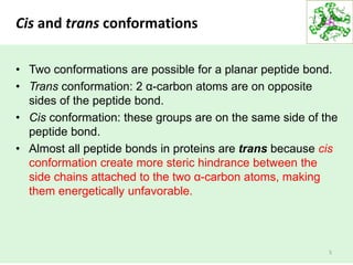
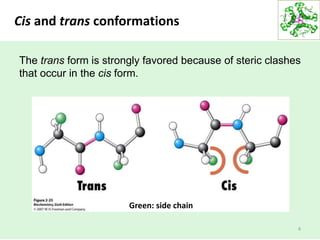
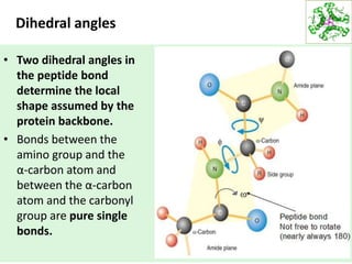
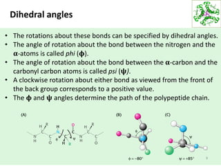
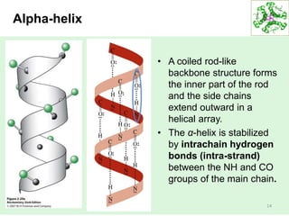
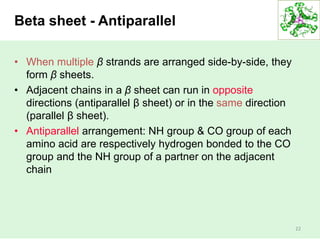
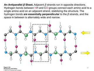
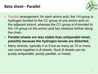
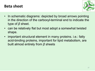
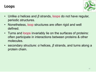
SlideShare a Scribd company logo
HBC1011 Biochemistry I
Trimester I, 2018/2019
Lecture 7 – Protein structure and
function
Ng Chong Han, PhD
MNAR1010, 06-2523751
chng@mmu.edu.my
1
Overview
• Conformation of the peptide group
• Secondary structures
– Alpha helix
– Beta sheet
– Turns and loops
2
The conformation of the peptide group
• The structure of peptides are flexible and yet
conformationally restricted
• Geometry of protein backbone:
– peptide bond is essentially planar
– 6 atoms lie in the same plane
• Nature of the chemical bonding:
– peptide bond has considerable double-bond character, it
is restricted to one of two possible conformations, either
trans or cis.
– constrains the conformation of the peptide backbone and
accounts for the bond's planarity.
3
Peptide Bonds Are Planar. In a pair of linked amino acids, six atoms (Cα, C, O, N, H,
and Cα) lie in a plane. Side chains are shown as green balls.
1
2
3
4
5
6
4
Cis and trans conformations
• Two conformations are possible for a planar peptide bond.
• Trans conformation: 2 α-carbon atoms are on opposite
sides of the peptide bond.
• Cis conformation: these groups are on the same side of the
peptide bond.
• Almost all peptide bonds in proteins are trans because cis
conformation create more steric hindrance between the
side chains attached to the two α-carbon atoms, making
them energetically unfavorable.
5
Cis and trans conformations
6
The trans form is strongly favored because of steric clashes
that occur in the cis form.
Green: side chain
Dihedral angles
• Two dihedral angles in
the peptide bond
determine the local
shape assumed by the
protein backbone.
• Bonds between the
amino group and the
α-carbon atom and
between the α-carbon
atom and the carbonyl
group are pure single
bonds.
7
Dihedral angles
• The two adjacent rigid
peptide units may
rotate about these
bonds, taking on
various orientations.
• This freedom of
rotation about two
bonds of each amino
acid allows proteins to
fold in many different
ways.
8
Dihedral angles
9
• The rotations about these bonds can be specified by dihedral angles.
• The angle of rotation about the bond between the nitrogen and the
𝛂-atoms is called phi (𝛟).
• The angle of rotation about the bond between the 𝛂-carbon and the
carbonyl carbon atoms is called psi (𝛙).
• A clockwise rotation about either bond as viewed from the front of
the back group corresponds to a positive value.
• The 𝛟 and 𝛙 angles determine the path of the polypeptide chain.
Ramachandran diagram
10
Are all combination of 𝛟 and 𝛙 possible?
G. N. Ramachandran recognized that many combinations are
forbidden because of steric collisions between atoms. The values
can be visualized on a 2D plot called Ramachandran diagram.
Three-quarters of the possible (𝛟 and 𝛙) combinations are
excluded simply by local steric clashes.
The most favorable regions are
shown in dark green; borderline
regions are shown in light
green. The structure on the
right is disfavored because of
steric clashes.
11
Secondary structure
• Protein secondary structure can be described by the hydrogen-
bonding pattern of the peptide backbone of the protein.
• The most common 2nd structures: alpha (α) helices and beta (β)
sheets/beta strands
• Other 2nd structures: β turn, omega (Ω) loop
• Alpha helices, beta strands and turns are formed by a regular
pattern of hydrogen bonds between the peptide N-H and C=O
groups of amino acids that near each one another in the linear
sequence.
12
• Linus Carl Pauling (February 28, 1901 –
August 19, 1994): American quantum chemist
and biochemist.
• Two Nobel prizes winner
• Awarded the Nobel Prize in chemistry for his
work describing the nature of chemical bonds.
The elucidation of the structure
of the α-helix is a landmark in
biochemistry because it
demonstrated that the
conformation of a polypeptide
chain can be predicted if the
properties of its components
are rigorously and precisely
known. 13
Alpha-helix
• A coiled rod-like
backbone structure forms
the inner part of the rod
and the side chains
extend outward in a
helical array.
• The α-helix is stabilized
by intrachain hydrogen
bonds (intra-strand)
between the NH and CO
groups of the main chain.
14
Alpha-helix
Hydrogen-Bonding Scheme for an α-helix. In the α
helix, the CO group of residue n forms a hydrogen
bond with the NH group of residue n+ 4.
15
Alpha-helix
• Each residue is related to the next one by
a rise of 1.5 Å (angstrom, 1Å = 0.1nm)
along the helix axis and a rotation of 100º,
which gives 3.6 amino acid residues per
turn of helix.
• Amino acids spaced three and four apart
in the sequence are spatially quite close to
one another in an α-helix.
16
Alpha-helix
• The Ramachandran diagram (for
visualization of dihedral angles)
reveals that both right-handed
and the left handed helices are
possible conformations.
• However, essentially all α helices
in proteins are right-handed.
• Right-handed helices are
energetically more favorable
because there is less steric clash
between the side chains and the
backbone.
17
Schematic Views of α-Helices. (A) A ball-and-stick model. (B) A ribbon
depiction. (C) A cylindrical depiction. 18
A Largely α-Helical Protein. Ferritin, an iron-storage protein,
is built from a bundle of α helices.
19
Beta structure : beta strand
• β structure : β strands and β sheets
• A polypeptide chain, called a β strand is almost fully
extended rather than being tightly coiled as in the α helix.
• Beta sheet are stabilized by hydrogen bonding between
polypeptide strands (inter-strand).
• The distance between adjacent amino acids along a β
strand is approximately 3.5 Å
• The side chains of adjacent amino acids point in opposite
directions.
• β strands are rare because they are conformationally less
stable. However, when two adjacent β strands line up they
can form hydrogen bonds. This creates a β sheet.
20
Structure of a β-Strand. The side chains are alternately
above and below the plane of the strand.
7 Å
21
Beta sheet - Antiparallel
• When multiple β strands are arranged side-by-side, they
form β sheets.
• Adjacent chains in a β sheet can run in opposite
directions (antiparallel β sheet) or in the same direction
(parallel β sheet).
• Antiparallel arrangement: NH group & CO group of each
amino acid are respectively hydrogen bonded to the CO
group and the NH group of a partner on the adjacent
chain
22
An Antiparallel β Sheet. Adjacent β strands run in opposite directions.
Hydrogen bonds between NH and CO groups connect each amino acid to a
single amino acid on an adjacent strand, stabilizing the structure. The
hydrogen bonds are essentially perpendicular to the β strands, and the
space in between is alternately wide and narrow.
23
Beta sheet - Parallel
• Parallel arrangement: for each amino acid, the NH group is
hydrogen bonded to the CO group of one amino acid on
the adjacent strand, whereas the CO group is H-bonded to
the NH group on the amino acid two residues farther along
the chain.
• Parallel sheets are less stable than antiparallel sheet,
possibly because the hydrogen bonds are distorted.
• Many strands, typically 4 or 5 but as many as 10 or more,
can come together in β sheets. Such β sheets can be
purely antiparallel, purely parallel, or mixed.
24
A Parallel β Sheet. Adjacent β strands run in the same direction.
Hydrogen bonds connect each amino acid on one strand with two
different amino acids on the adjacent strand. The hydrogen bonds are
evenly spaced but slanted.
25
Structure of a Mixed β Sheet 26
Beta sheet
• In schematic diagrams: depicted by broad arrows pointing
in the direction of the carboxyl-terminal end to indicate the
type of β sheet.
• can be relatively flat but most adopt a somewhat twisted
shape.
• important structural element in many proteins. i.e.: fatty
acid-binding proteins, important for lipid metabolism, are
built almost entirely from β sheets
27
A Twisted β Sheet. (A) A ball-and-stick model. (B) A schematic model. (C)
The schematic view rotated by 90 degrees to illustrate the twist more
clearly. 28
A Protein Rich in β Sheets.
The structure of a fatty acid-
binding protein.
29
Polypeptide chains can change
direction by making reverse turns & loops
• Most proteins required reversals in the direction of their
polypeptide chains.
• Reversals: reverse turn (also known as the β turn or hairpin
bend)
• Reverse turns: CO group of residue i of a polypeptide is H-
bonded to the NH group of residue i + 3.
• This interaction stabilizes abrupt changes in direction of the
polypeptide chain.
30
Structure of a
Reverse Turn.
The CO group of
residue i of the
polypeptide chain is
hydrogen bonded to
the NH group of
residue i + 3 to
stabilize the turn.
H bond stabilizes
the bend.
31
Loops
• Unlike α helices and β strands, loops do not have regular,
periodic structures.
• Nonetheless, loop structures are often rigid and well
defined.
• Turns and loops invariably lie on the surfaces of proteins:
often participate in interactions between proteins & other
molecules.
• secondary structure: α helices, β strands, and turns along a
protein chain.
32
Loops on a Protein Surface.
A part of an antibody molecule
has surface loops that mediate
interactions with other
molecules.
33
Amino acid have different propensities for
forming α helices, β sheets and turns
• Amino acids vary in their ability to form 2nd structure
elements.
• Proline and glycine are sometimes known as "helix
breakers" because they disrupt the regularity of the α
helical backbone conformation.
• Alanine, glutamate, and leucine: α helices
• Valine & isoleucine: β strands.
• Glycine, asparagine, and proline: turns
34
35
Amino acid
preference:
secondary
structure

More Related Content

What's hot

Biochemistry of DNA Structure
Biochemistry of DNA StructureBiochemistry of DNA Structure
Biochemistry of DNA Structure
Meghaj Mallick
 
Biochemistry - Ch4 protein structure , and function
Biochemistry - Ch4 protein structure , and function Biochemistry - Ch4 protein structure , and function
Biochemistry - Ch4 protein structure , and function
Areej Abu Hanieh
 
Membrane Proteins & its types
Membrane Proteins & its typesMembrane Proteins & its types
Membrane Proteins & its types
Sabahat Ali
 
Structural organisation of protiens
Structural organisation of protiensStructural organisation of protiens
Structural organisation of protiens
Vikas CJ Vikki
 
Lipids Chemistry Structure & Function (More Detailed)
Lipids Chemistry Structure & Function (More Detailed)Lipids Chemistry Structure & Function (More Detailed)
Lipids Chemistry Structure & Function (More Detailed)
hafizayyub
 
Ramachandran plot- biochemistry
Ramachandran plot- biochemistryRamachandran plot- biochemistry
Ramachandran plot- biochemistry
AanchalManchanda4
 
Protein folding
Protein foldingProtein folding
Protein folding
ANANT MOHAN SHAMA
 
Protein Structures
Protein StructuresProtein Structures
Protein Structures
LOGESWARAN KA
 
Beta
BetaBeta
Conformational properties of polysaccharide and polynucleotide
Conformational properties of polysaccharide and polynucleotide Conformational properties of polysaccharide and polynucleotide
Conformational properties of polysaccharide and polynucleotide
KAUSHAL SAHU
 
Protein structure
Protein structureProtein structure
Protein structure
ranjani n
 
Protein folding slids
Protein folding slidsProtein folding slids
Protein folding slids
anam tariq
 
RAMACHANDRAN PLOT
RAMACHANDRAN PLOTRAMACHANDRAN PLOT
RAMACHANDRAN PLOT
SabhyataChaubey
 
Secondary structural elements & ramachandran plot
Secondary structural elements & ramachandran plotSecondary structural elements & ramachandran plot
Secondary structural elements & ramachandran plot
Prasanthperceptron
 
Proetin Tertiary Structure
Proetin Tertiary StructureProetin Tertiary Structure
Proetin Tertiary Structure
Sabahat Ali
 
Glycoproteins
GlycoproteinsGlycoproteins
Glycoproteins
Dr Nishtha Wadhwa
 
Confirmation of nucleic acid
Confirmation of nucleic acidConfirmation of nucleic acid
Confirmation of nucleic acid
lashikam1
 
Ramachandran Plot
Ramachandran PlotRamachandran Plot
Ramachandran Plot
Nishanth S
 
Forces stabilising structure of proteins
Forces stabilising structure of proteinsForces stabilising structure of proteins
Forces stabilising structure of proteins
Raviz Prathyusha
 

What's hot (20)

Biochemistry of DNA Structure
Biochemistry of DNA StructureBiochemistry of DNA Structure
Biochemistry of DNA Structure
 
Biochemistry - Ch4 protein structure , and function
Biochemistry - Ch4 protein structure , and function Biochemistry - Ch4 protein structure , and function
Biochemistry - Ch4 protein structure , and function
 
Membrane Proteins & its types
Membrane Proteins & its typesMembrane Proteins & its types
Membrane Proteins & its types
 
Structural organisation of protiens
Structural organisation of protiensStructural organisation of protiens
Structural organisation of protiens
 
Lipids Chemistry Structure & Function (More Detailed)
Lipids Chemistry Structure & Function (More Detailed)Lipids Chemistry Structure & Function (More Detailed)
Lipids Chemistry Structure & Function (More Detailed)
 
Ramachandran plot- biochemistry
Ramachandran plot- biochemistryRamachandran plot- biochemistry
Ramachandran plot- biochemistry
 
Protein folding
Protein foldingProtein folding
Protein folding
 
Protein Structures
Protein StructuresProtein Structures
Protein Structures
 
Beta
BetaBeta
Beta
 
Ramachandran plot
Ramachandran plotRamachandran plot
Ramachandran plot
 
Conformational properties of polysaccharide and polynucleotide
Conformational properties of polysaccharide and polynucleotide Conformational properties of polysaccharide and polynucleotide
Conformational properties of polysaccharide and polynucleotide
 
Protein structure
Protein structureProtein structure
Protein structure
 
Protein folding slids
Protein folding slidsProtein folding slids
Protein folding slids
 
RAMACHANDRAN PLOT
RAMACHANDRAN PLOTRAMACHANDRAN PLOT
RAMACHANDRAN PLOT
 
Secondary structural elements & ramachandran plot
Secondary structural elements & ramachandran plotSecondary structural elements & ramachandran plot
Secondary structural elements & ramachandran plot
 
Proetin Tertiary Structure
Proetin Tertiary StructureProetin Tertiary Structure
Proetin Tertiary Structure
 
Glycoproteins
GlycoproteinsGlycoproteins
Glycoproteins
 
Confirmation of nucleic acid
Confirmation of nucleic acidConfirmation of nucleic acid
Confirmation of nucleic acid
 
Ramachandran Plot
Ramachandran PlotRamachandran Plot
Ramachandran Plot
 
Forces stabilising structure of proteins
Forces stabilising structure of proteinsForces stabilising structure of proteins
Forces stabilising structure of proteins
 

Similar to 219102 lecture 7

structureofproteins-161119045143.pptx
structureofproteins-161119045143.pptxstructureofproteins-161119045143.pptx
structureofproteins-161119045143.pptx
abdulahad563527
 
Structure of proteins
Structure of proteinsStructure of proteins
Structure of proteins
Devyani Joshi
 
Lec4 protein structure aimec
Lec4 protein structure aimecLec4 protein structure aimec
Lec4 protein structure aimec
Shamim Akram
 
Lec4 proteinstructure
Lec4 proteinstructureLec4 proteinstructure
Lec4 proteinstructure
DrShamimAkram
 
Protein_structure_2022.pdf
Protein_structure_2022.pdfProtein_structure_2022.pdf
Protein_structure_2022.pdf
MuhammadSalman453340
 
protein
protein protein
PROTEIN STRUCTURE PRESENTATION
PROTEIN STRUCTURE PRESENTATIONPROTEIN STRUCTURE PRESENTATION
PROTEIN STRUCTURE PRESENTATION
devadevi666
 
Proteins chp-4-bioc-361-version-oct-2012b
Proteins chp-4-bioc-361-version-oct-2012bProteins chp-4-bioc-361-version-oct-2012b
Proteins chp-4-bioc-361-version-oct-2012b
Jody Haddow
 
protein str good.ppt
protein str good.pptprotein str good.ppt
protein str good.ppt
DrJoginderSingh2
 
protin super sec strctr.pptx
protin super sec strctr.pptxprotin super sec strctr.pptx
protin super sec strctr.pptx
Kusumkumari37
 
Structure of protein By KK Sahu Sir
Structure of protein By KK Sahu SirStructure of protein By KK Sahu Sir
Structure of protein By KK Sahu Sir
KAUSHAL SAHU
 
Protein
ProteinProtein
Protein
Ramya A.S.
 
Secondary structure of proteins.pptx
Secondary structure of proteins.pptxSecondary structure of proteins.pptx
Secondary structure of proteins.pptx
NaveenKrishnaMK
 
Protein chemistry by Dr. Anurag Yadav
Protein chemistry by Dr. Anurag YadavProtein chemistry by Dr. Anurag Yadav
Protein chemistry by Dr. Anurag Yadav
Dr Anurag Yadav
 
Structure of Proteins
Structure of ProteinsStructure of Proteins
Structure of Proteins
Rahul SIR
 
Protein
ProteinProtein
Protein
sharmin14
 
Protein structure basics
Protein structure basicsProtein structure basics
Protein structure basics
MuhammadMukheed1
 
Atindra-protein.pptx
Atindra-protein.pptxAtindra-protein.pptx
Atindra-protein.pptx
Dr. Atindra Pandey
 
Structure of protiens and the applied aspects
Structure of protiens and the applied aspectsStructure of protiens and the applied aspects
Structure of protiens and the applied aspects
Mohit Adhikary
 
Amino Acid and Proteins.pdf
Amino Acid and Proteins.pdfAmino Acid and Proteins.pdf
Amino Acid and Proteins.pdf
Ranjeettaram
 

Similar to 219102 lecture 7 (20)

structureofproteins-161119045143.pptx
structureofproteins-161119045143.pptxstructureofproteins-161119045143.pptx
structureofproteins-161119045143.pptx
 
Structure of proteins
Structure of proteinsStructure of proteins
Structure of proteins
 
Lec4 protein structure aimec
Lec4 protein structure aimecLec4 protein structure aimec
Lec4 protein structure aimec
 
Lec4 proteinstructure
Lec4 proteinstructureLec4 proteinstructure
Lec4 proteinstructure
 
Protein_structure_2022.pdf
Protein_structure_2022.pdfProtein_structure_2022.pdf
Protein_structure_2022.pdf
 
protein
protein protein
protein
 
PROTEIN STRUCTURE PRESENTATION
PROTEIN STRUCTURE PRESENTATIONPROTEIN STRUCTURE PRESENTATION
PROTEIN STRUCTURE PRESENTATION
 
Proteins chp-4-bioc-361-version-oct-2012b
Proteins chp-4-bioc-361-version-oct-2012bProteins chp-4-bioc-361-version-oct-2012b
Proteins chp-4-bioc-361-version-oct-2012b
 
protein str good.ppt
protein str good.pptprotein str good.ppt
protein str good.ppt
 
protin super sec strctr.pptx
protin super sec strctr.pptxprotin super sec strctr.pptx
protin super sec strctr.pptx
 
Structure of protein By KK Sahu Sir
Structure of protein By KK Sahu SirStructure of protein By KK Sahu Sir
Structure of protein By KK Sahu Sir
 
Protein
ProteinProtein
Protein
 
Secondary structure of proteins.pptx
Secondary structure of proteins.pptxSecondary structure of proteins.pptx
Secondary structure of proteins.pptx
 
Protein chemistry by Dr. Anurag Yadav
Protein chemistry by Dr. Anurag YadavProtein chemistry by Dr. Anurag Yadav
Protein chemistry by Dr. Anurag Yadav
 
Structure of Proteins
Structure of ProteinsStructure of Proteins
Structure of Proteins
 
Protein
ProteinProtein
Protein
 
Protein structure basics
Protein structure basicsProtein structure basics
Protein structure basics
 
Atindra-protein.pptx
Atindra-protein.pptxAtindra-protein.pptx
Atindra-protein.pptx
 
Structure of protiens and the applied aspects
Structure of protiens and the applied aspectsStructure of protiens and the applied aspects
Structure of protiens and the applied aspects
 
Amino Acid and Proteins.pdf
Amino Acid and Proteins.pdfAmino Acid and Proteins.pdf
Amino Acid and Proteins.pdf
 

More from mohamedseyam13

Lecture 4 5
Lecture 4 5Lecture 4 5
Lecture 4 5
mohamedseyam13
 
Lecture 2 3
Lecture 2 3Lecture 2 3
Lecture 2 3
mohamedseyam13
 
Lecture 1
Lecture 1Lecture 1
Lecture 1
mohamedseyam13
 
229983 lecture 26
229983 lecture 26229983 lecture 26
229983 lecture 26
mohamedseyam13
 
Lecture 1
Lecture 1Lecture 1
Lecture 1
mohamedseyam13
 
229983 lecture 26
229983 lecture 26229983 lecture 26
229983 lecture 26
mohamedseyam13
 
Lecture 4 5
Lecture 4 5Lecture 4 5
Lecture 4 5
mohamedseyam13
 
Lecture 2 3
Lecture 2 3Lecture 2 3
Lecture 2 3
mohamedseyam13
 
212121 lecture 2 and 3
212121 lecture 2 and 3212121 lecture 2 and 3
212121 lecture 2 and 3
mohamedseyam13
 
229981 lecture 25
229981 lecture 25229981 lecture 25
229981 lecture 25
mohamedseyam13
 
228216 lec14 15 slide 64
228216 lec14 15 slide 64228216 lec14 15 slide 64
228216 lec14 15 slide 64
mohamedseyam13
 
228132 lecture 21 22
228132 lecture 21 22228132 lecture 21 22
228132 lecture 21 22
mohamedseyam13
 
225377 lecture 19 20
225377 lecture 19 20225377 lecture 19 20
225377 lecture 19 20
mohamedseyam13
 
225375 lecture 18
225375 lecture 18225375 lecture 18
225375 lecture 18
mohamedseyam13
 
222397 lecture 16 17
222397 lecture 16 17222397 lecture 16 17
222397 lecture 16 17
mohamedseyam13
 
222396 lecture 14 15
222396 lecture 14 15222396 lecture 14 15
222396 lecture 14 15
mohamedseyam13
 
220739 lecture 12 13
220739 lecture 12 13220739 lecture 12 13
220739 lecture 12 13
mohamedseyam13
 
219160 lecture 11
219160 lecture 11219160 lecture 11
219160 lecture 11
mohamedseyam13
 
219159 lecture 10
219159 lecture 10219159 lecture 10
219159 lecture 10
mohamedseyam13
 
219158 lecture 9
219158 lecture 9219158 lecture 9
219158 lecture 9
mohamedseyam13
 

More from mohamedseyam13 (20)

Lecture 4 5
Lecture 4 5Lecture 4 5
Lecture 4 5
 
Lecture 2 3
Lecture 2 3Lecture 2 3
Lecture 2 3
 
Lecture 1
Lecture 1Lecture 1
Lecture 1
 
229983 lecture 26
229983 lecture 26229983 lecture 26
229983 lecture 26
 
Lecture 1
Lecture 1Lecture 1
Lecture 1
 
229983 lecture 26
229983 lecture 26229983 lecture 26
229983 lecture 26
 
Lecture 4 5
Lecture 4 5Lecture 4 5
Lecture 4 5
 
Lecture 2 3
Lecture 2 3Lecture 2 3
Lecture 2 3
 
212121 lecture 2 and 3
212121 lecture 2 and 3212121 lecture 2 and 3
212121 lecture 2 and 3
 
229981 lecture 25
229981 lecture 25229981 lecture 25
229981 lecture 25
 
228216 lec14 15 slide 64
228216 lec14 15 slide 64228216 lec14 15 slide 64
228216 lec14 15 slide 64
 
228132 lecture 21 22
228132 lecture 21 22228132 lecture 21 22
228132 lecture 21 22
 
225377 lecture 19 20
225377 lecture 19 20225377 lecture 19 20
225377 lecture 19 20
 
225375 lecture 18
225375 lecture 18225375 lecture 18
225375 lecture 18
 
222397 lecture 16 17
222397 lecture 16 17222397 lecture 16 17
222397 lecture 16 17
 
222396 lecture 14 15
222396 lecture 14 15222396 lecture 14 15
222396 lecture 14 15
 
220739 lecture 12 13
220739 lecture 12 13220739 lecture 12 13
220739 lecture 12 13
 
219160 lecture 11
219160 lecture 11219160 lecture 11
219160 lecture 11
 
219159 lecture 10
219159 lecture 10219159 lecture 10
219159 lecture 10
 
219158 lecture 9
219158 lecture 9219158 lecture 9
219158 lecture 9
 

Recently uploaded

GBSN - Biochemistry (Unit 5) Chemistry of Lipids
GBSN - Biochemistry (Unit 5) Chemistry of LipidsGBSN - Biochemistry (Unit 5) Chemistry of Lipids
GBSN - Biochemistry (Unit 5) Chemistry of Lipids
Areesha Ahmad
 
Richard's entangled aventures in wonderland
Richard's entangled aventures in wonderlandRichard's entangled aventures in wonderland
Richard's entangled aventures in wonderland
Richard Gill
 
GBSN- Microbiology (Lab 3) Gram Staining
GBSN- Microbiology (Lab 3) Gram StainingGBSN- Microbiology (Lab 3) Gram Staining
GBSN- Microbiology (Lab 3) Gram Staining
Areesha Ahmad
 
Comparative structure of adrenal gland in vertebrates
Comparative structure of adrenal gland in vertebratesComparative structure of adrenal gland in vertebrates
Comparative structure of adrenal gland in vertebrates
sachin783648
 
RNA INTERFERENCE: UNRAVELING GENETIC SILENCING
RNA INTERFERENCE: UNRAVELING GENETIC SILENCINGRNA INTERFERENCE: UNRAVELING GENETIC SILENCING
RNA INTERFERENCE: UNRAVELING GENETIC SILENCING
AADYARAJPANDEY1
 
Predicting property prices with machine learning algorithms.pdf
Predicting property prices with machine learning algorithms.pdfPredicting property prices with machine learning algorithms.pdf
Predicting property prices with machine learning algorithms.pdf
binhminhvu04
 
Hemoglobin metabolism_pathophysiology.pptx
Hemoglobin metabolism_pathophysiology.pptxHemoglobin metabolism_pathophysiology.pptx
Hemoglobin metabolism_pathophysiology.pptx
muralinath2
 
Citrus Greening Disease and its Management
Citrus Greening Disease and its ManagementCitrus Greening Disease and its Management
Citrus Greening Disease and its Management
subedisuryaofficial
 
Circulatory system_ Laplace law. Ohms law.reynaults law,baro-chemo-receptors-...
Circulatory system_ Laplace law. Ohms law.reynaults law,baro-chemo-receptors-...Circulatory system_ Laplace law. Ohms law.reynaults law,baro-chemo-receptors-...
Circulatory system_ Laplace law. Ohms law.reynaults law,baro-chemo-receptors-...
muralinath2
 
Large scale production of streptomycin.pptx
Large scale production of streptomycin.pptxLarge scale production of streptomycin.pptx
Large scale production of streptomycin.pptx
Cherry
 
Cancer cell metabolism: special Reference to Lactate Pathway
Cancer cell metabolism: special Reference to Lactate PathwayCancer cell metabolism: special Reference to Lactate Pathway
Cancer cell metabolism: special Reference to Lactate Pathway
AADYARAJPANDEY1
 
extra-chromosomal-inheritance[1].pptx.pdfpdf
extra-chromosomal-inheritance[1].pptx.pdfpdfextra-chromosomal-inheritance[1].pptx.pdfpdf
extra-chromosomal-inheritance[1].pptx.pdfpdf
DiyaBiswas10
 
SCHIZOPHRENIA Disorder/ Brain Disorder.pdf
SCHIZOPHRENIA Disorder/ Brain Disorder.pdfSCHIZOPHRENIA Disorder/ Brain Disorder.pdf
SCHIZOPHRENIA Disorder/ Brain Disorder.pdf
SELF-EXPLANATORY
 
In silico drugs analogue design: novobiocin analogues.pptx
In silico drugs analogue design: novobiocin analogues.pptxIn silico drugs analogue design: novobiocin analogues.pptx
In silico drugs analogue design: novobiocin analogues.pptx
AlaminAfendy1
 
Body fluids_tonicity_dehydration_hypovolemia_hypervolemia.pptx
Body fluids_tonicity_dehydration_hypovolemia_hypervolemia.pptxBody fluids_tonicity_dehydration_hypovolemia_hypervolemia.pptx
Body fluids_tonicity_dehydration_hypovolemia_hypervolemia.pptx
muralinath2
 
Unveiling the Energy Potential of Marshmallow Deposits.pdf
Unveiling the Energy Potential of Marshmallow Deposits.pdfUnveiling the Energy Potential of Marshmallow Deposits.pdf
Unveiling the Energy Potential of Marshmallow Deposits.pdf
Erdal Coalmaker
 
erythropoiesis-I_mechanism& clinical significance.pptx
erythropoiesis-I_mechanism& clinical significance.pptxerythropoiesis-I_mechanism& clinical significance.pptx
erythropoiesis-I_mechanism& clinical significance.pptx
muralinath2
 
The ASGCT Annual Meeting was packed with exciting progress in the field advan...
The ASGCT Annual Meeting was packed with exciting progress in the field advan...The ASGCT Annual Meeting was packed with exciting progress in the field advan...
The ASGCT Annual Meeting was packed with exciting progress in the field advan...
Health Advances
 
Mammalian Pineal Body Structure and Also Functions
Mammalian Pineal Body Structure and Also FunctionsMammalian Pineal Body Structure and Also Functions
Mammalian Pineal Body Structure and Also Functions
YOGESH DOGRA
 
Viksit bharat till 2047 India@2047.pptx
Viksit bharat till 2047  India@2047.pptxViksit bharat till 2047  India@2047.pptx
Viksit bharat till 2047 India@2047.pptx
rakeshsharma20142015
 

Recently uploaded (20)

GBSN - Biochemistry (Unit 5) Chemistry of Lipids
GBSN - Biochemistry (Unit 5) Chemistry of LipidsGBSN - Biochemistry (Unit 5) Chemistry of Lipids
GBSN - Biochemistry (Unit 5) Chemistry of Lipids
 
Richard's entangled aventures in wonderland
Richard's entangled aventures in wonderlandRichard's entangled aventures in wonderland
Richard's entangled aventures in wonderland
 
GBSN- Microbiology (Lab 3) Gram Staining
GBSN- Microbiology (Lab 3) Gram StainingGBSN- Microbiology (Lab 3) Gram Staining
GBSN- Microbiology (Lab 3) Gram Staining
 
Comparative structure of adrenal gland in vertebrates
Comparative structure of adrenal gland in vertebratesComparative structure of adrenal gland in vertebrates
Comparative structure of adrenal gland in vertebrates
 
RNA INTERFERENCE: UNRAVELING GENETIC SILENCING
RNA INTERFERENCE: UNRAVELING GENETIC SILENCINGRNA INTERFERENCE: UNRAVELING GENETIC SILENCING
RNA INTERFERENCE: UNRAVELING GENETIC SILENCING
 
Predicting property prices with machine learning algorithms.pdf
Predicting property prices with machine learning algorithms.pdfPredicting property prices with machine learning algorithms.pdf
Predicting property prices with machine learning algorithms.pdf
 
Hemoglobin metabolism_pathophysiology.pptx
Hemoglobin metabolism_pathophysiology.pptxHemoglobin metabolism_pathophysiology.pptx
Hemoglobin metabolism_pathophysiology.pptx
 
Citrus Greening Disease and its Management
Citrus Greening Disease and its ManagementCitrus Greening Disease and its Management
Citrus Greening Disease and its Management
 
Circulatory system_ Laplace law. Ohms law.reynaults law,baro-chemo-receptors-...
Circulatory system_ Laplace law. Ohms law.reynaults law,baro-chemo-receptors-...Circulatory system_ Laplace law. Ohms law.reynaults law,baro-chemo-receptors-...
Circulatory system_ Laplace law. Ohms law.reynaults law,baro-chemo-receptors-...
 
Large scale production of streptomycin.pptx
Large scale production of streptomycin.pptxLarge scale production of streptomycin.pptx
Large scale production of streptomycin.pptx
 
Cancer cell metabolism: special Reference to Lactate Pathway
Cancer cell metabolism: special Reference to Lactate PathwayCancer cell metabolism: special Reference to Lactate Pathway
Cancer cell metabolism: special Reference to Lactate Pathway
 
extra-chromosomal-inheritance[1].pptx.pdfpdf
extra-chromosomal-inheritance[1].pptx.pdfpdfextra-chromosomal-inheritance[1].pptx.pdfpdf
extra-chromosomal-inheritance[1].pptx.pdfpdf
 
SCHIZOPHRENIA Disorder/ Brain Disorder.pdf
SCHIZOPHRENIA Disorder/ Brain Disorder.pdfSCHIZOPHRENIA Disorder/ Brain Disorder.pdf
SCHIZOPHRENIA Disorder/ Brain Disorder.pdf
 
In silico drugs analogue design: novobiocin analogues.pptx
In silico drugs analogue design: novobiocin analogues.pptxIn silico drugs analogue design: novobiocin analogues.pptx
In silico drugs analogue design: novobiocin analogues.pptx
 
Body fluids_tonicity_dehydration_hypovolemia_hypervolemia.pptx
Body fluids_tonicity_dehydration_hypovolemia_hypervolemia.pptxBody fluids_tonicity_dehydration_hypovolemia_hypervolemia.pptx
Body fluids_tonicity_dehydration_hypovolemia_hypervolemia.pptx
 
Unveiling the Energy Potential of Marshmallow Deposits.pdf
Unveiling the Energy Potential of Marshmallow Deposits.pdfUnveiling the Energy Potential of Marshmallow Deposits.pdf
Unveiling the Energy Potential of Marshmallow Deposits.pdf
 
erythropoiesis-I_mechanism& clinical significance.pptx
erythropoiesis-I_mechanism& clinical significance.pptxerythropoiesis-I_mechanism& clinical significance.pptx
erythropoiesis-I_mechanism& clinical significance.pptx
 
The ASGCT Annual Meeting was packed with exciting progress in the field advan...
The ASGCT Annual Meeting was packed with exciting progress in the field advan...The ASGCT Annual Meeting was packed with exciting progress in the field advan...
The ASGCT Annual Meeting was packed with exciting progress in the field advan...
 
Mammalian Pineal Body Structure and Also Functions
Mammalian Pineal Body Structure and Also FunctionsMammalian Pineal Body Structure and Also Functions
Mammalian Pineal Body Structure and Also Functions
 
Viksit bharat till 2047 India@2047.pptx
Viksit bharat till 2047  India@2047.pptxViksit bharat till 2047  India@2047.pptx
Viksit bharat till 2047 India@2047.pptx
 

219102 lecture 7

  • 1. HBC1011 Biochemistry I Trimester I, 2018/2019 Lecture 7 – Protein structure and function Ng Chong Han, PhD MNAR1010, 06-2523751 chng@mmu.edu.my 1
  • 2. Overview • Conformation of the peptide group • Secondary structures – Alpha helix – Beta sheet – Turns and loops 2
  • 3. The conformation of the peptide group • The structure of peptides are flexible and yet conformationally restricted • Geometry of protein backbone: – peptide bond is essentially planar – 6 atoms lie in the same plane • Nature of the chemical bonding: – peptide bond has considerable double-bond character, it is restricted to one of two possible conformations, either trans or cis. – constrains the conformation of the peptide backbone and accounts for the bond's planarity. 3
  • 4. Peptide Bonds Are Planar. In a pair of linked amino acids, six atoms (Cα, C, O, N, H, and Cα) lie in a plane. Side chains are shown as green balls. 1 2 3 4 5 6 4
  • 5. Cis and trans conformations • Two conformations are possible for a planar peptide bond. • Trans conformation: 2 α-carbon atoms are on opposite sides of the peptide bond. • Cis conformation: these groups are on the same side of the peptide bond. • Almost all peptide bonds in proteins are trans because cis conformation create more steric hindrance between the side chains attached to the two α-carbon atoms, making them energetically unfavorable. 5
  • 6. Cis and trans conformations 6 The trans form is strongly favored because of steric clashes that occur in the cis form. Green: side chain
  • 7. Dihedral angles • Two dihedral angles in the peptide bond determine the local shape assumed by the protein backbone. • Bonds between the amino group and the α-carbon atom and between the α-carbon atom and the carbonyl group are pure single bonds. 7
  • 8. Dihedral angles • The two adjacent rigid peptide units may rotate about these bonds, taking on various orientations. • This freedom of rotation about two bonds of each amino acid allows proteins to fold in many different ways. 8
  • 9. Dihedral angles 9 • The rotations about these bonds can be specified by dihedral angles. • The angle of rotation about the bond between the nitrogen and the 𝛂-atoms is called phi (𝛟). • The angle of rotation about the bond between the 𝛂-carbon and the carbonyl carbon atoms is called psi (𝛙). • A clockwise rotation about either bond as viewed from the front of the back group corresponds to a positive value. • The 𝛟 and 𝛙 angles determine the path of the polypeptide chain.
  • 10. Ramachandran diagram 10 Are all combination of 𝛟 and 𝛙 possible? G. N. Ramachandran recognized that many combinations are forbidden because of steric collisions between atoms. The values can be visualized on a 2D plot called Ramachandran diagram. Three-quarters of the possible (𝛟 and 𝛙) combinations are excluded simply by local steric clashes. The most favorable regions are shown in dark green; borderline regions are shown in light green. The structure on the right is disfavored because of steric clashes.
  • 11. 11
  • 12. Secondary structure • Protein secondary structure can be described by the hydrogen- bonding pattern of the peptide backbone of the protein. • The most common 2nd structures: alpha (α) helices and beta (β) sheets/beta strands • Other 2nd structures: β turn, omega (Ω) loop • Alpha helices, beta strands and turns are formed by a regular pattern of hydrogen bonds between the peptide N-H and C=O groups of amino acids that near each one another in the linear sequence. 12
  • 13. • Linus Carl Pauling (February 28, 1901 – August 19, 1994): American quantum chemist and biochemist. • Two Nobel prizes winner • Awarded the Nobel Prize in chemistry for his work describing the nature of chemical bonds. The elucidation of the structure of the α-helix is a landmark in biochemistry because it demonstrated that the conformation of a polypeptide chain can be predicted if the properties of its components are rigorously and precisely known. 13
  • 14. Alpha-helix • A coiled rod-like backbone structure forms the inner part of the rod and the side chains extend outward in a helical array. • The α-helix is stabilized by intrachain hydrogen bonds (intra-strand) between the NH and CO groups of the main chain. 14
  • 15. Alpha-helix Hydrogen-Bonding Scheme for an α-helix. In the α helix, the CO group of residue n forms a hydrogen bond with the NH group of residue n+ 4. 15
  • 16. Alpha-helix • Each residue is related to the next one by a rise of 1.5 Å (angstrom, 1Å = 0.1nm) along the helix axis and a rotation of 100º, which gives 3.6 amino acid residues per turn of helix. • Amino acids spaced three and four apart in the sequence are spatially quite close to one another in an α-helix. 16
  • 17. Alpha-helix • The Ramachandran diagram (for visualization of dihedral angles) reveals that both right-handed and the left handed helices are possible conformations. • However, essentially all α helices in proteins are right-handed. • Right-handed helices are energetically more favorable because there is less steric clash between the side chains and the backbone. 17
  • 18. Schematic Views of α-Helices. (A) A ball-and-stick model. (B) A ribbon depiction. (C) A cylindrical depiction. 18
  • 19. A Largely α-Helical Protein. Ferritin, an iron-storage protein, is built from a bundle of α helices. 19
  • 20. Beta structure : beta strand • β structure : β strands and β sheets • A polypeptide chain, called a β strand is almost fully extended rather than being tightly coiled as in the α helix. • Beta sheet are stabilized by hydrogen bonding between polypeptide strands (inter-strand). • The distance between adjacent amino acids along a β strand is approximately 3.5 Å • The side chains of adjacent amino acids point in opposite directions. • β strands are rare because they are conformationally less stable. However, when two adjacent β strands line up they can form hydrogen bonds. This creates a β sheet. 20
  • 21. Structure of a β-Strand. The side chains are alternately above and below the plane of the strand. 7 Å 21
  • 22. Beta sheet - Antiparallel • When multiple β strands are arranged side-by-side, they form β sheets. • Adjacent chains in a β sheet can run in opposite directions (antiparallel β sheet) or in the same direction (parallel β sheet). • Antiparallel arrangement: NH group & CO group of each amino acid are respectively hydrogen bonded to the CO group and the NH group of a partner on the adjacent chain 22
  • 23. An Antiparallel β Sheet. Adjacent β strands run in opposite directions. Hydrogen bonds between NH and CO groups connect each amino acid to a single amino acid on an adjacent strand, stabilizing the structure. The hydrogen bonds are essentially perpendicular to the β strands, and the space in between is alternately wide and narrow. 23
  • 24. Beta sheet - Parallel • Parallel arrangement: for each amino acid, the NH group is hydrogen bonded to the CO group of one amino acid on the adjacent strand, whereas the CO group is H-bonded to the NH group on the amino acid two residues farther along the chain. • Parallel sheets are less stable than antiparallel sheet, possibly because the hydrogen bonds are distorted. • Many strands, typically 4 or 5 but as many as 10 or more, can come together in β sheets. Such β sheets can be purely antiparallel, purely parallel, or mixed. 24
  • 25. A Parallel β Sheet. Adjacent β strands run in the same direction. Hydrogen bonds connect each amino acid on one strand with two different amino acids on the adjacent strand. The hydrogen bonds are evenly spaced but slanted. 25
  • 26. Structure of a Mixed β Sheet 26
  • 27. Beta sheet • In schematic diagrams: depicted by broad arrows pointing in the direction of the carboxyl-terminal end to indicate the type of β sheet. • can be relatively flat but most adopt a somewhat twisted shape. • important structural element in many proteins. i.e.: fatty acid-binding proteins, important for lipid metabolism, are built almost entirely from β sheets 27
  • 28. A Twisted β Sheet. (A) A ball-and-stick model. (B) A schematic model. (C) The schematic view rotated by 90 degrees to illustrate the twist more clearly. 28
  • 29. A Protein Rich in β Sheets. The structure of a fatty acid- binding protein. 29
  • 30. Polypeptide chains can change direction by making reverse turns & loops • Most proteins required reversals in the direction of their polypeptide chains. • Reversals: reverse turn (also known as the β turn or hairpin bend) • Reverse turns: CO group of residue i of a polypeptide is H- bonded to the NH group of residue i + 3. • This interaction stabilizes abrupt changes in direction of the polypeptide chain. 30
  • 31. Structure of a Reverse Turn. The CO group of residue i of the polypeptide chain is hydrogen bonded to the NH group of residue i + 3 to stabilize the turn. H bond stabilizes the bend. 31
  • 32. Loops • Unlike α helices and β strands, loops do not have regular, periodic structures. • Nonetheless, loop structures are often rigid and well defined. • Turns and loops invariably lie on the surfaces of proteins: often participate in interactions between proteins & other molecules. • secondary structure: α helices, β strands, and turns along a protein chain. 32
  • 33. Loops on a Protein Surface. A part of an antibody molecule has surface loops that mediate interactions with other molecules. 33
  • 34. Amino acid have different propensities for forming α helices, β sheets and turns • Amino acids vary in their ability to form 2nd structure elements. • Proline and glycine are sometimes known as "helix breakers" because they disrupt the regularity of the α helical backbone conformation. • Alanine, glutamate, and leucine: α helices • Valine & isoleucine: β strands. • Glycine, asparagine, and proline: turns 34