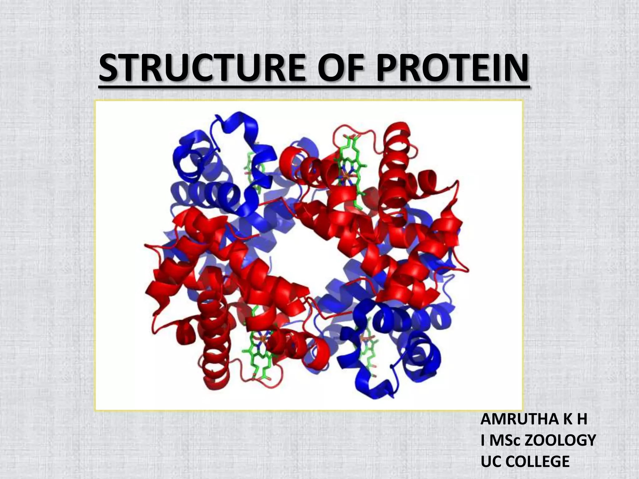
Secondary Structure Of Protein (Repeating structure of protein)
This document discusses the structure of proteins at various levels. It describes the primary, secondary, tertiary, and quaternary structures. The secondary structures discussed in detail include the alpha helix, beta pleated sheet, random coil, collagen helix, and beta turn. The alpha helix and beta pleated sheet are stabilized by hydrogen bonding between amino acids. The collagen helix structure provides strength and is the main component of connective tissues. Genetic disorders like Ehlers-Danlos syndrome and osteogenesis imperfecta result from defects in collagen structures. Ramachandran plots are used to visualize allowed backbone dihedral angles in protein structures.
INTRODUCTION
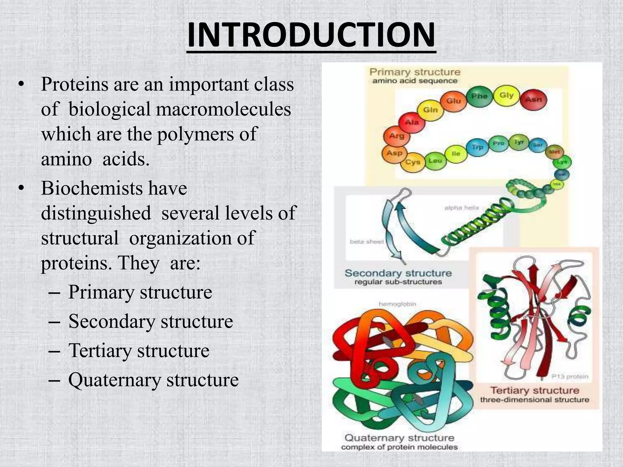
• Proteins arean important class
of biological macromolecules
which are the polymers of
amino acids.
• Biochemists have
distinguished several levels of
structural organization of
proteins. They are:
– Primary structure
– Secondary structure
– Tertiary structure
– Quaternary structure
4.
SECONDARY STRUCTURE
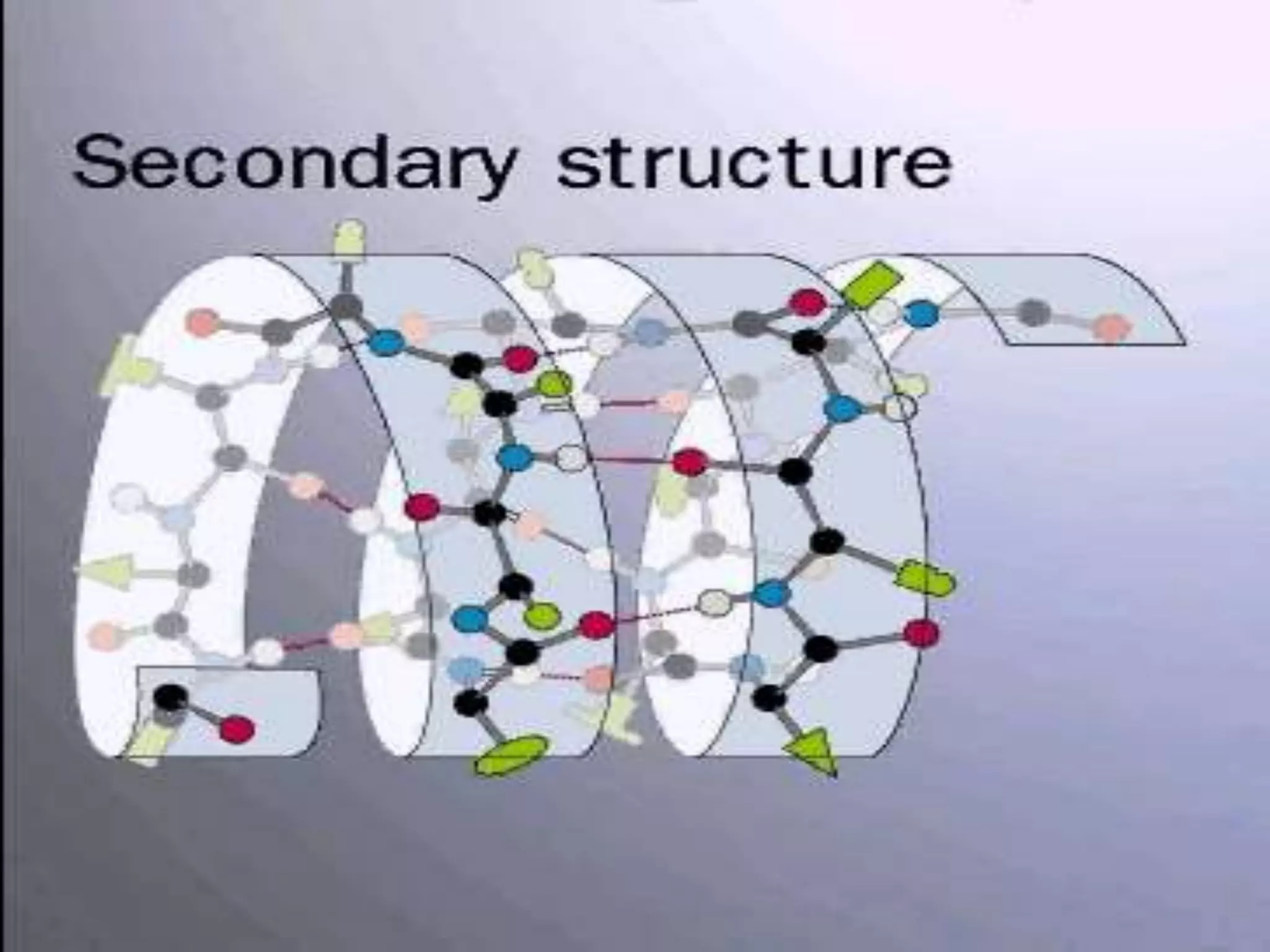
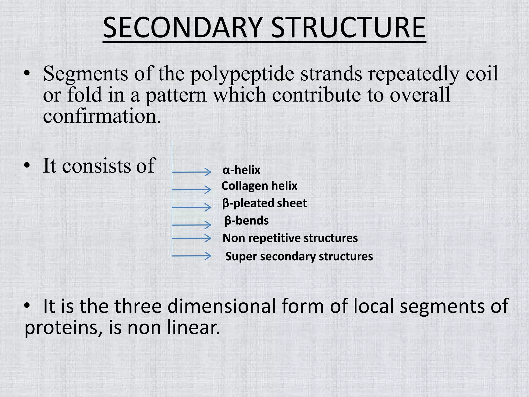
• Segmentsof the polypeptide strands repeatedly coil
or fold in a pattern which contribute to overall
confirmation.
• It consists of
• It is the three dimensional form of local segments of
proteins, is non linear.
α-helix
Collagen helix
β-pleated sheet
β-bends
Non repetitive structures
Super secondary structures
5.
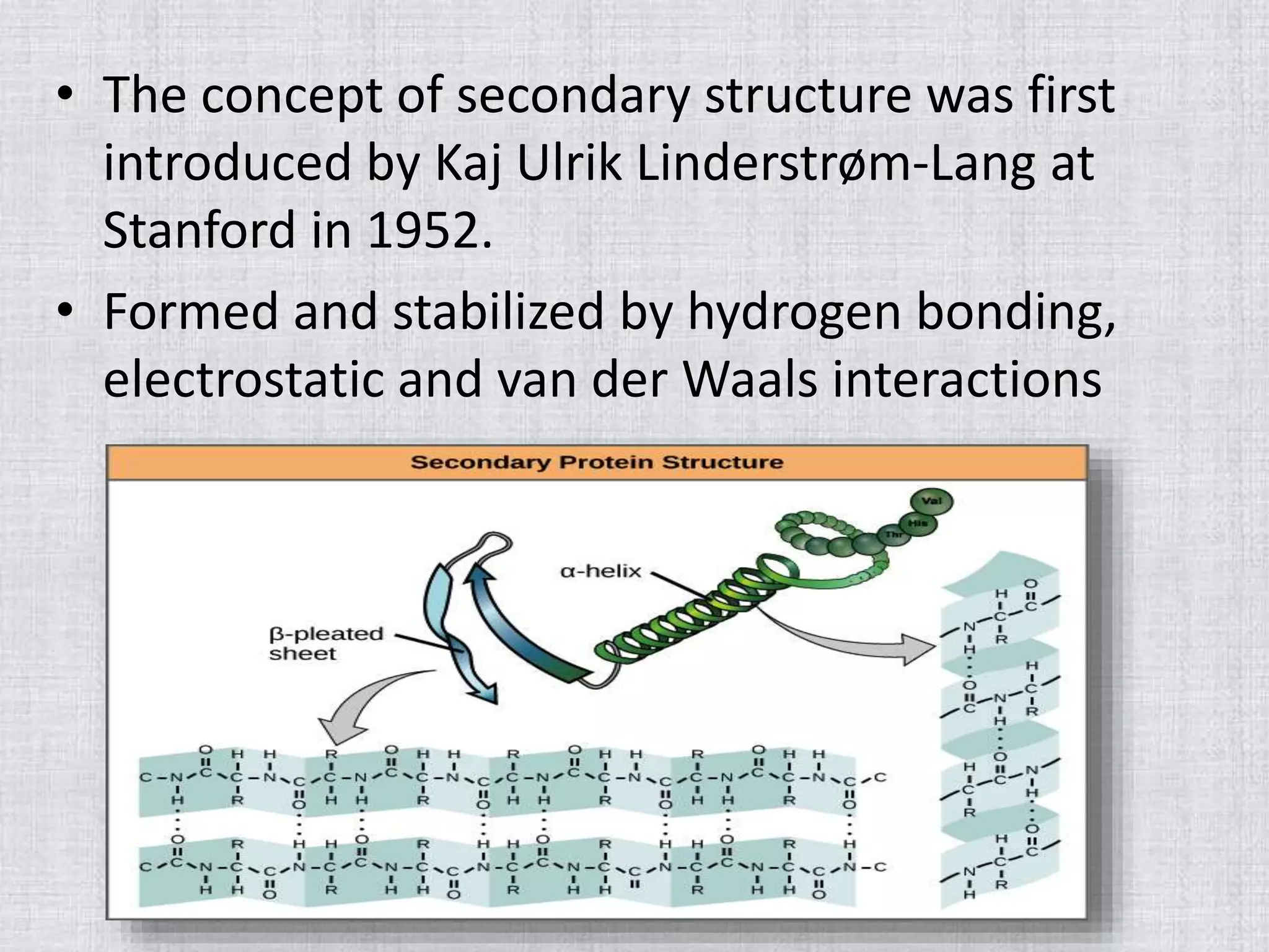
• The conceptof secondary structure was first
introduced by Kaj Ulrik Linderstrøm-Lang at
Stanford in 1952.
• Formed and stabilized by hydrogen bonding,
electrostatic and van der Waals interactions
6.
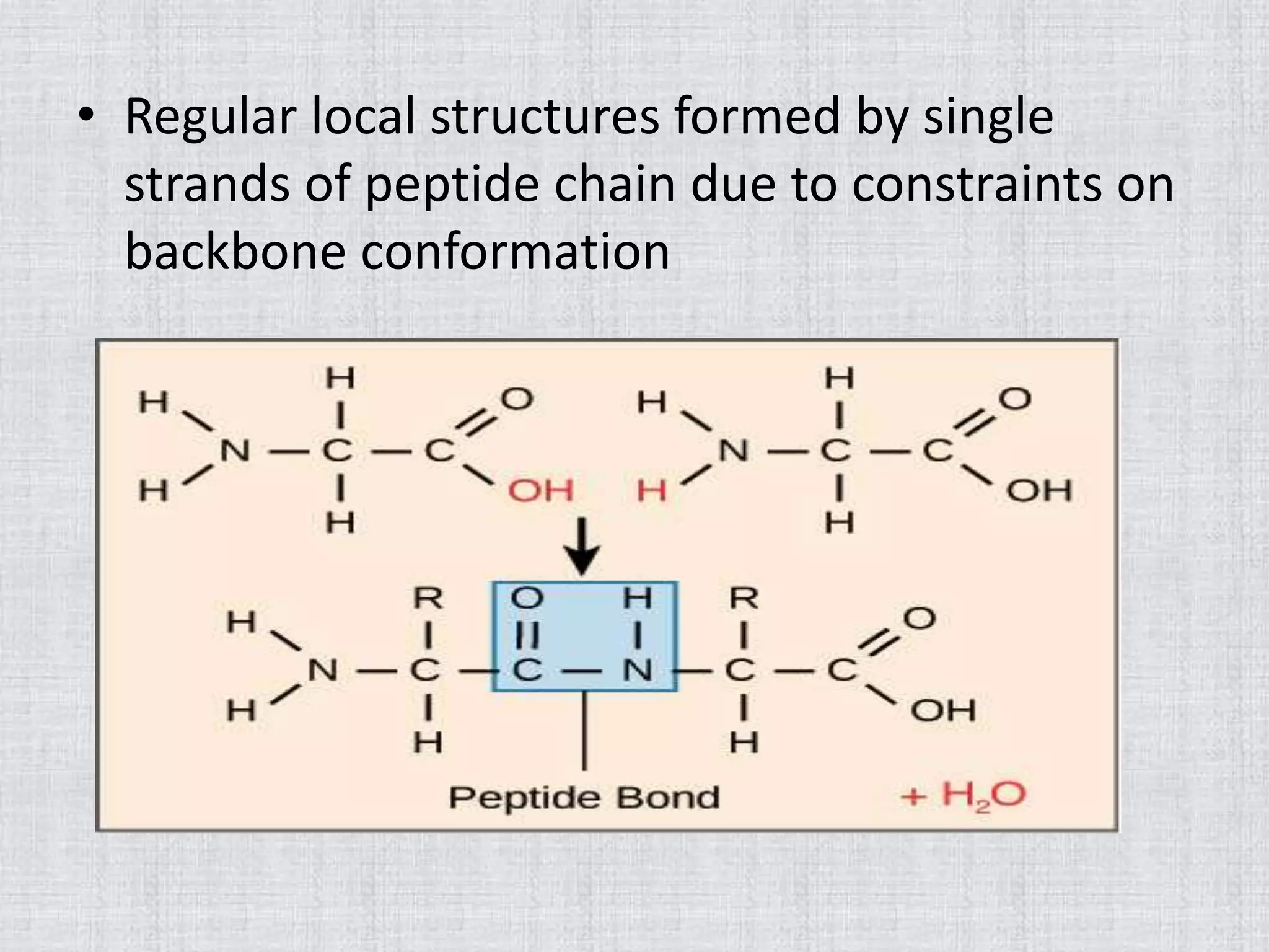
• Regular localstructures formed by single
strands of peptide chain due to constraints on
backbone conformation
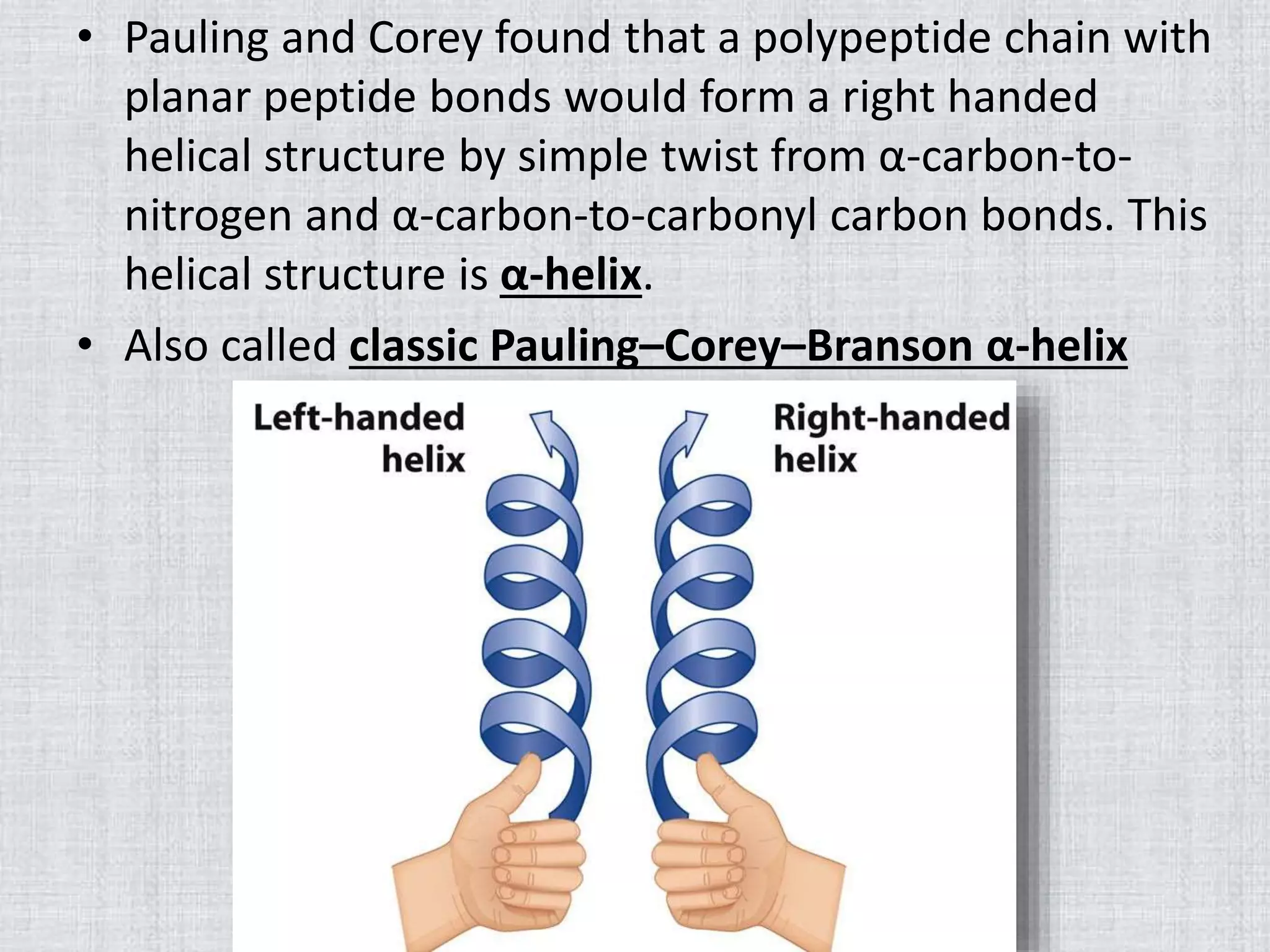
• Pauling andCorey found that a polypeptide chain with
planar peptide bonds would form a right handed
helical structure by simple twist from α-carbon-to-
nitrogen and α-carbon-to-carbonyl carbon bonds. This
helical structure is α-helix.
• Also called classic Pauling–Corey–Branson α-helix
9.
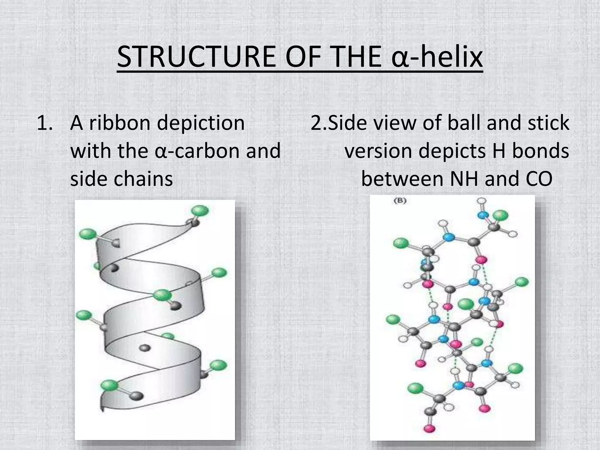
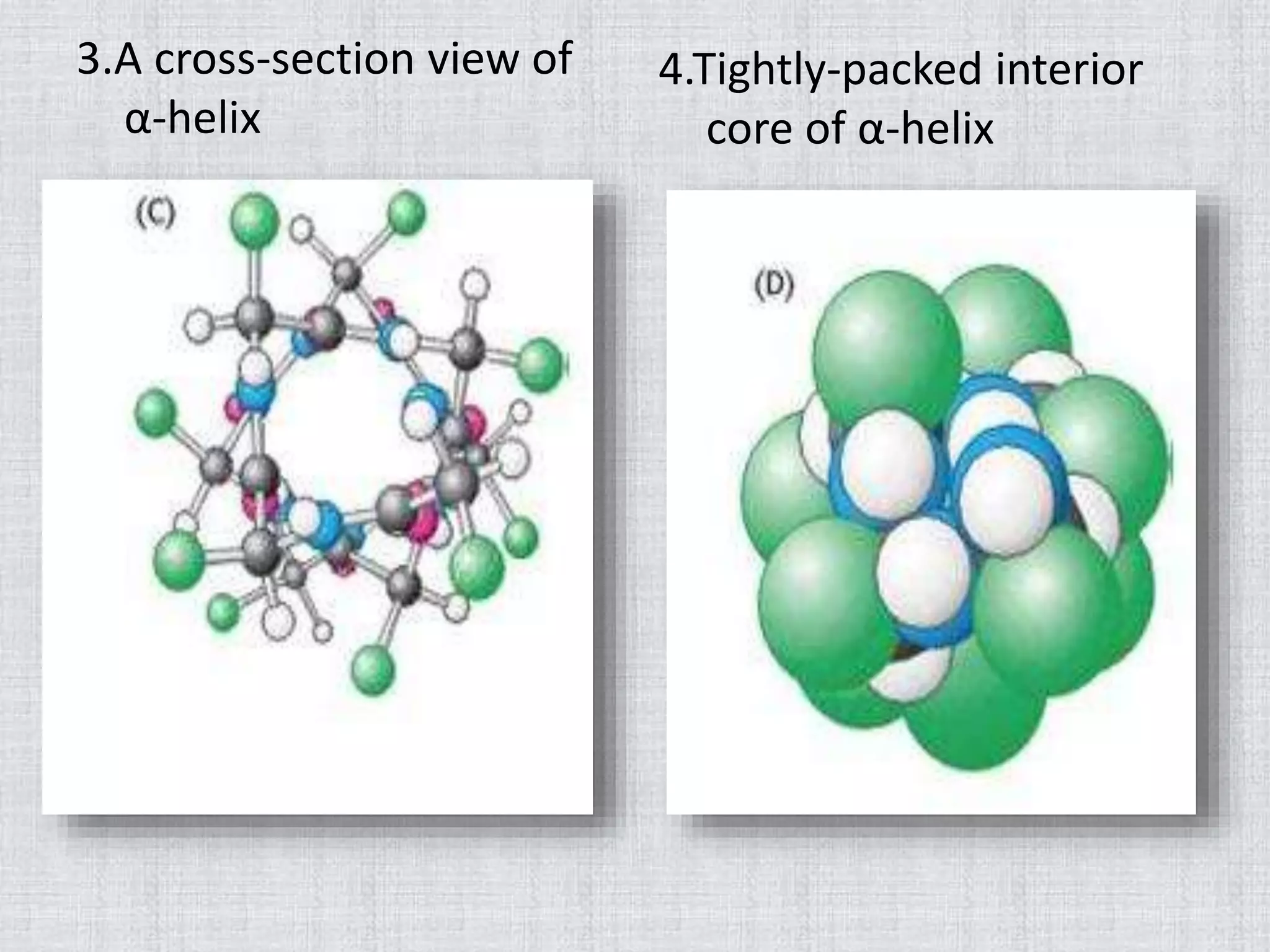
STRUCTURE OF THEα-helix
1. A ribbon depiction
with the α-carbon and
side chains
2.Side view of ball and stick
version depicts H bonds
between NH and CO
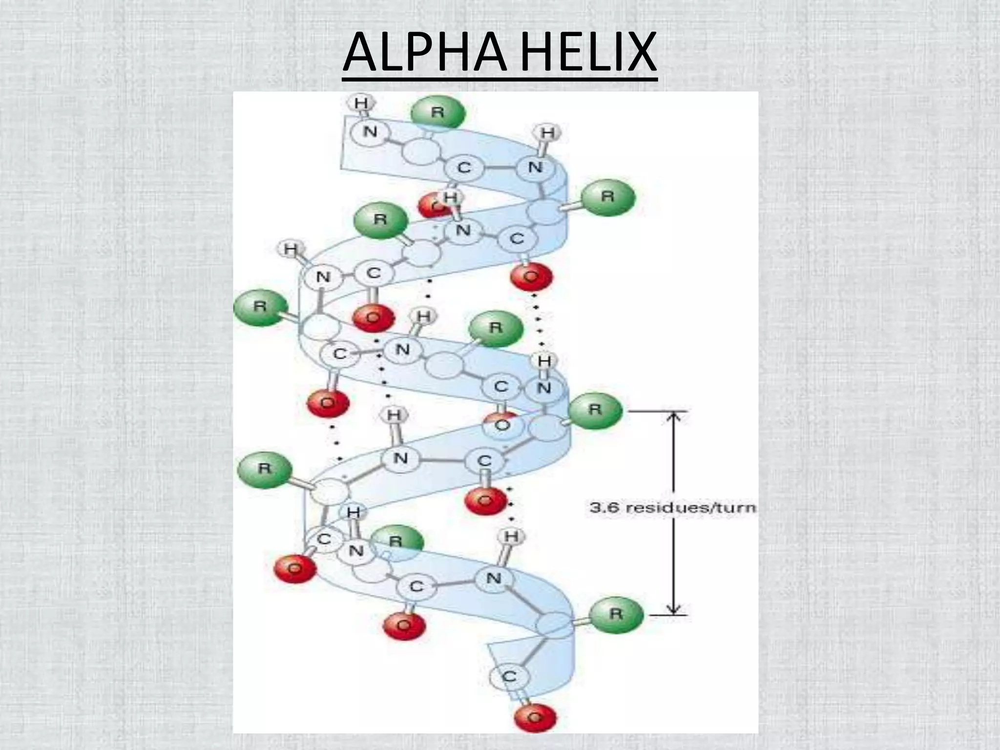
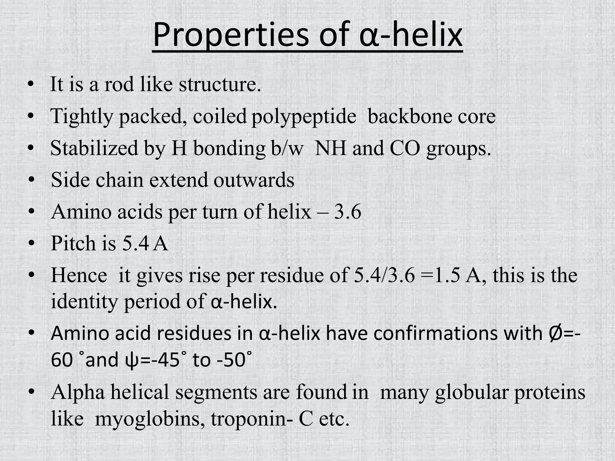
Properties of α-helix
•It is a rod like structure.
• Tightly packed, coiled polypeptide backbone core
• Stabilized by H bonding b/w NH and CO groups.
• Side chain extend outwards
• Amino acids per turn of helix – 3.6
• Pitch is 5.4A
• Hence it gives rise per residue of 5.4/3.6 =1.5 A, this is the
identity period of α-helix.
• Amino acid residues in α-helix have confirmations with Ø=-
60 ˚and ψ=-45˚ to -50˚
• Alpha helical segments are found in many globular proteins
like myoglobins, troponin- C etc.
13.
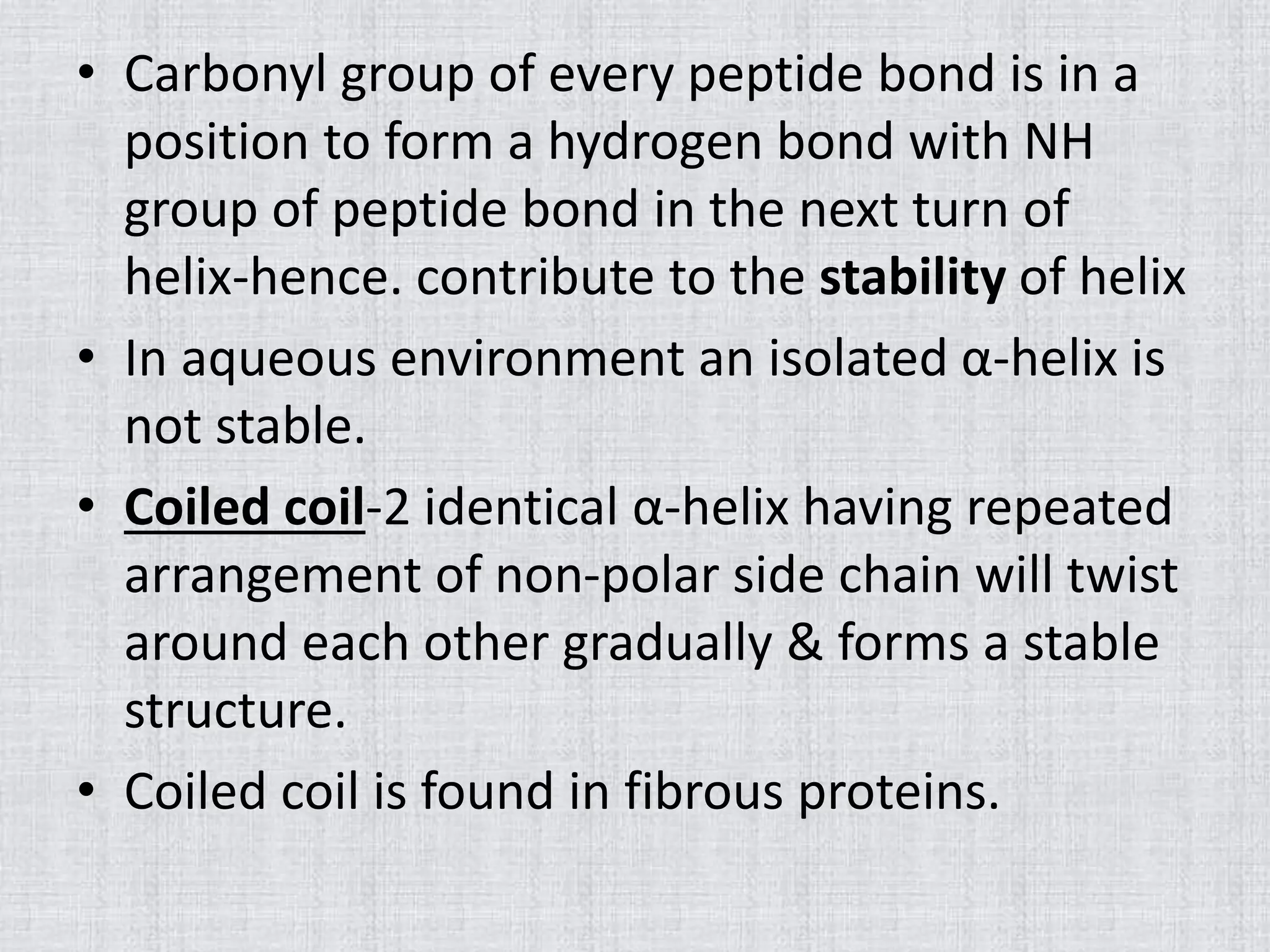
• Carbonyl groupof every peptide bond is in a
position to form a hydrogen bond with NH
group of peptide bond in the next turn of
helix-hence. contribute to the stability of helix
• In aqueous environment an isolated α-helix is
not stable.
• Coiled coil-2 identical α-helix having repeated
arrangement of non-polar side chain will twist
around each other gradually & forms a stable
structure.
• Coiled coil is found in fibrous proteins.
14.
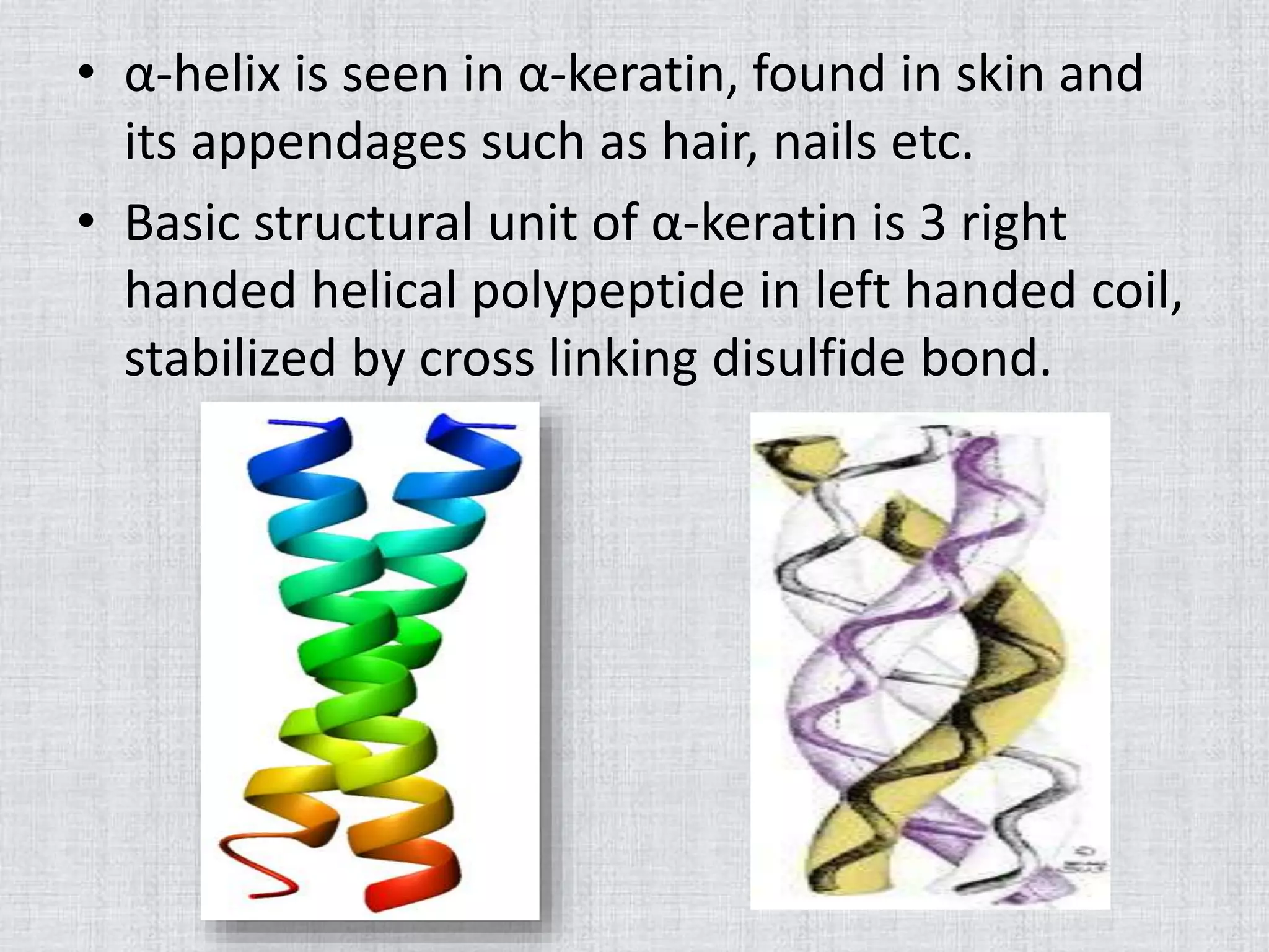
• α-helix isseen in α-keratin, found in skin and
its appendages such as hair, nails etc.
• Basic structural unit of α-keratin is 3 right
handed helical polypeptide in left handed coil,
stabilized by cross linking disulfide bond.
15.
• Destabilization ofα-helix can occur as follows :-
I. Prolyl residue cannot participate in α-helix
structure, so creates a sharp bend in helix
II. Negatively charged side chain repels one
another
III. Steric hindrance imposed by R-groups
IV. Lack of side chain on glycine allows great
degree of rotation about amino acids α-
carbon.
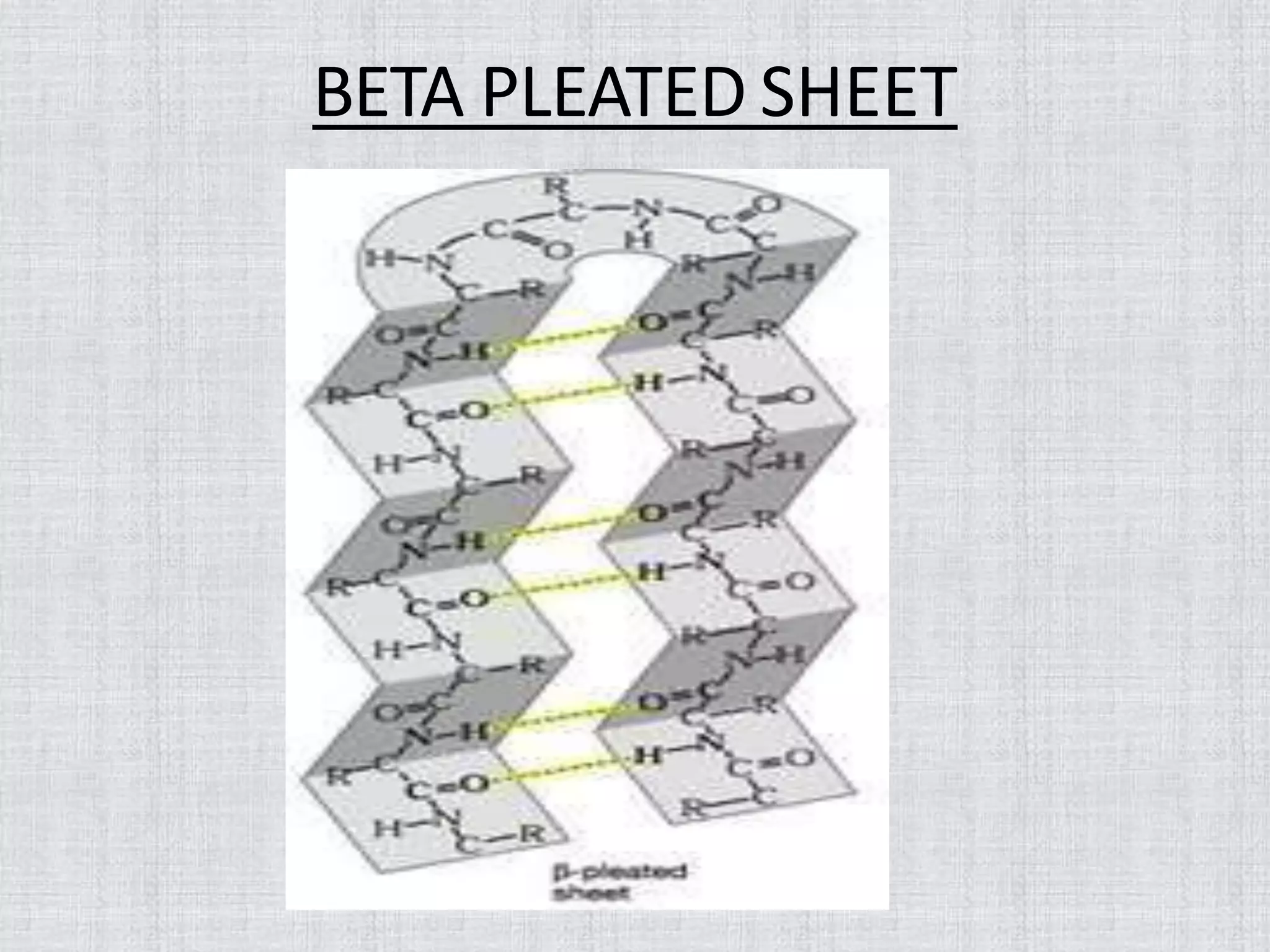
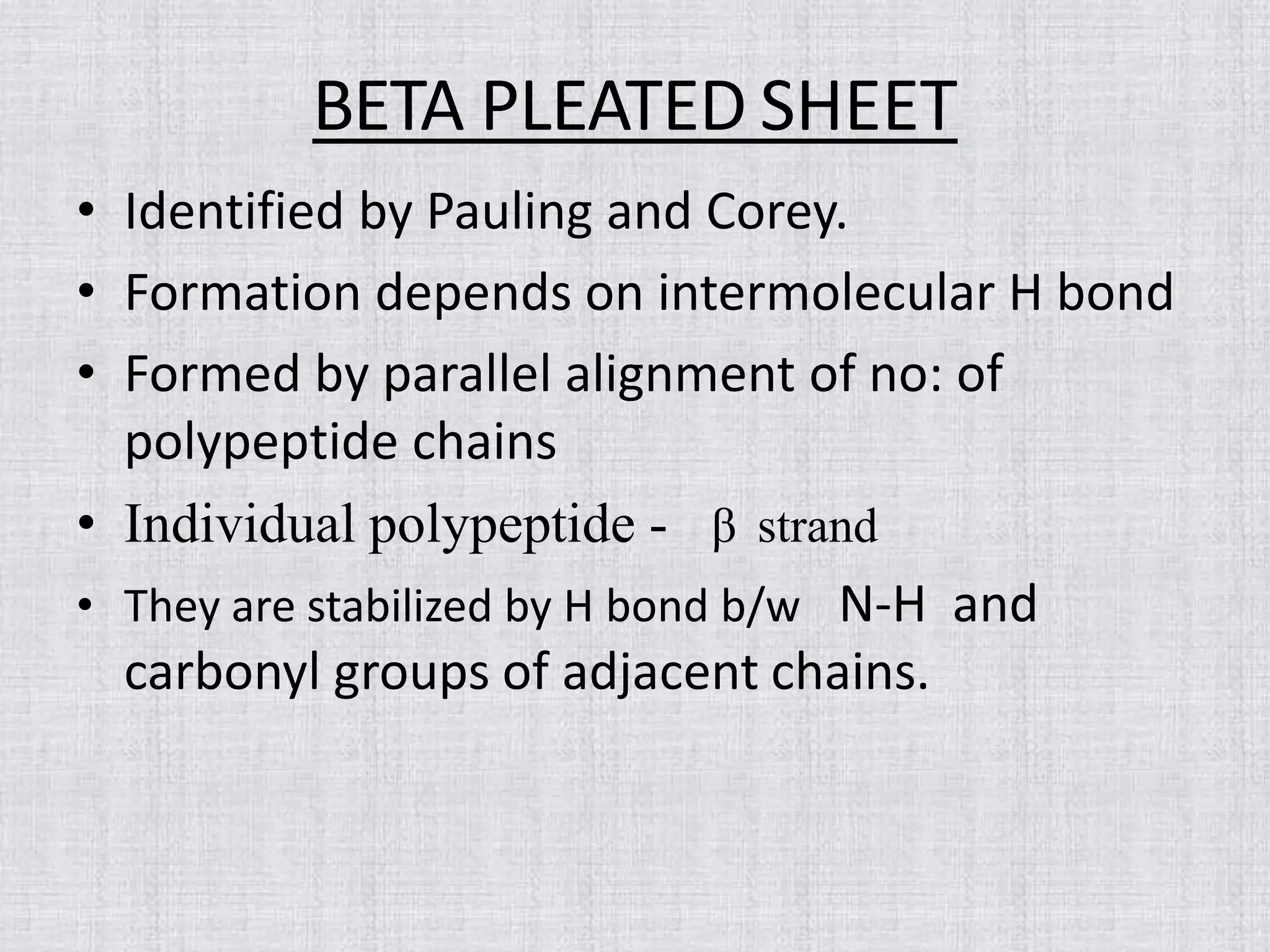
BETA PLEATED SHEET
•Identified by Pauling and Corey.
• Formation depends on intermolecular H bond
• Formed by parallel alignment of no: of
polypeptide chains
• Individual polypeptide - β strand
• They are stabilized by H bond b/w N-H and
carbonyl groups of adjacent chains.
18.
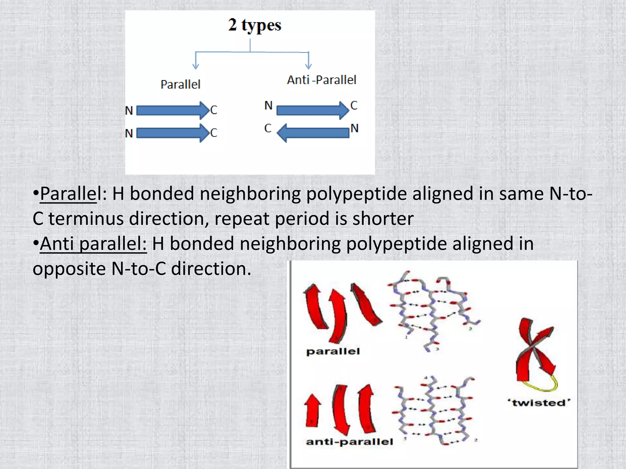
•Parallel: H bondedneighboring polypeptide aligned in same N-to-
C terminus direction, repeat period is shorter
•Anti parallel: H bonded neighboring polypeptide aligned in
opposite N-to-C direction.
19.
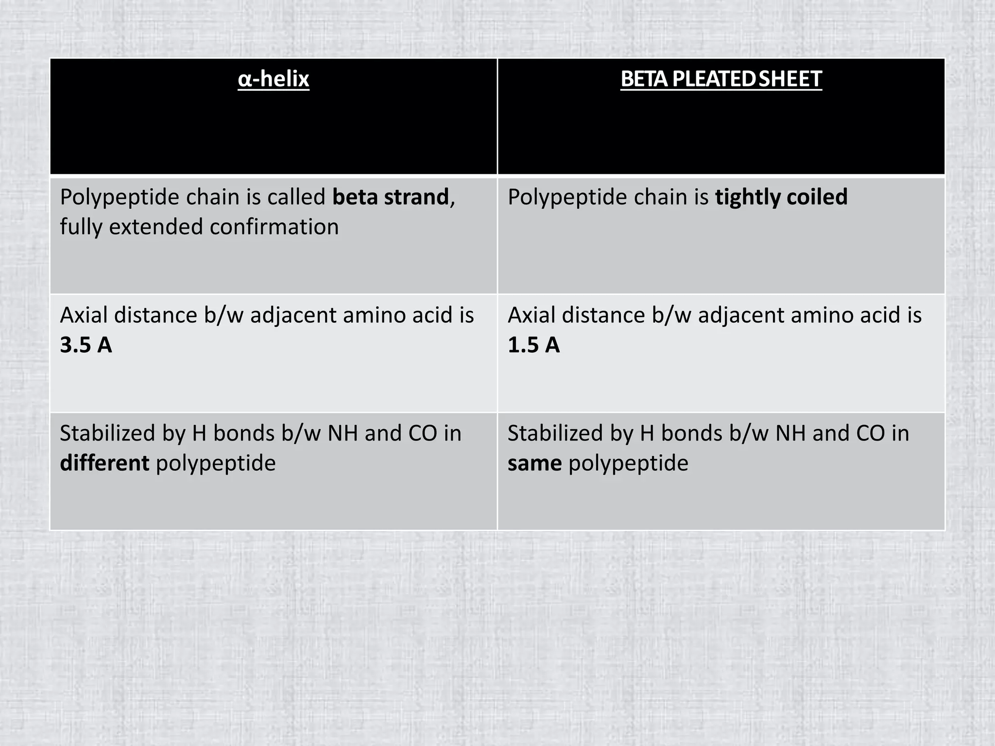
α-helix BETAPLEATEDSHEET
Polypeptide chainis called beta strand,
fully extended confirmation
Polypeptide chain is tightly coiled
Axial distance b/w adjacent amino acid is
3.5 A
Axial distance b/w adjacent amino acid is
1.5 A
Stabilized by H bonds b/w NH and CO in
different polypeptide
Stabilized by H bonds b/w NH and CO in
same polypeptide
20.
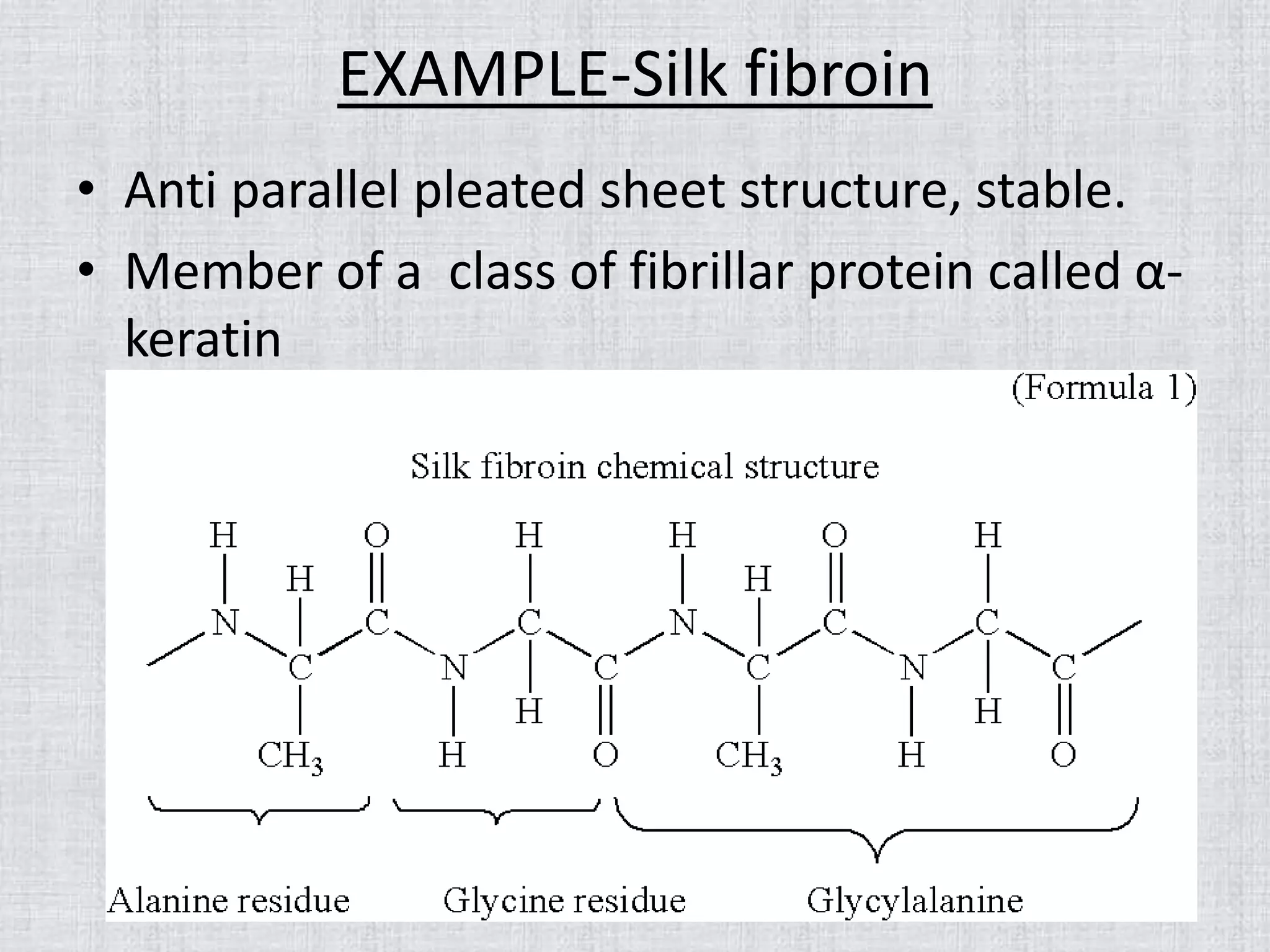
EXAMPLE-Silk fibroin
• Antiparallel pleated sheet structure, stable.
• Member of a class of fibrillar protein called α-
keratin
21.
RANDOM COIL
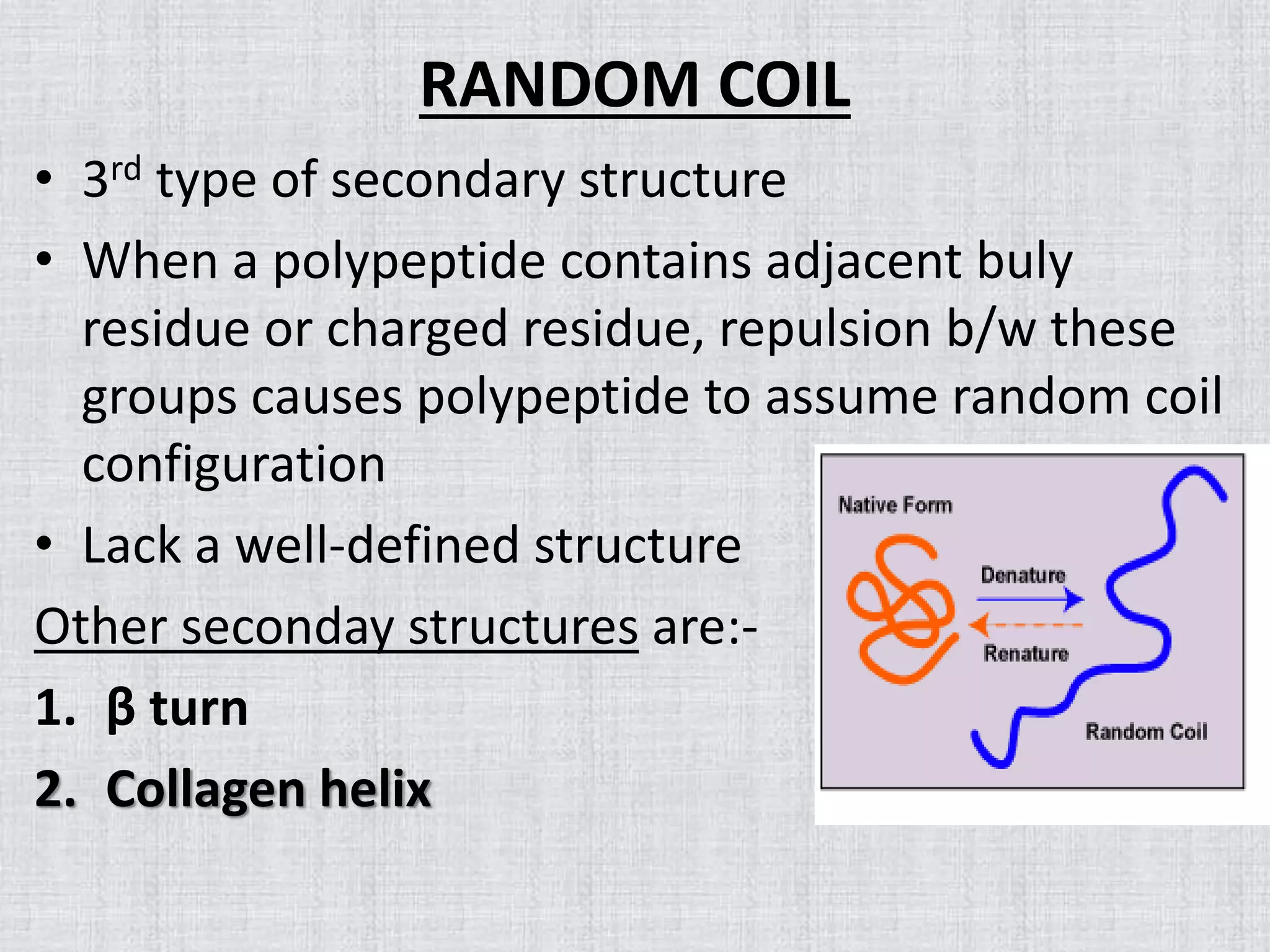
• 3rdtype of secondary structure
• When a polypeptide contains adjacent buly
residue or charged residue, repulsion b/w these
groups causes polypeptide to assume random coil
configuration
• Lack a well-defined structure
Other seconday structures are:-
1. β turn
2. Collagen helix
22.
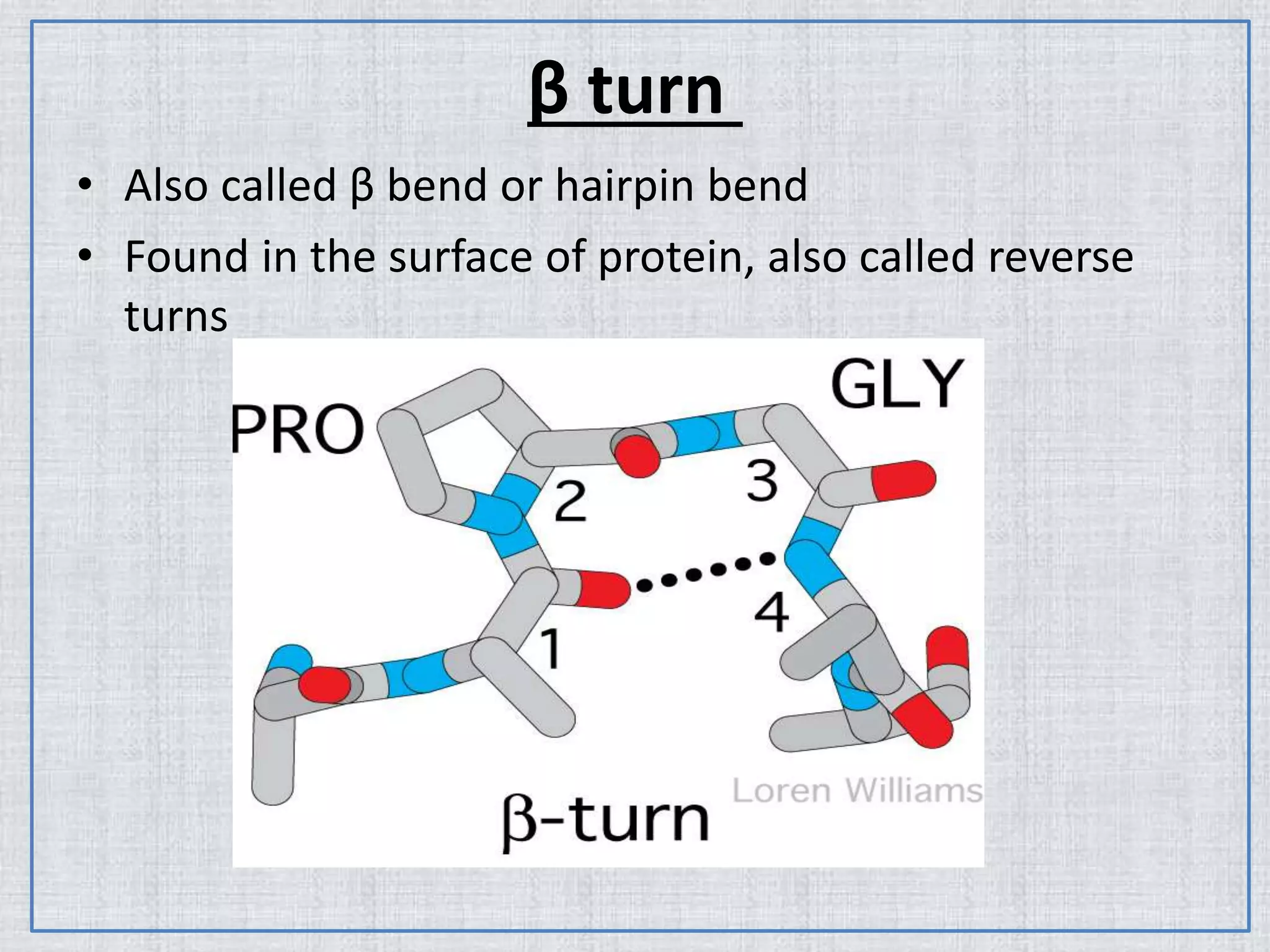
β turn
• Alsocalled β bend or hairpin bend
• Found in the surface of protein, also called reverse
turns
23.
• Permits thechange of direction of the peptide chain
to get a folded structure.
• It gives a protein globularity rather than linearity.
• H bond stabilizes the beta bend structure
• Proline and Glycine are frequently found in beta
turns.
• Beta turns often promote the formation of antiparallel
beta sheets.
• Occur at protein surfaces.
• Involve four successive amino acid residues
24.
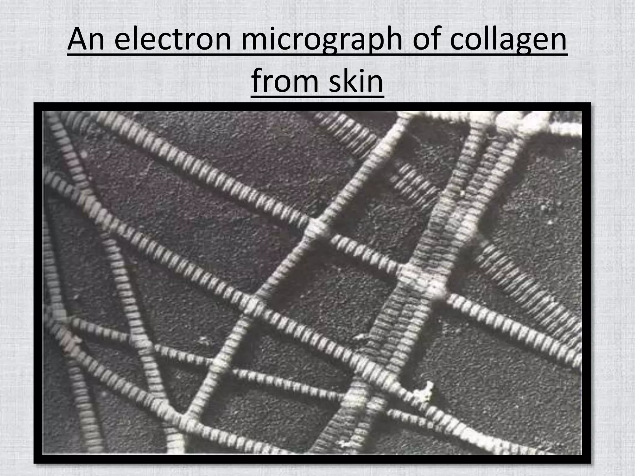
Collagen helix
• Alsocalled type-2 helix.
• Most abundant protein in mammals.
• Principal structural element of the human body makes up
25-30% of all the body protein.
• Found in connective tissues such as
tendons,cartilage,cornea of eye etc
• Contain 3 helical polypeptide nearly 1000 residues long.
• Amino acid sequence structure is remarkably regular,
nearly every 3rd residue is a glycine
• It contains 4-hydroxyproline(Hyp)
• The % composition is given as:-
Gly(35%),Ala(11%) & Pro+Hyp (25%)
25.
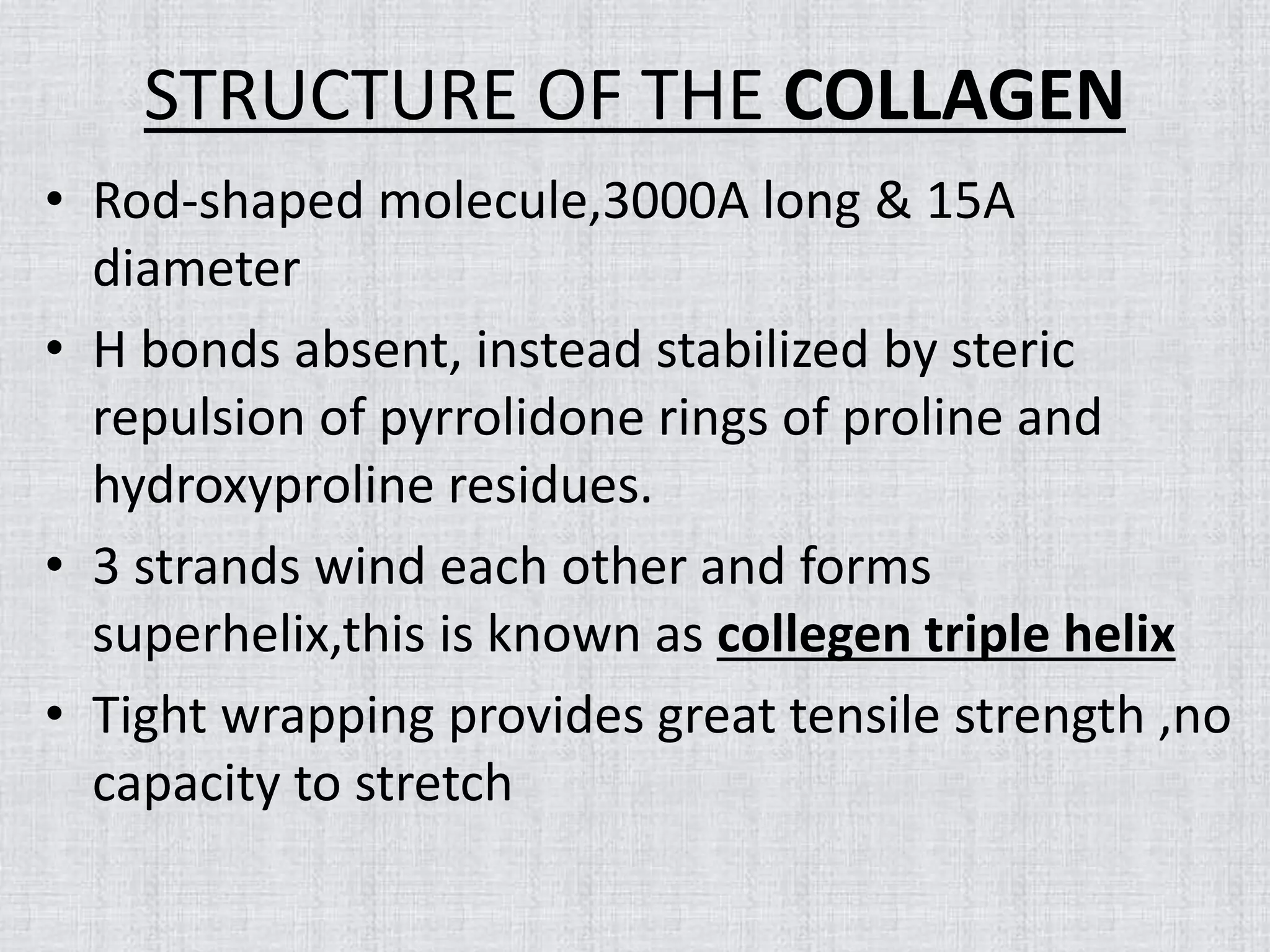
STRUCTURE OF THECOLLAGEN
• Rod-shaped molecule,3000A long & 15A
diameter
• H bonds absent, instead stabilized by steric
repulsion of pyrrolidone rings of proline and
hydroxyproline residues.
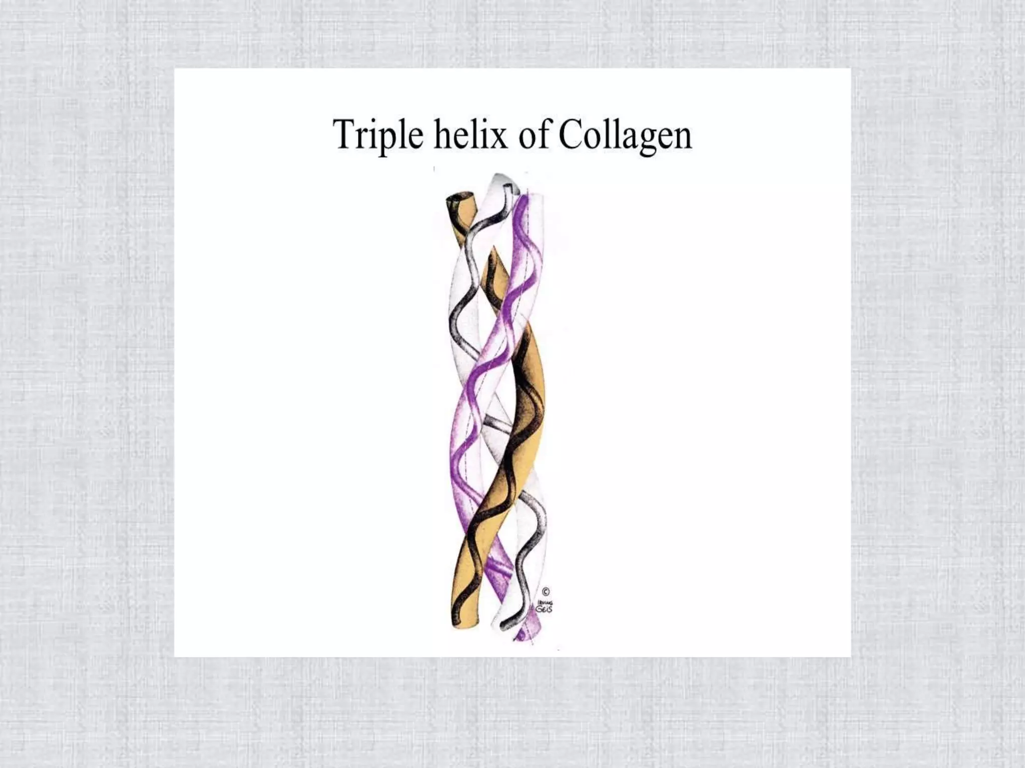
• 3 strands wind each other and forms
superhelix,this is known as collegen triple helix
• Tight wrapping provides great tensile strength ,no
capacity to stretch
27.
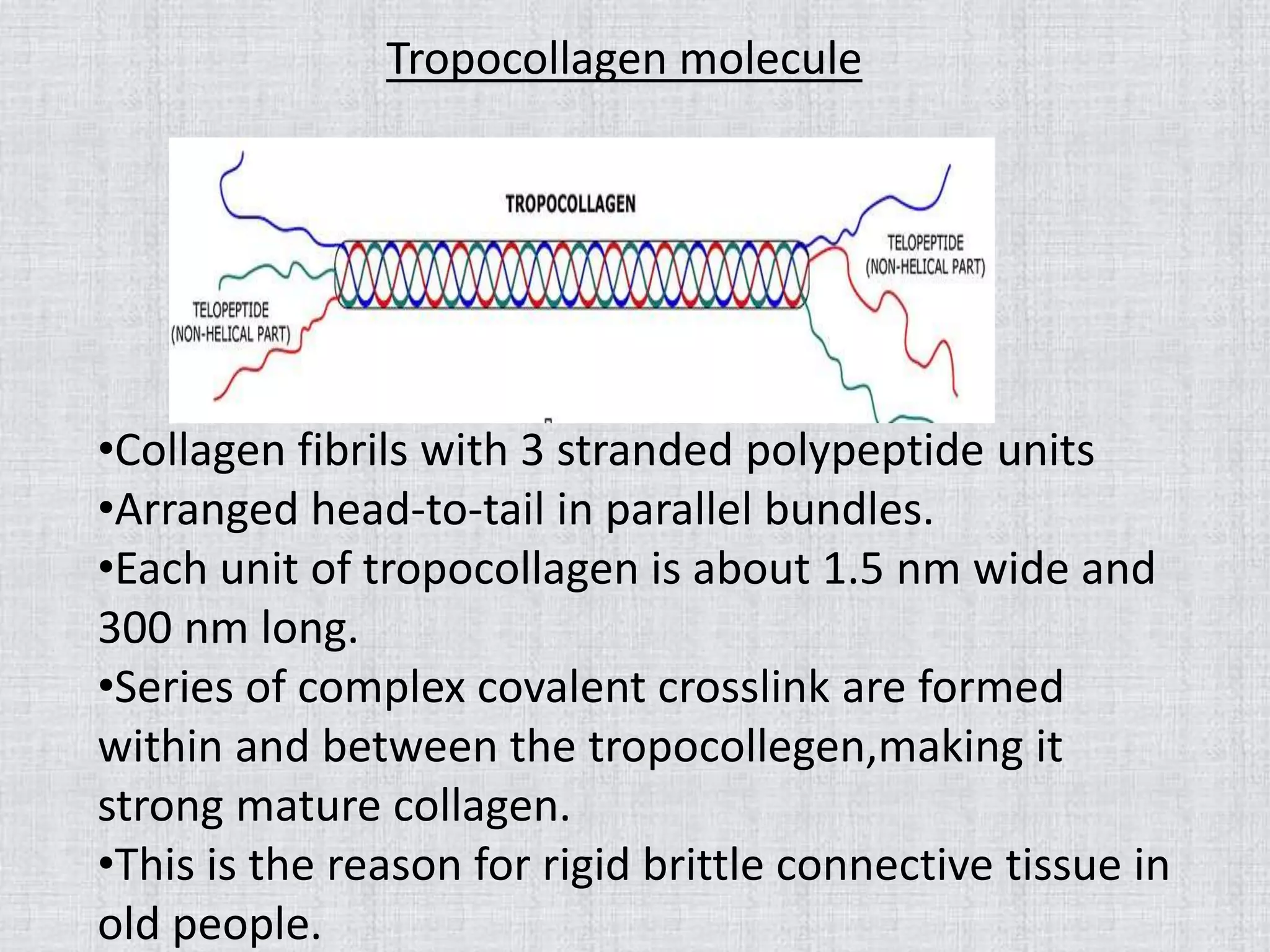
Tropocollagen molecule
•Collagen fibrilswith 3 stranded polypeptide units
•Arranged head-to-tail in parallel bundles.
•Each unit of tropocollagen is about 1.5 nm wide and
300 nm long.
•Series of complex covalent crosslink are formed
within and between the tropocollegen,making it
strong mature collagen.
•This is the reason for rigid brittle connective tissue in
old people.
28.
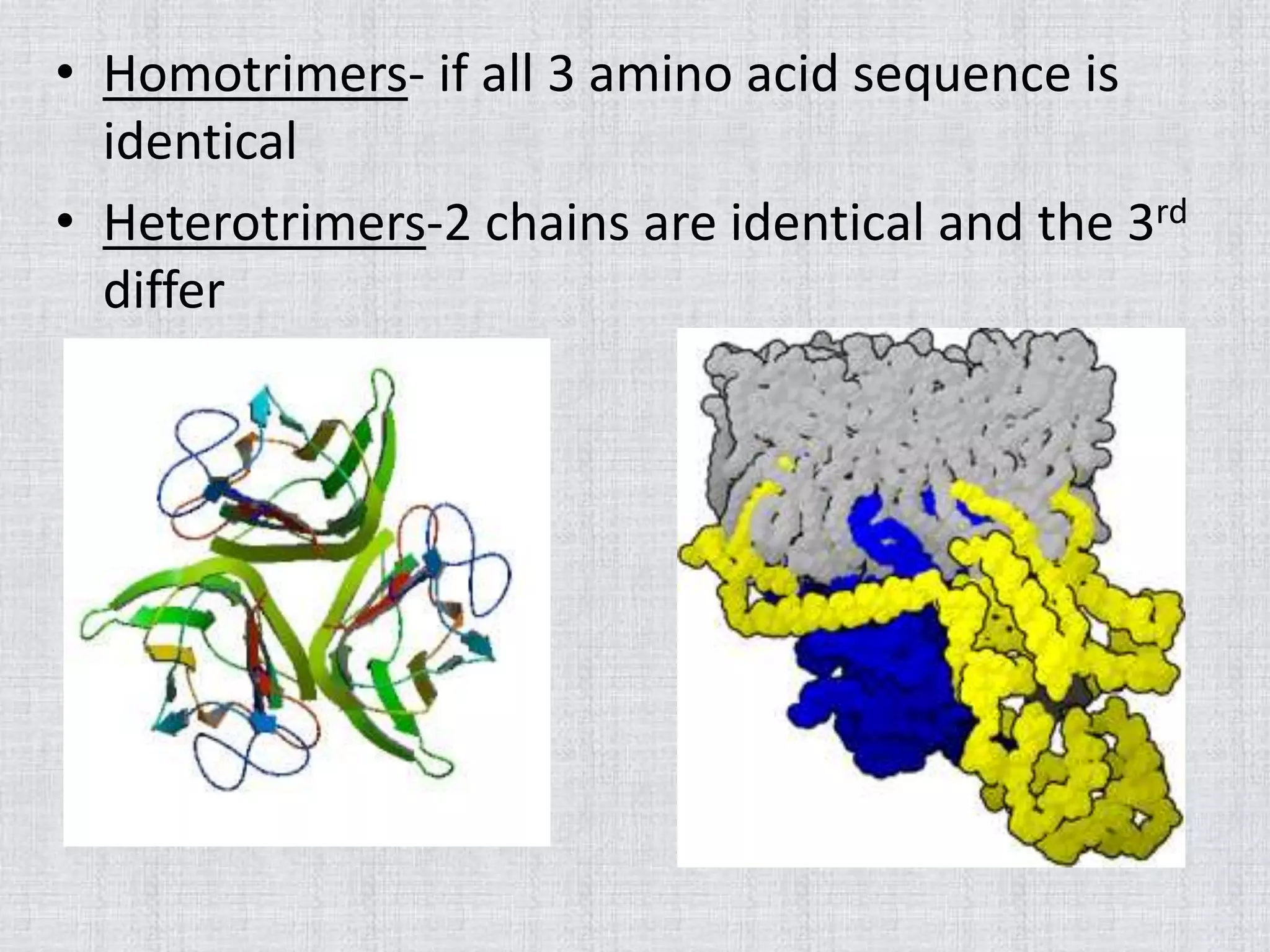
• Homotrimers- ifall 3 amino acid sequence is
identical
• Heterotrimers-2 chains are identical and the 3rd
differ
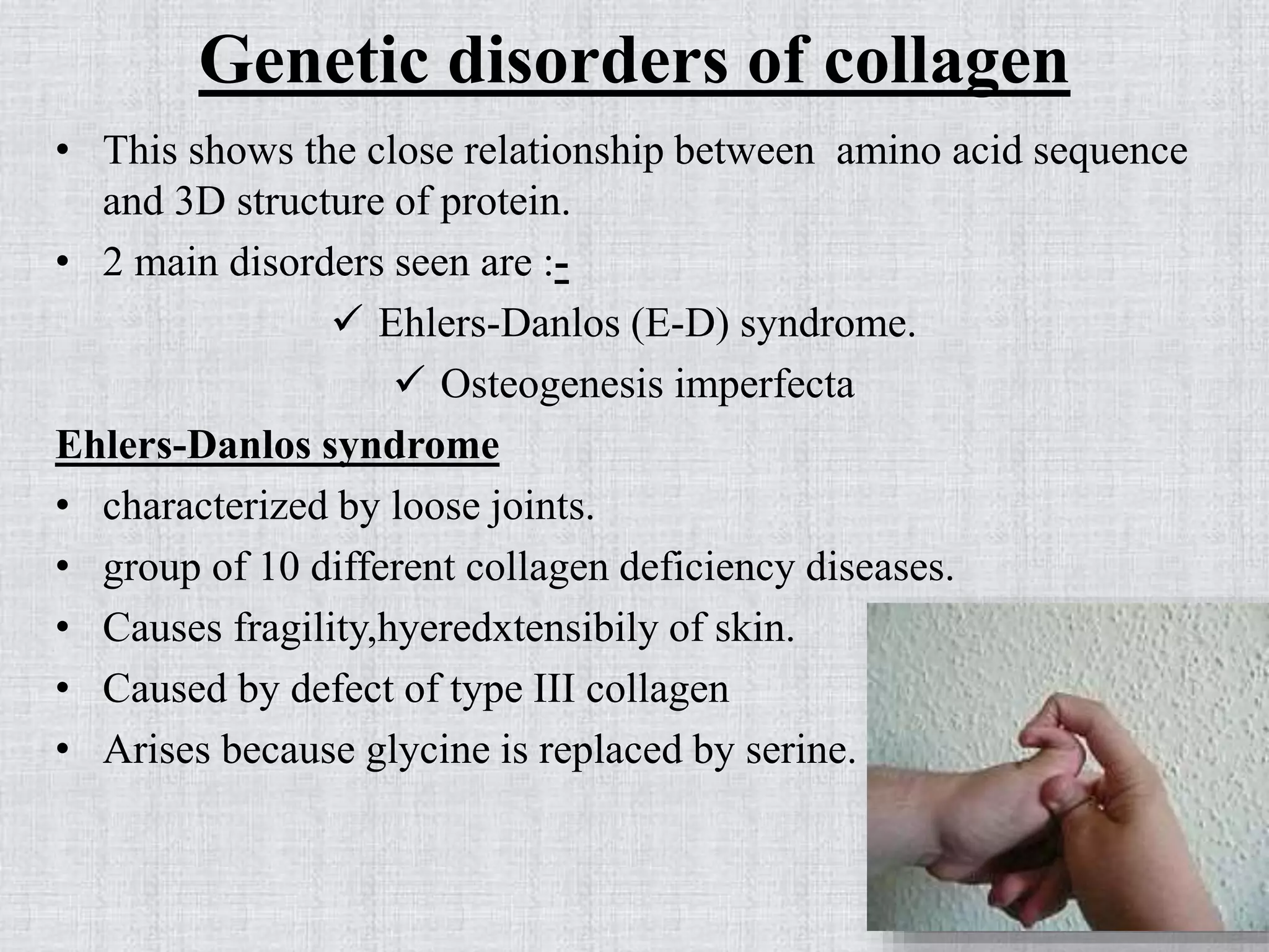
Genetic disorders ofcollagen
• This shows the close relationship between amino acid sequence
and 3D structure of protein.
• 2 main disorders seen are :-
Ehlers-Danlos (E-D) syndrome.
Osteogenesis imperfecta
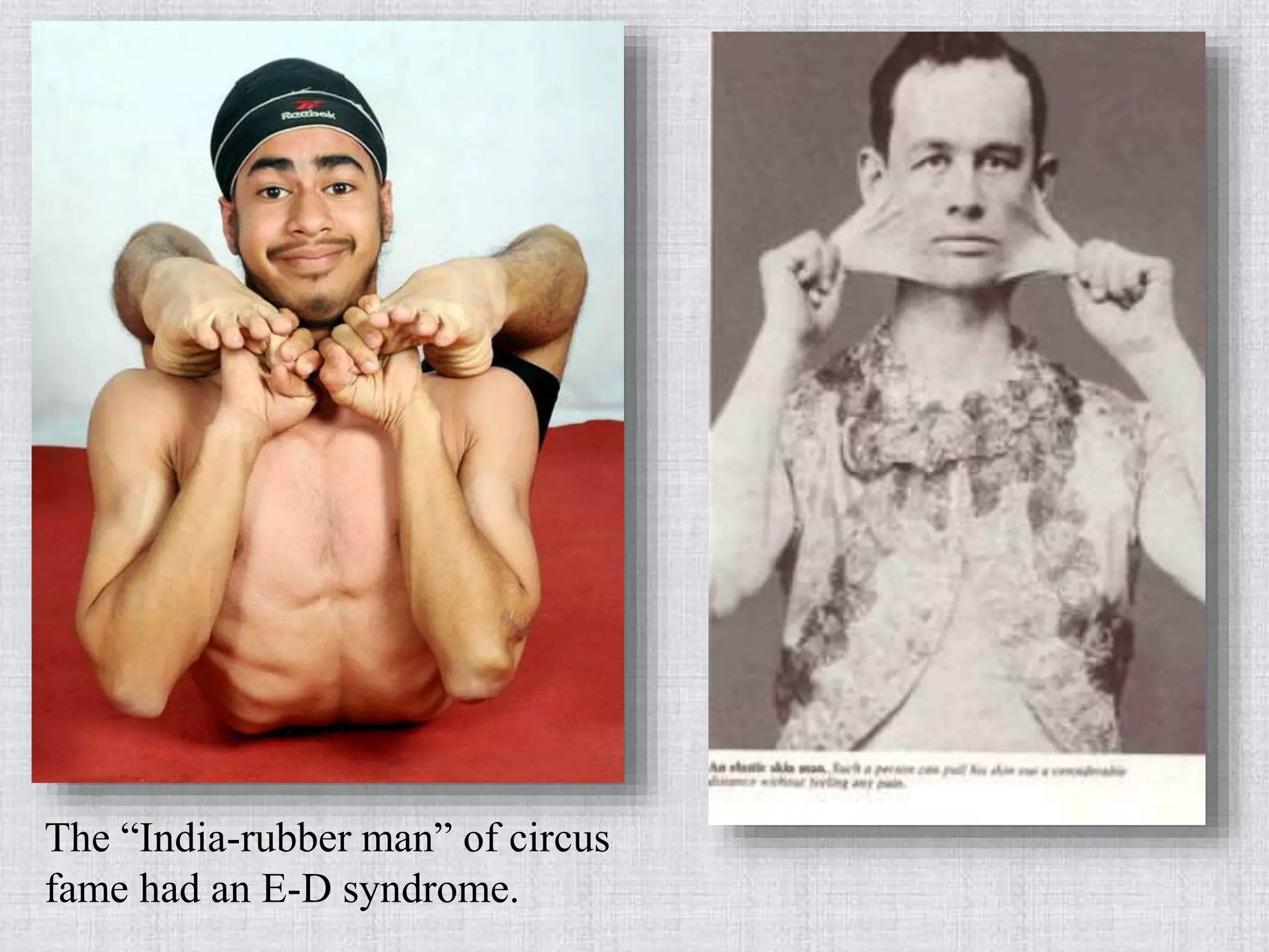
Ehlers-Danlos syndrome
• characterized by loose joints.
• group of 10 different collagen deficiency diseases.
• Causes fragility,hyeredxtensibily of skin.
• Caused by defect of type III collagen
• Arises because glycine is replaced by serine.
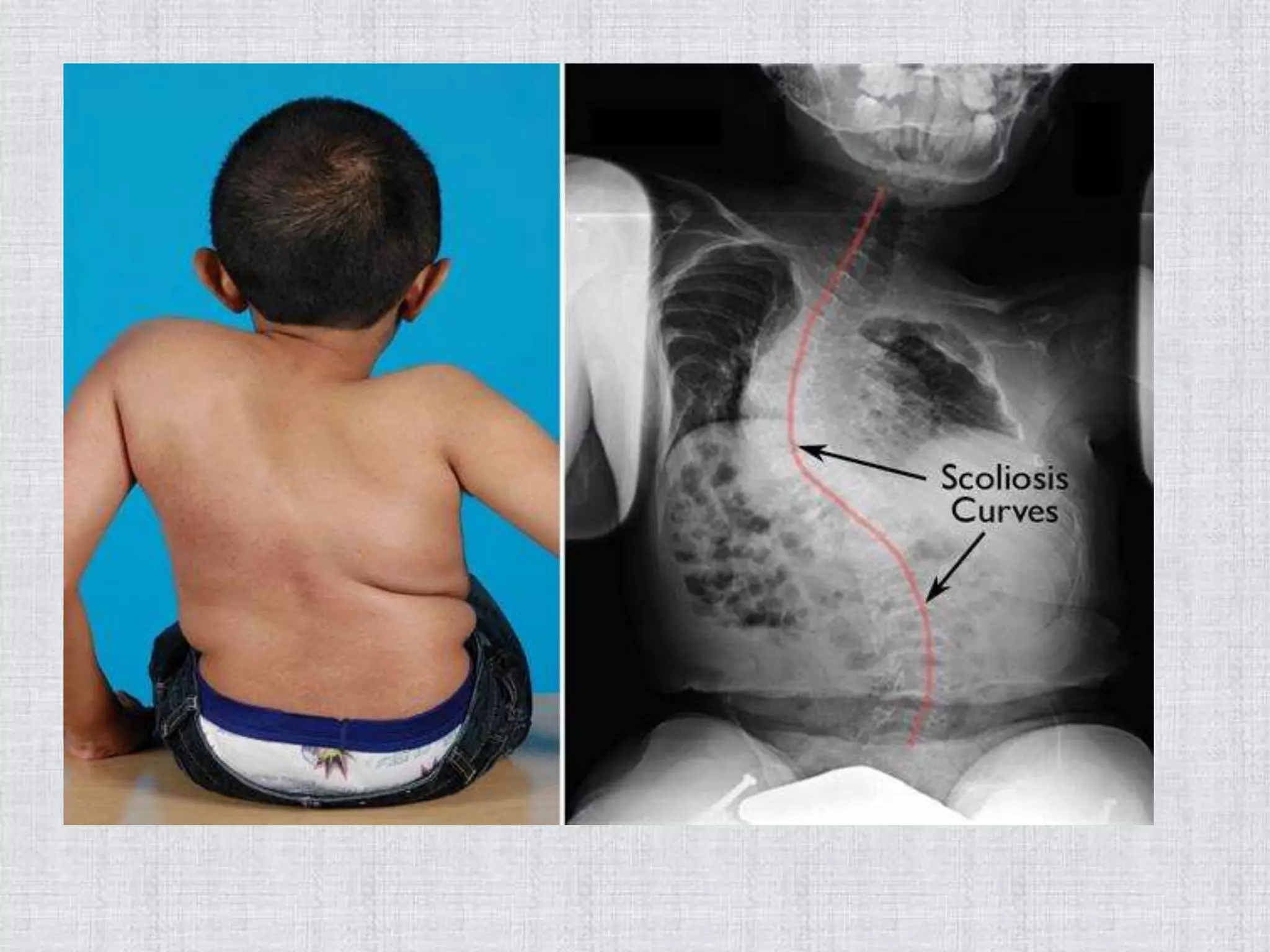
Osteogenesis imperfecta(OI)
• Alsocalled brittle bone disease.
• abnormal (fragile) bone formation in human babies.
• Arises because glycine is replaced by cysteine.
• Defect in the synthesis of type-I collagen
• Numerous fractures and severe bone deformity; small stature
with underdeveloped lungs.
35.
RAMACHANDRAN PLOT
• alsoknown as a Ramachandran diagram or a [φ,ψ]
plot
• originally developed in 1963 by G. N. Ramachandran
36.
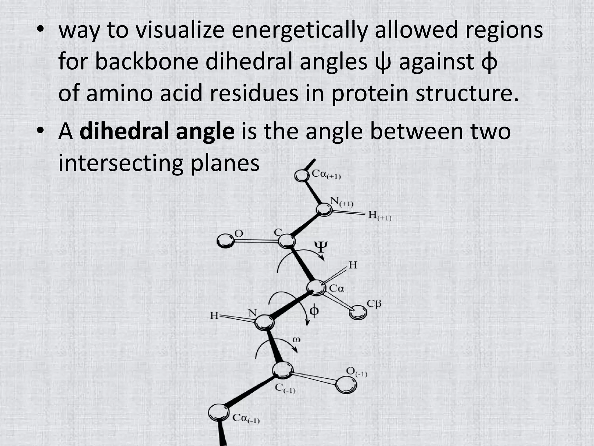
• way tovisualize energetically allowed regions
for backbone dihedral angles ψ against φ
of amino acid residues in protein structure.
• A dihedral angle is the angle between two
intersecting planes
37.
• The phiangle is the angle around the -N-CA-
bond (where 'CA' is the alpha-carbon)
• The psi angle is the angle around the -CA-C-
bond.
• The omega angle is the angle around the -C-N-
bond (i.e. the peptide bond)
38.
•
•
•
.
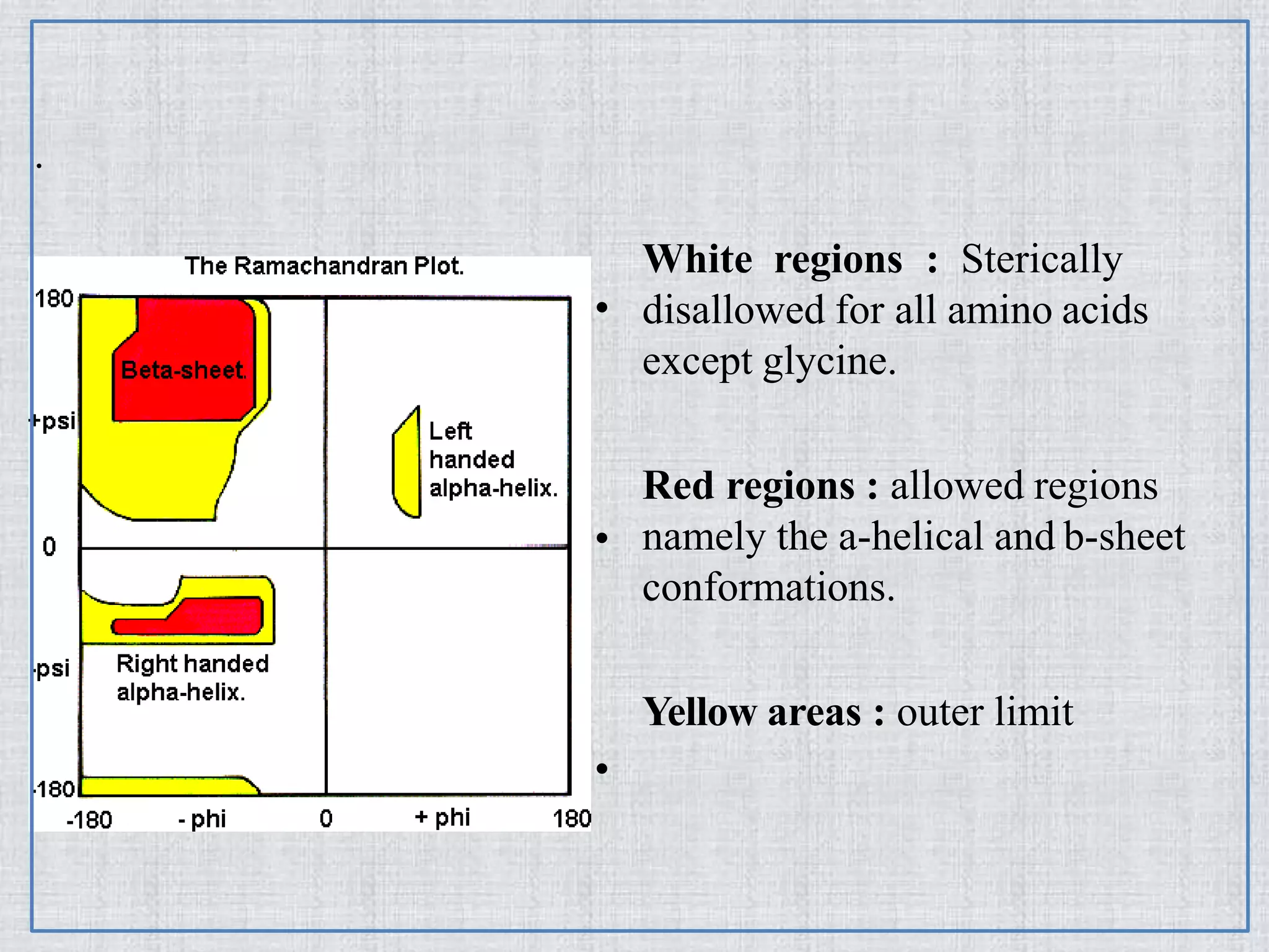
White regions :Sterically
disallowed for all amino acids
except glycine.
Red regions : allowed regions
namely the a-helical and b-sheet
conformations.
Yellow areas : outer limit
39.
Uses
• can beused in two somewhat different ways
• First to show in theory which values,
or conformation of the ψ and φ angles are
possible for an amino-acid residue in a
protein.
• second is to show the empirical distribution of
data points observed in a single structure


![RAMACHANDRAN PLOT
• also known as a Ramachandran diagram or a [φ,ψ]
plot
• originally developed in 1963 by G. N. Ramachandran](https://image.slidesharecdn.com/secondarystructureofprotein-180411194709/75/Secondary-Structure-Of-Protein-Repeating-structure-of-protein-35-2048.jpg)