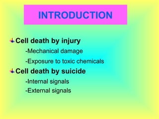
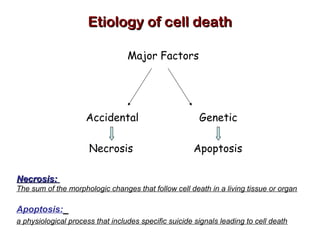
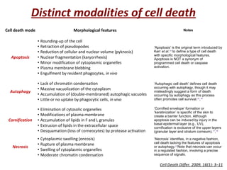
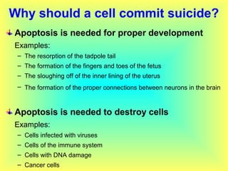
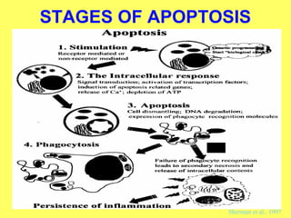
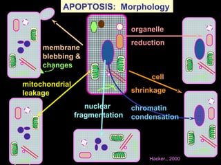
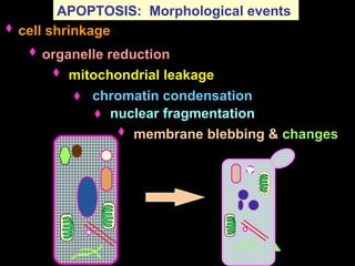
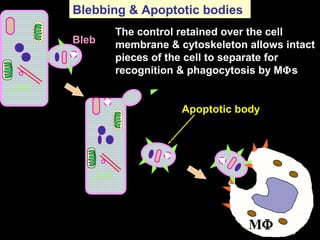
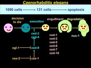
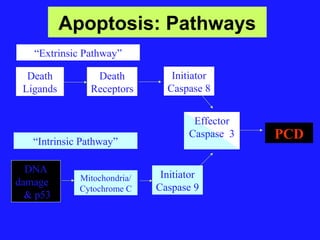
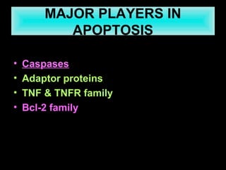
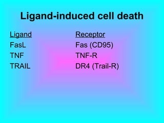
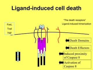
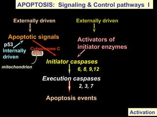
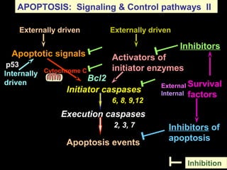
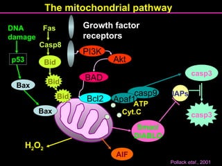
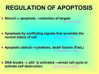
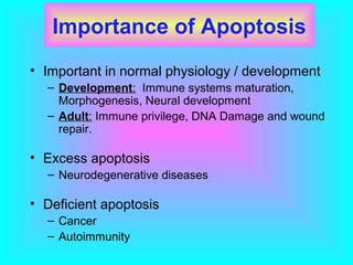
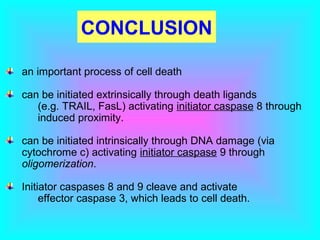
Delvadiya Indrajay
Ph.D. (Agri.)
Department of Genetics and Plant Breeding,
College of Agriculture, JAU, Junagadh
INTRODUCTION
Cell death by injury
-Mechanical damage
-Exposure to toxic chemicals
Cell death by suicide
-Internal signals
-External signals
Conted…..
Apoptosis or programmed cell death, is carefully
coordinated collapse of cell, protein degradation ,
DNA fragmentation followed by rapid engulfment
of corpses by neighbouring cells. (Tommi, 2002)
Essential part of life for every multicellular
organism from worms to humans. (Faddy et al.,1992)
Apoptosis plays a major role from embryonic
development to senescence.
Etiology of cell deathEtiology of cell death
Major Factors
Accidental Genetic
Necrosis Apoptosis
Necrosis:Necrosis:
The sum of the morphologic changes that follow cell death in a living tissue or organ
Apoptosis:
a physiological process that includes specific suicide signals leading to cell death
Cell death mode Morphological features Notes
Apoptosis
• Rounding-up of the cell
• Retraction of pseudopodes
• Reduction of cellular and nuclear volume (pyknosis)
• Nuclear fragmentation (karyorrhexis)
• Minor modification of cytoplasmic organelles
• Plasma membrane blebbing
• Engulfment by resident phagocytes, in vivo
‘Apoptosis’ is the original term introduced by
Kerr et al.14
to define a type of cell death
with specific morphological features.
Apoptosis is NOT a synonym of
programmed cell death or caspase
activation.
Autophagy
• Lack of chromatin condensation
• Massive vacuolization of the cytoplasm
• Accumulation of (double-membraned) autophagic vacuoles
• Little or no uptake by phagocytic cells, in vivo
‘Autophagic cell death’ defines cell death
occurring with autophagy, though it may
misleadingly suggest a form of death
occurring by autophagy as this process
often promotes cell survival.15
,16
Cornification
• Elimination of cytosolic organelles
• Modifications of plasma membrane
• Accumulation of lipids in F and L granules
• Extrusion of lipids in the extracellular space
• Desquamation (loss of corneocytes) by protease activation
‘Cornified envelope’ formation or
‘keratinization’ is specific of the skin to
create a barrier function. Although
apoptosis can be induced by injury in the
basal epidermal layer (e.g., UV),
cornification is exclusive of the upper layers
(granular layer and stratum corneum).17
,18
Necrosis
• Cytoplasmic swelling (oncosis)
• Rupture of plasma membrane
• Swelling of cytoplasmic organelles
• Moderate chromatin condensation
‘Necrosis’ identifies, in a negative fashion,
cell death lacking the features of apoptosis
or autophagy.4
Note that necrosis can occur
in a regulated fashion, involving a precise
sequence of signals.
Distinct modalities of cell death
Cell Death Differ. 2009, 16(1): 3–11
Why should a cell commit suicide?
Apoptosis is needed for proper development
Examples:
– The resorption of the tadpole tail
– The formation of the fingers and toes of the fetus
– The sloughing off of the inner lining of the uterus
– The formation of the proper connections between neurons in the brain
Apoptosis is needed to destroy cells
Examples:
– Cells infected with viruses
– Cells of the immune system
– Cells with DNA damage
– Cancer cells
What makes a cell decide to commit suicide?
Withdrawal of positive signals
examples :
– growth factors for neurons
– Interleukin-2 (IL-2)
Receipt of negative signals
examples :
– increased levels of oxidants within the cell
– damage to DNA by oxidants
– death activators :
• Tumor necrosis factor alpha (TNF-α)
• Lymphotoxin (TNF-β)
• Fas ligand (FasL)
History of cell death / apoptosis research
1800s Numerous observation of cell death
1908 Mechnikov wins Nobel prize (phagocytosis)
1930-40 Studies of metamorphosis
1948-49 Cell death in chick limb & exploration of NGF
1955 Beginning of studies of lysomes
1964-66 Necrosis & PCD described
1971 Term apoptosis coined
1977 Cell death genes in C. elegans
1980-82 DNA ladder observed & ced-3 identified
1989-91 Apoptosis genes identified, including bcl-2,
fas/apo1 & p53, ced-3 sequenced
(Richerd et.al., 2001)
Necrosis vs. Apoptosis
• Cellular condensation
• Membranes remain intact
• Requires ATP
• Cell is phagocytosed, no
tissue reaction
• Ladder-like DNA
fragmentation
• In vivo, individual cells
appear affected
• Cellular swelling
• Membranes are broken
• ATP is depleted
• Cell lyses, eliciting an
inflammatory reaction
• DNA fragmentation is
random, or smeared
• In vivo, whole areas of
the tissue are affected
Necrosis Apoptosis
NECROSIS Vs APOPTOSIS
Wilde, 1999
STAGES OF APOPTOSIS
Sherman et al., 1997
Induction of apoptosis related genes, signal transduction
membrane
blebbing &
changes
mitochondrial
leakage
organelle
reduction
cell
shrinkage
nuclear
fragmentation
chromatin
condensation
APOPTOSIS: Morphology
Hacker., 2000
membrane blebbing & changes
mitochondrial leakage
organelle reduction
cell shrinkage
nuclear fragmentation
chromatin condensation
APOPTOSIS: Morphological events
Bleb
Blebbing & Apoptotic bodies
The control retained over the cell
membrane & cytoskeleton allows intact
pieces of the cell to separate for
recognition & phagocytosis by MΦs
Apoptotic body
MΦ MΦ
Caenorhabditis elegans
1090 cells 131 cells apoptosis
ced-1
ced-2
ced-5
ced-6
ced-7
ced-10
ced-3
ced-4
ced-9egl-1
ces-1ces-2
nuc-1
execution
decision
to die
engulfment degradation
Apoptosis: Pathways
Death
Ligands
Effector
Caspase 3
Death
Receptors
Initiator
Caspase 8
PCD
DNA
damage
& p53
Mitochondria/
Cytochrome C
Initiator
Caspase 9
“Extrinsic Pathway”
“Intrinsic Pathway”
MAJOR PLAYERS IN
APOPTOSIS
• Caspases
• Adaptor proteins
• TNF & TNFR family
• Bcl-2 family
Ligand-induced cell death
Ligand Receptor
FasL Fas (CD95)
TNF TNF-R
TRAIL DR4 (Trail-R)
Ligand-induced cell death
“The death receptors”
Ligand-induced trimerization
Death Domains
Death Effectors
Induced proximity
of Caspase 8
Activation of
Caspase 8
FasL
Trail
TNF
p53
Apoptosis events
Initiator caspases
6, 8, 9,12
Activators of
initiator enzymes
Apoptotic signals
Execution caspases
2, 3, 7
APOPTOSIS: Signaling & Control pathways I
Externally driven
Internally
driven
Cytochrome C
Externally driven
Activation
mitochondrion
p53
External
Internal
Apoptosis events
Initiator caspases
6, 8, 9,12
Activators of
initiator enzymes
Apoptotic signals
Execution caspases
2, 3, 7 Inhibitors of
apoptosis
APOPTOSIS: Signaling & Control pathways II
Inhibitors
Externally driven
Internally
driven
Cytochrome C
Externally driven
Survival
factors
Bcl2
Inhibition
H2O2
Growth factor
receptors
casp9
Bcl2
PI3K
Akt
BAD
Apaf1
Cyt.C
ATP
The mitochondrial pathway
casp3
casp3
IAPs
Smac/
DIABLO
AIF
Bax
Bax
p53
Fas
Casp8
Bid
Bid
Bid
DNA
damage
Pollack etal., 2001
REGULATION OF APOPTOSIS
Stimuli→ apoptosis →selection of targets
(Rich et al., 2000)
Apoptosis by conflicting signals that scramble the
normal status of cell (Canlon & Raff, 1999)
Apoptotic stimuli→cytokines, death factors (FasL)
(Tabibzadeh et al., 1999)
DNA breaks → p53 is activated →arrest cell cycle or
activate self destruction (Blaint & Vousden, 2001)
Importance of Apoptosis
• Important in normal physiology / development
– Development: Immune systems maturation,
Morphogenesis, Neural development
– Adult: Immune privilege, DNA Damage and wound
repair.
• Excess apoptosis
– Neurodegenerative diseases
• Deficient apoptosis
– Cancer
– Autoimmunity
FUTURE PERSPECTIVES
The biological roles of newly identified death
receptors and ligands need to be studied
Need to know whether defects in these ligands
and receptors contribute to disease
CONCLUSION
an important process of cell death
can be initiated extrinsically through death ligands
(e.g. TRAIL, FasL) activating initiator caspase 8 through
induced proximity.
can be initiated intrinsically through DNA damage (via
cytochrome c) activating initiator caspase 9 through
oligomerization.
Initiator caspases 8 and 9 cleave and activate
effector caspase 3, which leads to cell death.
programed cell death

programed cell death

  • 1.
    Delvadiya Indrajay Ph.D. (Agri.) Departmentof Genetics and Plant Breeding, College of Agriculture, JAU, Junagadh
  • 3.
    INTRODUCTION Cell death byinjury -Mechanical damage -Exposure to toxic chemicals Cell death by suicide -Internal signals -External signals
  • 4.
    Conted….. Apoptosis or programmedcell death, is carefully coordinated collapse of cell, protein degradation , DNA fragmentation followed by rapid engulfment of corpses by neighbouring cells. (Tommi, 2002) Essential part of life for every multicellular organism from worms to humans. (Faddy et al.,1992) Apoptosis plays a major role from embryonic development to senescence.
  • 5.
    Etiology of celldeathEtiology of cell death Major Factors Accidental Genetic Necrosis Apoptosis Necrosis:Necrosis: The sum of the morphologic changes that follow cell death in a living tissue or organ Apoptosis: a physiological process that includes specific suicide signals leading to cell death
  • 6.
    Cell death modeMorphological features Notes Apoptosis • Rounding-up of the cell • Retraction of pseudopodes • Reduction of cellular and nuclear volume (pyknosis) • Nuclear fragmentation (karyorrhexis) • Minor modification of cytoplasmic organelles • Plasma membrane blebbing • Engulfment by resident phagocytes, in vivo ‘Apoptosis’ is the original term introduced by Kerr et al.14 to define a type of cell death with specific morphological features. Apoptosis is NOT a synonym of programmed cell death or caspase activation. Autophagy • Lack of chromatin condensation • Massive vacuolization of the cytoplasm • Accumulation of (double-membraned) autophagic vacuoles • Little or no uptake by phagocytic cells, in vivo ‘Autophagic cell death’ defines cell death occurring with autophagy, though it may misleadingly suggest a form of death occurring by autophagy as this process often promotes cell survival.15 ,16 Cornification • Elimination of cytosolic organelles • Modifications of plasma membrane • Accumulation of lipids in F and L granules • Extrusion of lipids in the extracellular space • Desquamation (loss of corneocytes) by protease activation ‘Cornified envelope’ formation or ‘keratinization’ is specific of the skin to create a barrier function. Although apoptosis can be induced by injury in the basal epidermal layer (e.g., UV), cornification is exclusive of the upper layers (granular layer and stratum corneum).17 ,18 Necrosis • Cytoplasmic swelling (oncosis) • Rupture of plasma membrane • Swelling of cytoplasmic organelles • Moderate chromatin condensation ‘Necrosis’ identifies, in a negative fashion, cell death lacking the features of apoptosis or autophagy.4 Note that necrosis can occur in a regulated fashion, involving a precise sequence of signals. Distinct modalities of cell death Cell Death Differ. 2009, 16(1): 3–11
  • 7.
    Why should acell commit suicide? Apoptosis is needed for proper development Examples: – The resorption of the tadpole tail – The formation of the fingers and toes of the fetus – The sloughing off of the inner lining of the uterus – The formation of the proper connections between neurons in the brain Apoptosis is needed to destroy cells Examples: – Cells infected with viruses – Cells of the immune system – Cells with DNA damage – Cancer cells
  • 8.
    What makes acell decide to commit suicide? Withdrawal of positive signals examples : – growth factors for neurons – Interleukin-2 (IL-2) Receipt of negative signals examples : – increased levels of oxidants within the cell – damage to DNA by oxidants – death activators : • Tumor necrosis factor alpha (TNF-α) • Lymphotoxin (TNF-β) • Fas ligand (FasL)
  • 9.
    History of celldeath / apoptosis research 1800s Numerous observation of cell death 1908 Mechnikov wins Nobel prize (phagocytosis) 1930-40 Studies of metamorphosis 1948-49 Cell death in chick limb & exploration of NGF 1955 Beginning of studies of lysomes 1964-66 Necrosis & PCD described 1971 Term apoptosis coined 1977 Cell death genes in C. elegans 1980-82 DNA ladder observed & ced-3 identified 1989-91 Apoptosis genes identified, including bcl-2, fas/apo1 & p53, ced-3 sequenced (Richerd et.al., 2001)
  • 10.
    Necrosis vs. Apoptosis •Cellular condensation • Membranes remain intact • Requires ATP • Cell is phagocytosed, no tissue reaction • Ladder-like DNA fragmentation • In vivo, individual cells appear affected • Cellular swelling • Membranes are broken • ATP is depleted • Cell lyses, eliciting an inflammatory reaction • DNA fragmentation is random, or smeared • In vivo, whole areas of the tissue are affected Necrosis Apoptosis
  • 11.
  • 12.
    STAGES OF APOPTOSIS Shermanet al., 1997 Induction of apoptosis related genes, signal transduction
  • 13.
  • 14.
    membrane blebbing &changes mitochondrial leakage organelle reduction cell shrinkage nuclear fragmentation chromatin condensation APOPTOSIS: Morphological events
  • 15.
    Bleb Blebbing & Apoptoticbodies The control retained over the cell membrane & cytoskeleton allows intact pieces of the cell to separate for recognition & phagocytosis by MΦs Apoptotic body MΦ MΦ
  • 16.
    Caenorhabditis elegans 1090 cells131 cells apoptosis ced-1 ced-2 ced-5 ced-6 ced-7 ced-10 ced-3 ced-4 ced-9egl-1 ces-1ces-2 nuc-1 execution decision to die engulfment degradation
  • 17.
    Apoptosis: Pathways Death Ligands Effector Caspase 3 Death Receptors Initiator Caspase8 PCD DNA damage & p53 Mitochondria/ Cytochrome C Initiator Caspase 9 “Extrinsic Pathway” “Intrinsic Pathway”
  • 18.
    MAJOR PLAYERS IN APOPTOSIS •Caspases • Adaptor proteins • TNF & TNFR family • Bcl-2 family
  • 19.
    Ligand-induced cell death LigandReceptor FasL Fas (CD95) TNF TNF-R TRAIL DR4 (Trail-R)
  • 20.
    Ligand-induced cell death “Thedeath receptors” Ligand-induced trimerization Death Domains Death Effectors Induced proximity of Caspase 8 Activation of Caspase 8 FasL Trail TNF
  • 21.
    p53 Apoptosis events Initiator caspases 6,8, 9,12 Activators of initiator enzymes Apoptotic signals Execution caspases 2, 3, 7 APOPTOSIS: Signaling & Control pathways I Externally driven Internally driven Cytochrome C Externally driven Activation mitochondrion
  • 22.
    p53 External Internal Apoptosis events Initiator caspases 6,8, 9,12 Activators of initiator enzymes Apoptotic signals Execution caspases 2, 3, 7 Inhibitors of apoptosis APOPTOSIS: Signaling & Control pathways II Inhibitors Externally driven Internally driven Cytochrome C Externally driven Survival factors Bcl2 Inhibition
  • 23.
    H2O2 Growth factor receptors casp9 Bcl2 PI3K Akt BAD Apaf1 Cyt.C ATP The mitochondrialpathway casp3 casp3 IAPs Smac/ DIABLO AIF Bax Bax p53 Fas Casp8 Bid Bid Bid DNA damage Pollack etal., 2001
  • 24.
    REGULATION OF APOPTOSIS Stimuli→apoptosis →selection of targets (Rich et al., 2000) Apoptosis by conflicting signals that scramble the normal status of cell (Canlon & Raff, 1999) Apoptotic stimuli→cytokines, death factors (FasL) (Tabibzadeh et al., 1999) DNA breaks → p53 is activated →arrest cell cycle or activate self destruction (Blaint & Vousden, 2001)
  • 25.
    Importance of Apoptosis •Important in normal physiology / development – Development: Immune systems maturation, Morphogenesis, Neural development – Adult: Immune privilege, DNA Damage and wound repair. • Excess apoptosis – Neurodegenerative diseases • Deficient apoptosis – Cancer – Autoimmunity
  • 26.
    FUTURE PERSPECTIVES The biologicalroles of newly identified death receptors and ligands need to be studied Need to know whether defects in these ligands and receptors contribute to disease
  • 27.
    CONCLUSION an important processof cell death can be initiated extrinsically through death ligands (e.g. TRAIL, FasL) activating initiator caspase 8 through induced proximity. can be initiated intrinsically through DNA damage (via cytochrome c) activating initiator caspase 9 through oligomerization. Initiator caspases 8 and 9 cleave and activate effector caspase 3, which leads to cell death.