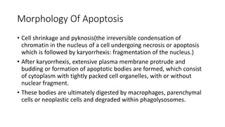
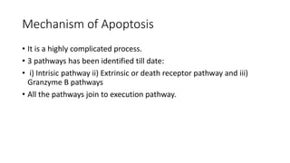
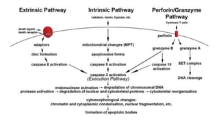
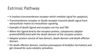
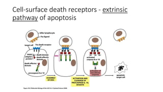
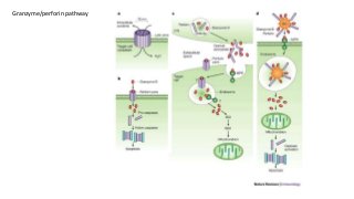
Apoptosis
A review on Programmed Cell Death
Introduction
• Apoptosis is a vital part of various processes including :-
i)normal cell turnover,
ii)proper development and functioning of the immune system,
iii)embryonic development and
iv)chemical-induced cell death.
• The process of programmed cell death, or apoptosis, is generally
characterized by distinct morphological characteristics and energy-
dependent biochemical mechanisms.
Introduction
• Apoptosis is defined as a distinctive and important mode of
“programmed” cell death, which involves the genetically determined
elimination of cells.
• The term apoptosis (a-po-toe-sis) was first used to describe a
morphologically distinct form of cell death.
• Apoptosis is a coordinated and often energy-driven process that
involves the activation of a group of cysteine proteases called
“caspases” and a complex cascade of events that link the initiating
stimuli to the final demise of the cell.
Introduction
• Apoptosis occurs normally in the body through development and
ageing to maintain the cell population.
• And also when cell is damaged by disease or toxins.
• It is opposite to mitosis.
• Protein triggered cell death occurs when certain specific protein is
attacked by activated “caspases” which are activated by external
stimuli like cell insults: DNA damage.
• Some cell have Fas or TNF binding receptor on cell membrane that
lead to apoptosis by ligand binding and protein cross-linking.
• Default death pathway of apoptosis are blocked by survival factor.
Necrosis Vs Apoptosis
• A variety of injurious stimulus such as heat, toxic, radiation can induce
apoptosis at low intensity but at higher dosage of such trigger can cause
necrosis.
• Necrosis is not organized cell death, affected cells burst to die and affect
the neighboring cells are affected.
• This eruption triggers a potentially damaging inflammatory response.
• But a cell that undergoes apoptosis dies neatly, without inflammatory
response.
• Oncosis leads to necrosis with karyolysis and cell swelling whereas
apoptosis leads to cell death with cell shrinkage, pyknosis, and
karyorrhexis.
Necrosis
6
Apoptosis
7
Morphology Of Apoptosis
• Cell shrinkage and pyknosis(the irreversible condensation of
chromatin in the nucleus of a cell undergoing necrosis or apoptosis
which is followed by karyorrhexis: fragmentation of the nucleus.)
• After karyorrhexis, extensive plasma membrane protrude and
budding or formation of apoptotic bodies are formed, which consist
of cytoplasm with tightly packed cell organelles, with or without
nuclear fragment.
• These bodies are ultimately digested by macrophages, parenchymal
cells or neoplastic cells and degraded within phagolysosomes.
Necrosis and Apoptosis
• Necrosis and Apoptosis can occur simultaneously depending upon
intensity and duration of stimulus, Caspases availability and extent of
ATP depletion.
• Apoptosis changes to necrosis if less caspases and ATP are available.
• Necrosis include cell swelling, disrupted cell organelle membrane and
lysosome membrane which leads to disruption of cell membrane
releasing intracellular matter into surrounding interstitial tissues.
Mechanism of Apoptosis
• It is a highly complicated process.
• 3 pathways has been identified till date:
• i) Intrisic pathway ii) Extrinsic or death receptor pathway and iii)
Granzyme B pathways
• All the pathways join to execution pathway.
Extrinsic Pathway
• It involves transmembrane receptor which mediate signal for apoptosis.
• Transmembrane receptor or Death receptor transmit death signal from
extracellular matrix to intracellular signaling.
• Example of death ligand and receptor are Fas and TNF.
• When this ligand bind to the receptor protein, cytoplasmic adaptor
protein(FADD) bind with the death domain of the receptor protein.
• Adaptor protein consist of two domain, death domain and death effector
domain.
• On death effector domain, inactive procaspases-8 bind(disc formation) and
get cleaved for auto-catalytic activation.
Cell-surface death receptors - extrinsic
pathway of apoptosis
Extrinsic pathway
• Activated caspase-8 activates execution caspase-3.
• After this it enters execution pathway.
• Execution pathway is similar for all the pathways except granzyme A
which doesn’t need caspase -3 for DNA cleavage.
• This activation by death receptor can be inhibited by c-FLIP which
binds to FADD and caspase-8.
Intrinsic Pathway
• Involves non-receptor mediated cell signaling that are produced inside cell,
with the help of Bak and Bax protein of Bcl-2 family and cytochrome c.
• Bak and Bax protein are activated by death promoting member of the Bcl-2
family, which are activated by various insults to the cell like DNA damage.
• These Bak and Bax protein reside in the mitochondrial outer
transmembrane, when activated, facilitates release of cytochrome c to
cytosol.
• These cytochrome c promote assembly of large adaptor protein(Apaf-1)
with procaspase-9 into wheel like structure called apoptosome, this
apoptosome is activated with caspase-9 which finally activates caspase-3
for execution pathway.
Intrinsic Pathway
Perforin/granzyme Pathway
• This is T-cell mediated cytotoxicity where sensitized CD8+ cells kill
antigen bearing cells.
• Cytotoxic effect on tumor bearing cell and virus infected cells.
• Involves secretion of transmembrane pore-forming
molecule(perforin) and cytoplasmic granules(granzyme A and B).
• Granzyme B cleaves protein at aspartate residue and activates
procaspase-10. It can utilize mitochondrial pathway(cleave Bid) or
directly activate caspase-3 for execution pathway.
• Granzyme A induce caspase independent apoptosis. It cleaves SET
complex(inhibits gene that causes DNA nicking), gene is not inhibited
by SET complex and thus DNA nicking takes place.
Granzyme/perforin pathway
Execution Pathway
• All the pathway leads to activation of caspase-3.
• Caspase-3 activate cytoplasmic endonuclease, which degrades nuclear
material, and proteases that degrade the nuclear and cytoskeletal proteins.
• Caspase-3 also induces cytoskeletal reorganization and disintegration of
the cell into apoptotic bodies.
• Phagocytic uptake of apoptotic cells is the last component of apoptosis.
• Phosphotidylserine on the outer leaflet of apoptotic cells facilitates
noninflammatory phagocytic recognition, allowing for their early uptake
and disposal.
Thank you

Slide share

  • 1.
    Apoptosis A review onProgrammed Cell Death
  • 2.
    Introduction • Apoptosis isa vital part of various processes including :- i)normal cell turnover, ii)proper development and functioning of the immune system, iii)embryonic development and iv)chemical-induced cell death. • The process of programmed cell death, or apoptosis, is generally characterized by distinct morphological characteristics and energy- dependent biochemical mechanisms.
  • 3.
    Introduction • Apoptosis isdefined as a distinctive and important mode of “programmed” cell death, which involves the genetically determined elimination of cells. • The term apoptosis (a-po-toe-sis) was first used to describe a morphologically distinct form of cell death. • Apoptosis is a coordinated and often energy-driven process that involves the activation of a group of cysteine proteases called “caspases” and a complex cascade of events that link the initiating stimuli to the final demise of the cell.
  • 4.
    Introduction • Apoptosis occursnormally in the body through development and ageing to maintain the cell population. • And also when cell is damaged by disease or toxins. • It is opposite to mitosis. • Protein triggered cell death occurs when certain specific protein is attacked by activated “caspases” which are activated by external stimuli like cell insults: DNA damage. • Some cell have Fas or TNF binding receptor on cell membrane that lead to apoptosis by ligand binding and protein cross-linking. • Default death pathway of apoptosis are blocked by survival factor.
  • 5.
    Necrosis Vs Apoptosis •A variety of injurious stimulus such as heat, toxic, radiation can induce apoptosis at low intensity but at higher dosage of such trigger can cause necrosis. • Necrosis is not organized cell death, affected cells burst to die and affect the neighboring cells are affected. • This eruption triggers a potentially damaging inflammatory response. • But a cell that undergoes apoptosis dies neatly, without inflammatory response. • Oncosis leads to necrosis with karyolysis and cell swelling whereas apoptosis leads to cell death with cell shrinkage, pyknosis, and karyorrhexis.
  • 6.
  • 7.
  • 8.
    Morphology Of Apoptosis •Cell shrinkage and pyknosis(the irreversible condensation of chromatin in the nucleus of a cell undergoing necrosis or apoptosis which is followed by karyorrhexis: fragmentation of the nucleus.) • After karyorrhexis, extensive plasma membrane protrude and budding or formation of apoptotic bodies are formed, which consist of cytoplasm with tightly packed cell organelles, with or without nuclear fragment. • These bodies are ultimately digested by macrophages, parenchymal cells or neoplastic cells and degraded within phagolysosomes.
  • 10.
    Necrosis and Apoptosis •Necrosis and Apoptosis can occur simultaneously depending upon intensity and duration of stimulus, Caspases availability and extent of ATP depletion. • Apoptosis changes to necrosis if less caspases and ATP are available. • Necrosis include cell swelling, disrupted cell organelle membrane and lysosome membrane which leads to disruption of cell membrane releasing intracellular matter into surrounding interstitial tissues.
  • 11.
    Mechanism of Apoptosis •It is a highly complicated process. • 3 pathways has been identified till date: • i) Intrisic pathway ii) Extrinsic or death receptor pathway and iii) Granzyme B pathways • All the pathways join to execution pathway.
  • 13.
    Extrinsic Pathway • Itinvolves transmembrane receptor which mediate signal for apoptosis. • Transmembrane receptor or Death receptor transmit death signal from extracellular matrix to intracellular signaling. • Example of death ligand and receptor are Fas and TNF. • When this ligand bind to the receptor protein, cytoplasmic adaptor protein(FADD) bind with the death domain of the receptor protein. • Adaptor protein consist of two domain, death domain and death effector domain. • On death effector domain, inactive procaspases-8 bind(disc formation) and get cleaved for auto-catalytic activation.
  • 14.
    Cell-surface death receptors- extrinsic pathway of apoptosis
  • 15.
    Extrinsic pathway • Activatedcaspase-8 activates execution caspase-3. • After this it enters execution pathway. • Execution pathway is similar for all the pathways except granzyme A which doesn’t need caspase -3 for DNA cleavage. • This activation by death receptor can be inhibited by c-FLIP which binds to FADD and caspase-8.
  • 16.
    Intrinsic Pathway • Involvesnon-receptor mediated cell signaling that are produced inside cell, with the help of Bak and Bax protein of Bcl-2 family and cytochrome c. • Bak and Bax protein are activated by death promoting member of the Bcl-2 family, which are activated by various insults to the cell like DNA damage. • These Bak and Bax protein reside in the mitochondrial outer transmembrane, when activated, facilitates release of cytochrome c to cytosol. • These cytochrome c promote assembly of large adaptor protein(Apaf-1) with procaspase-9 into wheel like structure called apoptosome, this apoptosome is activated with caspase-9 which finally activates caspase-3 for execution pathway.
  • 17.
  • 18.
    Perforin/granzyme Pathway • Thisis T-cell mediated cytotoxicity where sensitized CD8+ cells kill antigen bearing cells. • Cytotoxic effect on tumor bearing cell and virus infected cells. • Involves secretion of transmembrane pore-forming molecule(perforin) and cytoplasmic granules(granzyme A and B). • Granzyme B cleaves protein at aspartate residue and activates procaspase-10. It can utilize mitochondrial pathway(cleave Bid) or directly activate caspase-3 for execution pathway. • Granzyme A induce caspase independent apoptosis. It cleaves SET complex(inhibits gene that causes DNA nicking), gene is not inhibited by SET complex and thus DNA nicking takes place.
  • 19.
  • 20.
    Execution Pathway • Allthe pathway leads to activation of caspase-3. • Caspase-3 activate cytoplasmic endonuclease, which degrades nuclear material, and proteases that degrade the nuclear and cytoskeletal proteins. • Caspase-3 also induces cytoskeletal reorganization and disintegration of the cell into apoptotic bodies. • Phagocytic uptake of apoptotic cells is the last component of apoptosis. • Phosphotidylserine on the outer leaflet of apoptotic cells facilitates noninflammatory phagocytic recognition, allowing for their early uptake and disposal.
  • 21.