The document summarizes key aspects of apoptosis including:
- The origins and definition of the term apoptosis from Greek meaning "falling leaves".
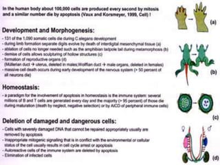
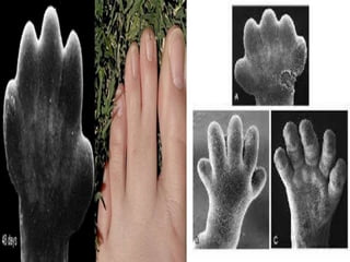
- The significance of apoptosis in development and maintenance of tissues by removing excess or damaged cells.
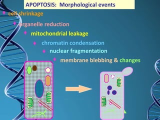
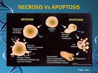
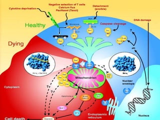
- The morphological features of apoptosis including membrane blebbing, nuclear fragmentation, and formation of apoptotic bodies.
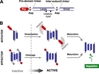
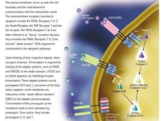
- The molecular mechanisms including caspase signaling pathways like the intrinsic pathway involving mitochondria and the extrinsic pathway involving death receptors.
- Regulatory mechanisms involving proteins like Bcl-2 that balance survival and death signals.
- Dysregulation of apoptosis can lead to diseases like cancer, autoimmune disorders, and HIV infection.


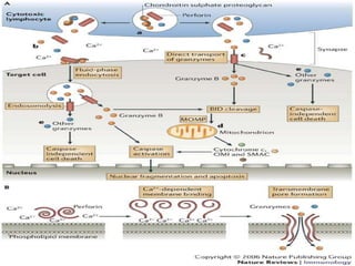
![Granzyme mediated apoptosis
Granzymes are serine proteases that are released by cytoplasmic
granules within cytotoxic T cells and natural killer (NK) cells.
They induce programmed cell death in the target cell, thus
eliminating cells that have become cancerous or are infected
with viruses or bacteria. The granzymes also kill bacteria[ and
inhibit viral replication. In NK cells and T cells, the granzymes are
packaged in cytotoxic granules with perforin Other locations that
granzymes can be detected are in the rough endoplasmic
reticulum, golgi complex, and the trans-golgi reticulum. The
contents of the cytotoxic granules function to permit entry of
the granzymes into the target cell cytosol. The granules are
released into an immune synapse formed with a target cell,
where perforin mediates the delivery of the granzymes
into endosomes in the target cell, and finally into the target cell
cytosol. Granzymes are identified as being part of the serine
esterase family.[3]](https://image.slidesharecdn.com/apoptosis-160304131254/85/Apoptosis-32-320.jpg)


![Apoptosis and Cancer
• Apoptosis does not occur in
Cancer
• Cancerous cells trick and skip
Apoptosis in number of ways
Inactivation of p53 [shooting the
guard]
Produce Bcl-2 or a protein which
mimics Bcl-2
Inhibits expression of Apaf-1](https://image.slidesharecdn.com/apoptosis-160304131254/85/Apoptosis-43-320.jpg)
![Apoptosis and Autoimmune
Disease
Autoimmune Lymph
Proliferative
Syndrome[ALPS]
Apoptosis doesnot
occur in self
reactive T & B cells
RBC
Hemolytic
Anemia
Neutrophil
Neutrope
nia
Platelets
Thromboc
ytopenia](https://image.slidesharecdn.com/apoptosis-160304131254/85/Apoptosis-44-320.jpg)

![Overview
Apoptosis is a good thing
Too little of a good thing
is bad [Cancer]
Too much of a good
thing is also bad [HIV]](https://image.slidesharecdn.com/apoptosis-160304131254/85/Apoptosis-46-320.jpg)


