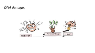
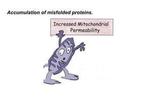
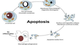
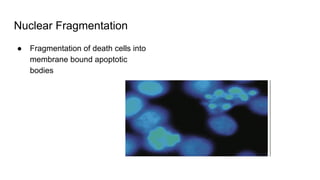
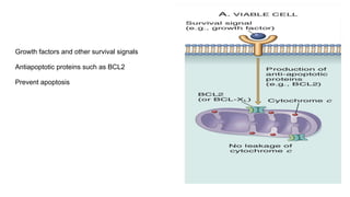
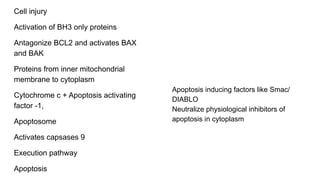
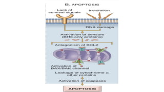
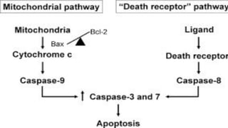
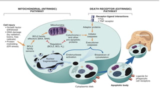
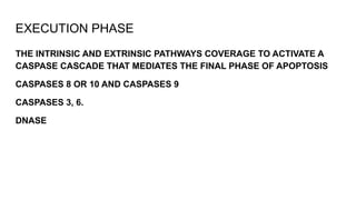
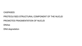
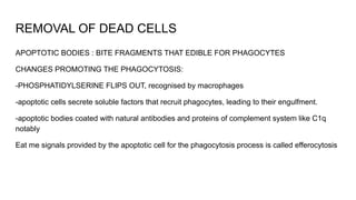
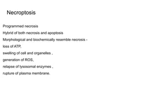
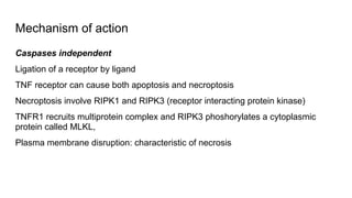
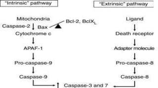
APOPTOSIS
Dr. Keerthika
SHORT TOPIC:
DEFINITION
Apoptosis is a type of cell death that is induced by a tightly
regulated suicide program in which cells destined to die
activate intrinsic enzymes that degrade the cells’ genomic
DNA and nuclear and cytoplasmic proteins.
PROGRAMMED CELL DEATH
ETYMOLOGY
APOPTOSIS: IN GREEK - FALLING OFF of leaves from a tree.
HIPPOCRATES- used the term to mean FALLING OFF THE BONES
GALEN- extended the term to mean DROPPING OF THE SCABS
James CORMACK- reintroduced the term for medical use after two decades
Pronunciation debate between second P being silenced or pronounced, John kerr
in his paper thanked comrack and mentioned the second of p is silent like in ptosis
and the stress should be given in the penultimate word.
DISCOVERIES
1842- German scientist CARL VOGT - principle of apoptosis
1885- Anatomist WALTHER FLEMMING -precise the process of programmed cell
death
1965- JOHN KERR was able to distinguish apoptosis from traumatic cell death.
JOHN KEER received the PAUL EHRLICH AND LUDWIG DARMSTAEDTER PRIZE
ON MARCH 14, 2000, shared with Biologist H.ROBERT HORVITZ
2002- NOBEL PRIZE IN MEDICINE was awarded to SYDNEY BRENNER, H.ROBERT
HORVITZ and JOHN SULSTON for their work identifying GENES THAT CONTROL
APOPTOSIS in nematode c.elegans and homologous of these genes function in
humans to regulate apoptosis.
APOPTOSIS
CAUSES
PHYSIOLOGICAL PATHOLOGICAL
● Death by apoptosis is a normal phenomenon that serves to
eliminate cells that are no longer needed, or as a
mechanism to maintain a constant number of various cell
populations in tissues.
● Cells undergo apoptosis because they are deprived of
necessary survival signals,
● or they receive pro-apoptotic signals from other cells
● And they are removed by phagocytosis
PHYSIOLOGICAL CAUSES
The removal of supernumerary cells during development:
Involution of hormone-dependent tissues on hormone withdrawal
Cell turnover in proliferating cell populations-HOMEOSTASIS
Elimination of potentially harmful self-reactive LYMPHOCYTES
DDeath of host cells that have served their useful purpose
PATHOLOGICAL CAUSES
Apoptosis eliminates cells that are injured beyond repair
without eliciting a host reaction, thus limiting collateral tissue damage.
Death by apoptosis is responsible for loss of cells in a variety of
pathologic states:
DNA damage.
Accumulation of misfolded proteins.
Cytotoxic T Lymphocytes-mediated mechanism
apoptosis of infected cells in an attempt to eliminate reservoirs of infection.
Pathologic atrophy in parenchymal organs
Morphologic and Biochemical
Changes in Apoptosis
Cell shrinkage
● Cell size reduced
● Cytoplasm dense and
eosinophilic
● Organelles : relatively normal are
more tightly packed.
Chromatin condensation
● MOST CHARACTERISTIC
FEATURE OF APOPTOSIS
● Chromatin aggregates
peripherally, under nuclear
membrane
Cytoplasmic blebs and Apoptotic bodies
● Extensive surface membrane
blebbing
Nuclear Fragmentation
● Fragmentation of death cells into
membrane bound apoptotic
bodies
Phagocytosis
Apoptotic bodies are rapidly ingested by phagocytes and degraded by the
phagocyte’s lysosomal enzymes.
MECHANISM OF APOPTOSIS
APOPTOSIS results from the activation of enzymes called CASPASES
CASPASES
● Protases
● Containing a Cysteine in their active site
● Cleave proteins after Aspartic residues
Presence of activated caspase is the marker for apoptosis
Process of Apoptosis
1.Initiation phase :
Some caspases become catalytically
active and unleash a cascade of
other caspases
2.Execution phase :
Terminal caspases trigger cellular
fragmentation
Regulation of these enzymes depends on a finely tuned balance b/w
the abundance and activity of apoptotic and anti-apoptotic proteins.
-CYTOCHROME C
-PRO APOPTOTIC CELLS
EXTRINSIC PATHWAY
INITIATED BY ENGAGEMENT OF PLASMA MEMBRANE DEATH RECEPTORS
Death receptors:
TNF that contain a cytoplasmic domain involved in protein protein interaction.
Important in delivering apoptotic signalling
Some TNFr don’t have cytoplasmic death domains, their function is to activate inflammatory cascade
system.
Best known death receptor: TNFr1 and its related protein called Fas (CD 95). The ligand of Fas is
called Fas L (Fas Ligand).
FasL is expressed in T cells that recognise self antigens and on some CTLs that kill virus infected and
tumor cells.
DEATH INDUCING MOLECULE (FasL)
DEATH RECEPTORS (DEATH BUTTONS)-
TNF RECEPTOR
Fas associated death domain (FADD)
Autocatalytic caspase activation
INITIATOR CASPASE:
Procaspase 8 OR 10 (INACTIVE)
Caspase 8 OR 10 (ACTIVE)
EXECUTIONER CASPASE (CASPASE 3,6,7)
DNAase
APOPTOSIS
INTRINSIC PATHWAY
Responsible for apoptosis in most physiological and pathological
situations.
Increased permeability of the mitochondrial outer membrane with consequent
release of death inducing molecules from the mitochondrial intermembrane space
into cytoplasm.
Mitochondria is a double edged sword:
-produce energy that sustains cell viability
-when released into cytoplasm, initiates suicide program apoptosis
Bcl family
Release of pro-apoptotic proteins such as cytochrome C is determined by the
integrity of the outer mitochondrial membrane which is tightly controlled by bcl2
family of the proteins.
There are more than 20 members in the bcl family and they divided into three
groups based upon their pro or anti apoptotic effect
bcl 2 is commonly expressed gene.
Anti- apoptotic proteins:
Also known as pro-life
BCL2, BCLX and MCL1
Posses BH(1-4 domains)
Prevent cleavage of cytochrome C by
keeping the mitochondrial membrane
impermeable
Pro-apototic proteins:
Also known as Pro-death proteins
BAX, BAK
Posses BH(1-3 domains)
assumed , they form a channel in the
outer membrane that allows
cytochrome C leakage from
intramembranous space
Regulated apoptosis initators:
BAD, BIM, BID, PUMA, NOXA
One BH domain, the third of the four BH domain- BH3 only domain
Following cellular stress and damage, upregulated and activated they can initiate
apoptosis.
Growth factors and other survival signals
Antiapoptotic proteins such as BCL2
Prevent apoptosis
Cell injury
Activation of BH3 only proteins
Antagonize BCL2 and activates BAX
and BAK
Proteins from inner mitochondrial
membrane to cytoplasm
Cytochrome c + Apoptosis activating
factor -1,
Apoptosome
Activates capsases 9
Execution pathway
Apoptosis
Apoptosis inducing factors like Smac/
DIABLO
Neutralize physiological inhibitors of
apoptosis in cytoplasm
EXECUTION PHASE
THE INTRINSIC AND EXTRINSIC PATHWAYS COVERAGE TO ACTIVATE A
CASPASE CASCADE THAT MEDIATES THE FINAL PHASE OF APOPTOSIS
CASPASES 8 OR 10 AND CASPASES 9
CASPASES 3, 6.
DNASE
CASPASES:
PROTEOLYSES STRUCTURAL COMPONENT OF THE NUCLEI
PROMOTES FRAGMENTATION OF NUCLEI
DNAse
DNA degradation
REMOVAL OF DEAD CELLS
APOPTOTIC BODIES : BITE FRAGMENTS THAT EDIBLE FOR PHAGOCYTES
CHANGES PROMOTING THE PHAGOCYTOSIS:
-PHOSPHATIDYLSERINE FLIPS OUT, recognised by macrophages
-apoptotic cells secrete soluble factors that recruit phagocytes, leading to their engulfment.
-apoptotic bodies coated with natural antibodies and proteins of complement system like C1q
notably
Eat me signals provided by the apoptotic cell for the phagocytosis process is called efferocytosis
Efferocyotsis:
Efficient
Dead cells disappears within minutes without leaving a trace
Production of pro inflammatory cytokines is reduced
Necroptpsis
Pyroptosis
Ferroptosis
Other mechanism of cell death
Necroptosis
Programmed necrosis
Hybrid of both necrosis and apoptosis
Morphological and biochemically resemble necrosis -
loss of ATP,
swelling of cell and organelles ,
generation of ROS,
relapse of lysosomal enzymes ,
rupture of plasma membrane.
Mechanism of action
Caspases independent
Ligation of a receptor by ligand
TNF receptor can cause both apoptosis and necroptosis
Necroptosis involve RIPK1 and RIPK3 (receptor interacting protein kinase)
TNFR1 recruits multiprotein complex and RIPK3 phoshorylates a cytoplasmic
protein called MLKL,
Plasma membrane disruption: characteristic of necrosis
Causes of necroptosis
PHYSIOLOGICAL NECROPTOSIS:
Mammalian bone growth
PATHOLOGICAL NECROPTOSIS :
Cell death in ischemia-repressusion injury, neurodegerative disease such as
Parkinsonism disease
Necroptosis also acts as a backup mechanism in host defence against certain
viruses that encode caspase inhibitors. Eg: cytomegalovirus
Pyroptosis
Form of apoptosis that is accompanied by the release of the fever inducing
cytokines IL-1
Microbial products that enter infected cells are recognised by cytoplasmic innate
immune receptors and can activate multiprotein complex called inflamasome.
Activation of caspase -1, which cleaves IL-1 and release the biological active form
Caspase 4 & 5 also induce cell death.
Ferroproteins
Discovered in 2012
Cell death triggered when excessive intracellular levels of iron or reactive oxygen
species overwhelming the gulathione dependent antioxidant defences to cause a
unchecked membrane lipid peroxidation
Lipid peroxidation
Loss of plasma permeability
Cell death resembling necrosis
However regulated by specific signals unlike necrosis, can be prevented by
reducing iron levels.
Characteristic features:
Loss of mitochondrial Cristae
Ruptured outer mitochondrial membrane
Causes:
Linked to cell death in variety of human pathologies including cancer,
neurodegenerative disease and stroke.
Apoptosis.pdf
Apoptosis.pdf

Apoptosis.pdf

  • 1.
  • 2.
    DEFINITION Apoptosis is atype of cell death that is induced by a tightly regulated suicide program in which cells destined to die activate intrinsic enzymes that degrade the cells’ genomic DNA and nuclear and cytoplasmic proteins. PROGRAMMED CELL DEATH
  • 3.
    ETYMOLOGY APOPTOSIS: IN GREEK- FALLING OFF of leaves from a tree. HIPPOCRATES- used the term to mean FALLING OFF THE BONES GALEN- extended the term to mean DROPPING OF THE SCABS James CORMACK- reintroduced the term for medical use after two decades Pronunciation debate between second P being silenced or pronounced, John kerr in his paper thanked comrack and mentioned the second of p is silent like in ptosis and the stress should be given in the penultimate word.
  • 4.
    DISCOVERIES 1842- German scientistCARL VOGT - principle of apoptosis 1885- Anatomist WALTHER FLEMMING -precise the process of programmed cell death 1965- JOHN KERR was able to distinguish apoptosis from traumatic cell death. JOHN KEER received the PAUL EHRLICH AND LUDWIG DARMSTAEDTER PRIZE ON MARCH 14, 2000, shared with Biologist H.ROBERT HORVITZ 2002- NOBEL PRIZE IN MEDICINE was awarded to SYDNEY BRENNER, H.ROBERT HORVITZ and JOHN SULSTON for their work identifying GENES THAT CONTROL APOPTOSIS in nematode c.elegans and homologous of these genes function in humans to regulate apoptosis.
  • 5.
  • 6.
    ● Death byapoptosis is a normal phenomenon that serves to eliminate cells that are no longer needed, or as a mechanism to maintain a constant number of various cell populations in tissues. ● Cells undergo apoptosis because they are deprived of necessary survival signals, ● or they receive pro-apoptotic signals from other cells ● And they are removed by phagocytosis PHYSIOLOGICAL CAUSES
  • 7.
    The removal ofsupernumerary cells during development:
  • 8.
    Involution of hormone-dependenttissues on hormone withdrawal
  • 9.
    Cell turnover inproliferating cell populations-HOMEOSTASIS
  • 10.
    Elimination of potentiallyharmful self-reactive LYMPHOCYTES
  • 11.
    DDeath of hostcells that have served their useful purpose
  • 12.
    PATHOLOGICAL CAUSES Apoptosis eliminatescells that are injured beyond repair without eliciting a host reaction, thus limiting collateral tissue damage. Death by apoptosis is responsible for loss of cells in a variety of pathologic states:
  • 13.
  • 14.
  • 15.
    Cytotoxic T Lymphocytes-mediatedmechanism apoptosis of infected cells in an attempt to eliminate reservoirs of infection.
  • 16.
    Pathologic atrophy inparenchymal organs
  • 17.
  • 19.
    Cell shrinkage ● Cellsize reduced ● Cytoplasm dense and eosinophilic ● Organelles : relatively normal are more tightly packed.
  • 20.
    Chromatin condensation ● MOSTCHARACTERISTIC FEATURE OF APOPTOSIS ● Chromatin aggregates peripherally, under nuclear membrane
  • 21.
    Cytoplasmic blebs andApoptotic bodies ● Extensive surface membrane blebbing
  • 22.
    Nuclear Fragmentation ● Fragmentationof death cells into membrane bound apoptotic bodies
  • 23.
    Phagocytosis Apoptotic bodies arerapidly ingested by phagocytes and degraded by the phagocyte’s lysosomal enzymes.
  • 24.
  • 25.
    APOPTOSIS results fromthe activation of enzymes called CASPASES CASPASES ● Protases ● Containing a Cysteine in their active site ● Cleave proteins after Aspartic residues Presence of activated caspase is the marker for apoptosis
  • 26.
    Process of Apoptosis 1.Initiationphase : Some caspases become catalytically active and unleash a cascade of other caspases 2.Execution phase : Terminal caspases trigger cellular fragmentation Regulation of these enzymes depends on a finely tuned balance b/w the abundance and activity of apoptotic and anti-apoptotic proteins.
  • 27.
  • 28.
    EXTRINSIC PATHWAY INITIATED BYENGAGEMENT OF PLASMA MEMBRANE DEATH RECEPTORS Death receptors: TNF that contain a cytoplasmic domain involved in protein protein interaction. Important in delivering apoptotic signalling Some TNFr don’t have cytoplasmic death domains, their function is to activate inflammatory cascade system. Best known death receptor: TNFr1 and its related protein called Fas (CD 95). The ligand of Fas is called Fas L (Fas Ligand). FasL is expressed in T cells that recognise self antigens and on some CTLs that kill virus infected and tumor cells.
  • 29.
    DEATH INDUCING MOLECULE(FasL) DEATH RECEPTORS (DEATH BUTTONS)- TNF RECEPTOR Fas associated death domain (FADD) Autocatalytic caspase activation INITIATOR CASPASE: Procaspase 8 OR 10 (INACTIVE) Caspase 8 OR 10 (ACTIVE) EXECUTIONER CASPASE (CASPASE 3,6,7) DNAase APOPTOSIS
  • 30.
    INTRINSIC PATHWAY Responsible forapoptosis in most physiological and pathological situations. Increased permeability of the mitochondrial outer membrane with consequent release of death inducing molecules from the mitochondrial intermembrane space into cytoplasm. Mitochondria is a double edged sword: -produce energy that sustains cell viability -when released into cytoplasm, initiates suicide program apoptosis
  • 31.
    Bcl family Release ofpro-apoptotic proteins such as cytochrome C is determined by the integrity of the outer mitochondrial membrane which is tightly controlled by bcl2 family of the proteins. There are more than 20 members in the bcl family and they divided into three groups based upon their pro or anti apoptotic effect bcl 2 is commonly expressed gene.
  • 32.
    Anti- apoptotic proteins: Alsoknown as pro-life BCL2, BCLX and MCL1 Posses BH(1-4 domains) Prevent cleavage of cytochrome C by keeping the mitochondrial membrane impermeable Pro-apototic proteins: Also known as Pro-death proteins BAX, BAK Posses BH(1-3 domains) assumed , they form a channel in the outer membrane that allows cytochrome C leakage from intramembranous space
  • 33.
    Regulated apoptosis initators: BAD,BIM, BID, PUMA, NOXA One BH domain, the third of the four BH domain- BH3 only domain Following cellular stress and damage, upregulated and activated they can initiate apoptosis.
  • 34.
    Growth factors andother survival signals Antiapoptotic proteins such as BCL2 Prevent apoptosis
  • 35.
    Cell injury Activation ofBH3 only proteins Antagonize BCL2 and activates BAX and BAK Proteins from inner mitochondrial membrane to cytoplasm Cytochrome c + Apoptosis activating factor -1, Apoptosome Activates capsases 9 Execution pathway Apoptosis Apoptosis inducing factors like Smac/ DIABLO Neutralize physiological inhibitors of apoptosis in cytoplasm
  • 39.
    EXECUTION PHASE THE INTRINSICAND EXTRINSIC PATHWAYS COVERAGE TO ACTIVATE A CASPASE CASCADE THAT MEDIATES THE FINAL PHASE OF APOPTOSIS CASPASES 8 OR 10 AND CASPASES 9 CASPASES 3, 6. DNASE
  • 40.
    CASPASES: PROTEOLYSES STRUCTURAL COMPONENTOF THE NUCLEI PROMOTES FRAGMENTATION OF NUCLEI DNAse DNA degradation
  • 41.
    REMOVAL OF DEADCELLS APOPTOTIC BODIES : BITE FRAGMENTS THAT EDIBLE FOR PHAGOCYTES CHANGES PROMOTING THE PHAGOCYTOSIS: -PHOSPHATIDYLSERINE FLIPS OUT, recognised by macrophages -apoptotic cells secrete soluble factors that recruit phagocytes, leading to their engulfment. -apoptotic bodies coated with natural antibodies and proteins of complement system like C1q notably Eat me signals provided by the apoptotic cell for the phagocytosis process is called efferocytosis
  • 42.
    Efferocyotsis: Efficient Dead cells disappearswithin minutes without leaving a trace Production of pro inflammatory cytokines is reduced
  • 43.
  • 44.
    Necroptosis Programmed necrosis Hybrid ofboth necrosis and apoptosis Morphological and biochemically resemble necrosis - loss of ATP, swelling of cell and organelles , generation of ROS, relapse of lysosomal enzymes , rupture of plasma membrane.
  • 45.
    Mechanism of action Caspasesindependent Ligation of a receptor by ligand TNF receptor can cause both apoptosis and necroptosis Necroptosis involve RIPK1 and RIPK3 (receptor interacting protein kinase) TNFR1 recruits multiprotein complex and RIPK3 phoshorylates a cytoplasmic protein called MLKL, Plasma membrane disruption: characteristic of necrosis
  • 46.
    Causes of necroptosis PHYSIOLOGICALNECROPTOSIS: Mammalian bone growth PATHOLOGICAL NECROPTOSIS : Cell death in ischemia-repressusion injury, neurodegerative disease such as Parkinsonism disease Necroptosis also acts as a backup mechanism in host defence against certain viruses that encode caspase inhibitors. Eg: cytomegalovirus
  • 48.
    Pyroptosis Form of apoptosisthat is accompanied by the release of the fever inducing cytokines IL-1 Microbial products that enter infected cells are recognised by cytoplasmic innate immune receptors and can activate multiprotein complex called inflamasome. Activation of caspase -1, which cleaves IL-1 and release the biological active form Caspase 4 & 5 also induce cell death.
  • 50.
    Ferroproteins Discovered in 2012 Celldeath triggered when excessive intracellular levels of iron or reactive oxygen species overwhelming the gulathione dependent antioxidant defences to cause a unchecked membrane lipid peroxidation
  • 51.
    Lipid peroxidation Loss ofplasma permeability Cell death resembling necrosis However regulated by specific signals unlike necrosis, can be prevented by reducing iron levels.
  • 53.
    Characteristic features: Loss ofmitochondrial Cristae Ruptured outer mitochondrial membrane Causes: Linked to cell death in variety of human pathologies including cancer, neurodegenerative disease and stroke.