Sl.no Apoptosis Necrosis
1.
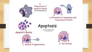
Apoptosisis a regular process of death of the cell that occurs in the
body where the cell itself takes part in the death
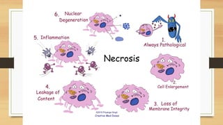
Necrosis is a cellular process of death occurring when the cells are
highly exposed to extreme external conditions
2. It is a natural process and not caused by external factors It is caused by external agents such as infection, trauma, and toxins.
3. The organelles are still functional even after the death of the cell The organelles are not functional after the death of the cell
4. The cell membrane breaks into several apoptotic bodies The cell membrane breaks and releases the cell contents
5.
This process is said to be a bit beneficial. But, is known to be
abnormal if in case the cellular processes that keep the body balanced
cause many cell deaths or even too few.
It is always damaging or harmful
6. No symptoms are observed during the process of apoptosis
The symptoms like Inflammation, tissue death and decreased blood
flow at the infected site are observed during the process
7. It is caused due to the self-generated signals within a cell.
It is caused by Bacterial and fungal infections, mycobacterial
infections, denatured proteins, pancreatitis, or by the deposits of
antibodies and antigens
8. Not much treatment is required in the case of apoptosis
Necrosis, when untreated, is said to be very dangerous and can prove
to be fatal. Hence, medical treatment is very much necessary in case
of necrosis.
9.
This process does not require energy since the enzymes carry out the
process.
This process requires energy to carry out the process
Difference between Apoptosis and Necrosis
5.
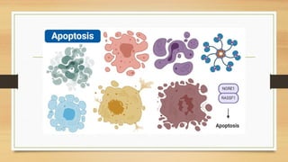
• Apoptosis isa normal genetically programmed cell death
where an aging cell at the end of its life cycle shrinks and
its remaining fragments are phagocytosed without any
inflammatory reaction.
• The term apoptosis was first introduced in a paper in
1972 by Kerr, Wyllie, and Currie to describe a
morphologically distinct type of cell death.
• It consists of a series of biochemical changes that lead to
changes in the cell’s morphology or death.
• It results in the death of 50 to 70 billion cells per day in
an average adult human being.
• It is also termed as ‘cellular suicide’ as cells undergo a
highly regulated process for the programmed removal of
cells from the body.
7.
• Most cellsare provided with an in-built mechanism of apoptosis as a part of the cell cycle.
• This mechanism allows the body to get rid of unnecessary cells or infected cells.
• Apoptosis is considered a vital part of various processes including normal cell cycle,
proper development and functioning of the immune system, embryonic development,
and chemical-induced cell death.
• Apoptosis is a part of development as it is essential in the differentiation of a mass of
tissue into various groups.
• Apoptosis occurs in cells that might have been infected with viruses or might even be
cancerous.
• This process usually takes place when the cell detects defects in the DNA and is not able
to repair it.
• Apoptosis is also an essential part of the immune system as it clears the pathogen-specific
immune cells once the foreign particle is removed from the body.
• This also helps to remove the immune cells that might react against the body’s cells and
cause autoimmune diseases.
• Another reason for apoptosis is to maintain homeostasis in the body by removing old cells
to make space for the new ones.
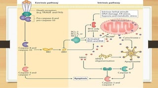
9.
• Three differentpathways work on different mechanisms to
achieve apoptosis.
• All three of these pathways converge at the same terminal
pathway, which results in the sequential degradation of cellular
organelles.
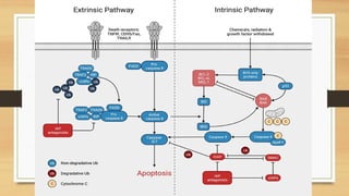
10.
1. Extrinsic ordeath receptor pathway
• The extrinsic pathway that initiates apoptosis involves transmembrane receptor-
mediated interactions.
• These interactions take place between ligands and their corresponding death receptors
that are all part of the tumor necrosis factor (TNF) family.
• All members of the TNF receptor family share a common cysteine-rich extracellular
domain with about 80 amino acids called the “death domain”.
• The death domain plays a vital role in transmitting the death signal from the cell
surface to the intracellular signaling pathways.
12.
• The eventsor interactions that take place in the extrinsic phase of apoptosis involve
two models; FasL/FasR and TNF-α/TNFR1 models, both of which include the
clustering and binding of receptors and their ligands.
• Upon ligand binding, cytoplasmic adapter proteins are activated, which causes the
receptors to exhibit death domains.
• The binding of FasL to FasR results in the activation of the adapter protein FADD
whereas the binding of TNF ligand (TNF α) to TNF receptor (TNFR1) results in the
binding of the adapter protein TRADD with activation of FADD and RIP.
• These events cause the dimerization of the death effector domain, causing FADD to
bind with procaspase-8.
• As a result of the binding, a death-inducing signaling complex (DISC) is formed,
resulting in the auto-catalytic activation of procaspase-8.
• Once caspase-8 is activated, the terminal phase or execution phase of apoptosis is
triggered.
13.
2. The intrinsicor mitochondrial pathway
• The intrinsic pathway that initiates apoptosis involves a series of non-receptor-mediated
processes that produce intracellular signals and act directly on targets within the cell.
• This pathway involves mitochondrial-initiated events.
• The factors that initiate the intrinsic pathway produce intracellular signals that might act in
either a positive or negative fashion.
• Negative signals include the absence of certain growth factors, cytokines, and hormones
that can lead to failure of inhibition of death programs, thereby triggering apoptosis.
• In simple words, the withdrawal of factors causes loss of apoptotic suppression and
subsequent activation of apoptosis.
• The factors that act positively include, radiation, toxins, hypoxia, hyperthermia, viral
infections, free radicals, among others.
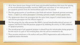
14.
• All ofthese factors cause changes in the inner mitochondrial membrane that causes the opening
of the mitochondrial permeability transition (MPT) pore and release of two main groups of
pro-apoptotic proteins from the intermembrane space into the cytosol.
• The first group consists of cytochrome c that binds and activates Apoptotic protease-activating
factor – 1(Apaf-1) as well as procaspase-9, forming a protein complex termed, apoptosome.
• The apoptosome cleaves the procaspase into the active form, caspase 9, which further cleaves
and activates procaspase into the effector caspase 3.
• The first group also has other proteins like SMACs (second mitochondria-derived activator of
caspases) and HtrA2/Omi that promote apoptosis by inhibiting the activity of IAPs (inhibitors
of apoptosis proteins).
• The second group of pro-apoptotic proteins is released from the mitochondria during apoptosis,
but this occurs as a part of the terminal phase after the cell has committed to die.
• These proteins translocate to the nucleus and cause DNA fragmentation and condensation of
peripheral nuclear chromatin.
16.
3. Perforin/granzyme pathway
•Perforin/granzyme pathway is a novel pathway employed by cytotoxic T lymphocytes that exert their
cytotoxic effects on tumor cells and virus-infected cells.
• This involves secretion of the transmembrane pore-forming molecule, perforin, with a subsequent
release of cytoplasmic granules through the pore and towards the target cell.
• The granules consist of two crucial serine proteases; granzyme A and granzyme B that activate
different proteins in the pathway.
• Granzyme B cleaves proteins at aspartate residues and therefore activates procaspase-10 and can cleave
factors like ICAD (Inhibitor of Caspase Activated DNAse.
• It has also been observed that granzyme B can utilize the mitochondrial pathway for amplification of
the death signal by induction of cytochrome c release.
17.
• But granzymeB can also directly activate caspase-3. In this pathway, there is a direct
induction of the execution phase of apoptosis.
• Granzyme A also has an essential role in cytotoxic T cell-induced apoptosis and
activates caspase-independent pathways.
• As granzyme A reaches the cell, it activates DNA nicking by DNAse enzyme that
prevents cancer through the induction of tumor cell apoptosis.
• Granzyme A protease cleaves the SET complex that inhibits the production of the
DNAse enzyme.
• The proteins that make up the SET complex together protect chromatin and DNA
structure.
• Thus, the inactivation of the SET complex by granzyme A contributes to apoptosis
by blocking the maintenance of DNA and chromatin structure integrity.
18.
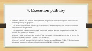
4. Execution pathway
•Both the extrinsic and intrinsic pathways end at the point of the execution phase, considered the
terminal pathway of apoptosis.
• This phase of apoptosis is initiated by the activation of various caspases that activate cytoplasmic
endonucleases and proteases.
• The cytoplasmic endonucleases degrade the nuclear material, whereas the proteases degrade the
nuclear and cytoskeletal proteins.
• Caspase-3 is the most important protein of the executioner caspases and is activated by any of the
initiator caspases (caspase-8, caspase-9, or caspase-10).
• Caspase-3 precisely activates the endonuclease Caspase-activated DNase (CAD). CAD then causes
chromatin condensation by degrading chromosomal DNA within the nuclei.
19.
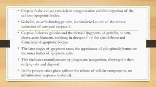
• Caspase-3 alsocauses cytoskeletal reorganization and disintegration of the
cell into apoptotic bodies.
• Gelsolin, an actin-binding protein, is considered as one of the critical
substrates of activated caspase-3.
• Caspase-3 cleaves gelsolin and the cleaved fragments of gelsolin, in turn,
cleave actin filaments, resulting in disruption of the cytoskeleton and
formation of apoptotic bodies.
• The later stages of apoptosis cause the appearance of phosphatidylserine on
the outer leaflet of apoptotic cells.
• This facilitates noninflammatory phagocytic recognition, allowing for their
early uptake and disposal.
• As the process takes place without the release of cellular components, no
inflammatory response is elicited.
20.
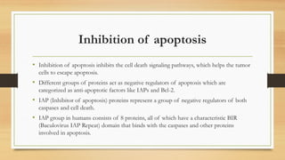
Inhibition of apoptosis
•Inhibition of apoptosis inhibits the cell death signaling pathways, which helps the tumor
cells to escape apoptosis.
• Different groups of proteins act as negative regulators of apoptosis which are
categorized as anti-apoptotic factors like IAPs and Bcl-2.
• IAP (Inhibitor of apoptosis) proteins represent a group of negative regulators of both
caspases and cell death.
• IAP group in humans consists of 8 proteins, all of which have a characteristic BIR
(Baculovirus IAP Repeat) domain that binds with the caspases and other proteins
involved in apoptosis.
21.
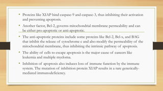
• Proteins likeXIAP bind caspase-9 and caspase-3, thus inhibiting their activation
and preventing apoptosis.
• Another factor, Bcl-2, governs mitochondrial membrane permeability and can
be either pro-apoptotic or anti-apoptotic.
• The anti-apoptotic proteins include some proteins like Bcl-2, Bcl-x, and BAG
that inhibit the release of cytochrome c and also modify the permeability of the
mitochondrial membrane, thus inhibiting the intrinsic pathway of apoptosis.
• The ability of cells to escape apoptosis is the major cause of cancers like
leukemia and multiple myeloma.
• Inhibition of apoptosis also induces loss of immune function by the immune
system. The mutation of inhibition protein XIAP results in a rare genetically-
mediated immunodeficiency.
22.
Regulation of apoptosis
•Several proteins and genes regulate apoptosis. Specific families of proteins are involved
in the regulation of apoptosis in various steps.
• Among all the factors, IAPs and Bcl-2 are two of the most important proteins involved
that decide whether the apoptosis is going to complete or inhibit.
• The extrinsic pathway of apoptosis is inhibited by a protein called c-FLIP which will
bind to FADD and caspase-8, rendering them ineffective.
• Another mechanism of apoptosis regulation in the extrinsic pathway involves a protein
called Toso, which blocks Fas-induced apoptosis in T cells by inhibition of caspase-8
activation.
23.
• In theintrinsic pathway, members of the Bcl-2 family play an important role in
the regulation and control of the pathway.
• The Bcl-2 family of proteins controls mitochondrial membrane permeability, and
the proteins can be either be pro-apoptotic or anti-apoptotic.
• The proteins of the Bcl-2 family regulate apoptosis by regulating the release of
cytochrome c from the mitochondria via alteration of the mitochondrial
membrane permeability.
• Proteins like Puma and Noxa are members of pro-apoptotic factors that facilitate
the activation of apoptosis by preventing the action of anti-apoptotic factors.
• A group of proteins released from the mitochondria, Smac, promote apoptosis
by inhibiting the action of IAPs in the mitochondrial pathway.