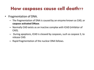
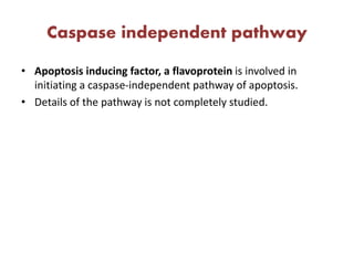
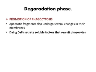
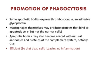
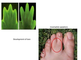
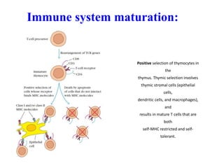
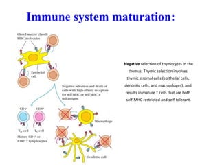
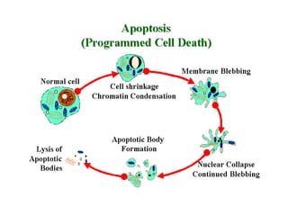
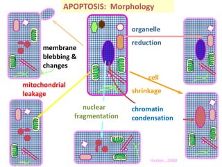
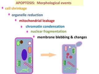
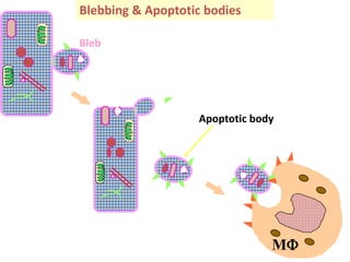
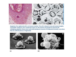
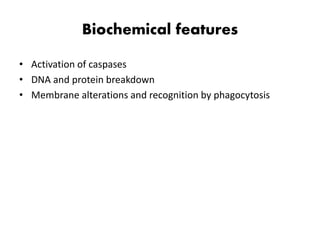
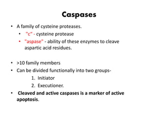
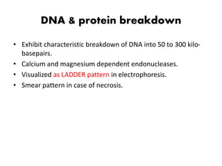
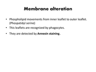
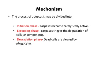
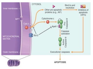
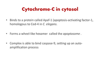
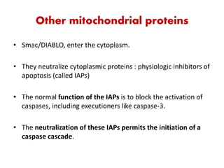
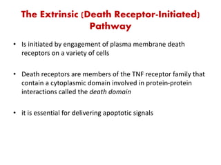
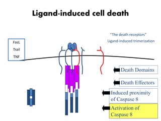
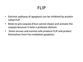
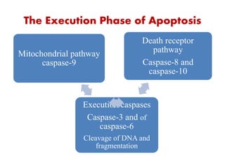
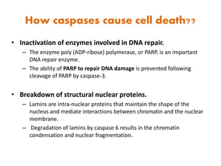
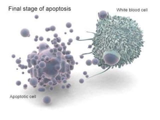
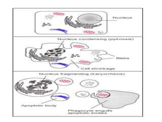
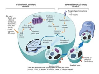
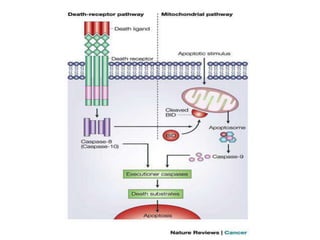
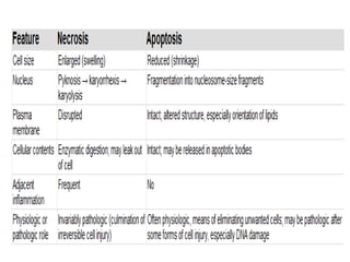
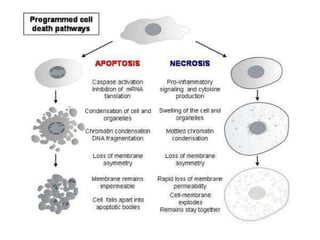
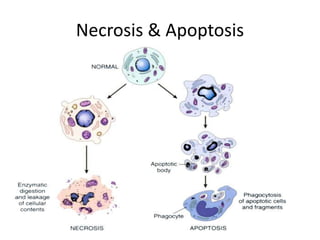
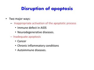
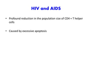
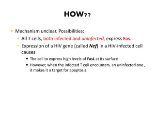
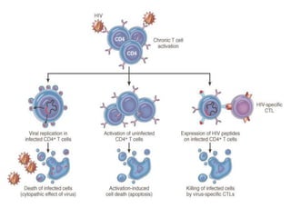
APOPTOSIS
Venkatesh Dhanasekaran
History!!
1800s Numerous observation of cell death
1908 Mechnikov wins Nobel prize (phagocytosis)
1930-40 Studies of metamorphosis
1948-49 Cell death in chick limb & exploration of NGF
1955 Beginning of studies of lysomes
1965 Necrosis & PCD described
1972 Term apoptosis coined
1977 Cell death genes in C. elegans
1980-82 DNA ladder observed & ced-3 identified
1990 Apoptosis genes identified, including bcl-2,
fas/apo1 & p53, ced-3 sequenced
• Named after the Greek designation for “falling off”.
• Process recognized in 1972 by distinct morphologic
appearance of membrane bound fragments derived from
cells.
• Quickly appreciated that it was a unique mechanism of cell
death, distinct from Necrosis.
What is apoptosis??
• Apoptosis is a pathway of cell death that is induced by tightly
regulated suicide program in which cells destined to die
activate enzymes that degrade the cells’s own nuclear DNA
and nuclear & cytoplasmic proteins. (Robbins)
• Apoptosis or programmed cell death, is carefully coordinated
collapse of cell, protein degradation , DNA fragmentation
followed by rapid engulfment of corpses by neighbouring
cells. (Tommi, 2002)
• Essential part of life for every multicellular organism from
worms to humans. (Faddy et al.,1992)
What causes it??
Physiological:
• Death by apoptosis is a normal phenomenon that
eliminates cells no longer needed and maintain steady cell
number in various populations.
– Immune system maturation
– Involution of hormone dependent tissues upon
hormone withdrawal
– Elimination of self reactive lymphocytes
– Embryogenesis
What causes it??
Pathological:
• Apoptosis eliminates cells that are injured beyond repair
without eliciting a host reaction.
– Immune privilege
– Elimination of misfolded proteins.
– Pathological atrophy after duct obstruction
What triggers it??
 Withdrawal of positive (growth) signals:
 Growth factors
 Interleukins(IL-2)
 Receipt of negative(death) signals:
 Increased levels of oxidants in the cell
 DNA damage
 Death activators
• TNF alpha
• TNF beta ( lymphotoxin )
• Fas ligand
Development of toes
Incomplete apoptosis
Immune system maturation:
Positive selection of thymocytes in
the
thymus. Thymic selection involves
thymic stromal cells (epithelial
cells,
dendritic cells, and macrophages),
and
results in mature T cells that are
both
self-MHC restricted and self-
tolerant.
Immune system maturation:
Negative selection of thymocytes in the
thymus. Thymic selection involves
thymic stromal cells (epithelial cells,
dendritic cells, and macrophages), and
results in mature T cells that are both
self-MHC restricted and self-tolerant.
Infections
• Cell death in certain infections, particularly viral infections, in
which loss of infected cells is largely due to apoptosis that
may be induced by the virus (as in adenovirus and HIV
infections) or by the host immune response (as in viral
hepatitis).
Morphological changes
• Cell shrinkage.
• Tightly packed organells.
• Chromatin condensation (Characteristic).
• Formation of cytoplasmic blebs and apoptotic bodies.
• Phagocytosis of apoptotic cells or cell bodies, usually by
macrophages.
• Plasma membranes are thought to remain intact during
apoptosis.
How it looks??
 Light microscopy:
• Apoptotic cells – oval or round mass of intensely
eosinophilic cytoplasm with dense nuclear chromatin.
• Because of rapid clearing, becomes apparent in
histology in late stage.
• No inflammation, so difficult to identify
Electron microscopy:
Cell shrinkage
Chromatin condensation
Cytoplasmic blebs and apoptotic bodies
Phagocytosis by macrophages
membrane
blebbing &
changes
mitochondrial
leakage
organelle
reduction
cell
shrinkage
nuclear
fragmentation
chromatin
condensation
APOPTOSIS: Morphology
Hacker., 2000
membrane blebbing & changes
mitochondrial leakage
organelle reduction
cell shrinkage
nuclear fragmentation
chromatin condensation
APOPTOSIS: Morphological events
Bleb
Blebbing & Apoptotic bodies
The control retained over the cell membrane
& cytoskeleton allows intact pieces of the cell
to separate for recognition & phagocytosis by
MFs
Apoptotic body
MF
Apoptosis of an epidermal cell in an immune reaction. The cell is reduced in size and contains brightly
eosinophilic cytoplasm and a condensed nucleus. B, This electron micrograph of cultured cells
undergoing apoptosis shows some nuclei with peripheral crescents of compacted chromatin, and others
that are uniformly dense or fragmented
Biochemical features
• Activation of caspases
• DNA and protein breakdown
• Membrane alterations and recognition by phagocytosis
Caspases
• A family of cysteine proteases.
• "c“ - cysteine protease
• "aspase" - ability of these enzymes to cleave
aspartic acid residues.
• >10 family members
• Can be divided functionally into two groups-
1. Initiator
2. Executioner.
• Cleaved and active caspases is a marker of active
apoptosis.
DNA & protein breakdown
• Exhibit characteristic breakdown of DNA into 50 to 300 kilo-
basepairs.
• Calcium and magnesium dependent endonucleases.
• Visualized as LADDER pattern in electrophoresis.
• Smear pattern in case of necrosis.
Membrane alteration
• Phospholipid movements from inner leaflet to outer leaflet.
(Phospatidyl serine)
• This leaflets are recognized by phagocytes.
• They are detected by Annexin staining.
 Others biochemical features:
Cell dehydration
Loss of mitochondrial membrane potential
Increase in free calcium ions
Protein cross-linking
Bcl2 – Bax interaction
How to recognize Apoptosis
 Morphological assessment by microscopy.
 DNA fragmentation by Gel Electrophoresis.
 Flow cytometry.
 Specific probes for apoptotic cells – Annexin V.
 Measurement of tissue transglutaminase by ELISA (in vivo)
 TUNEL (tdt biotin-dUTP nick end labelling)- detects apoptosis
in situ
Anti-apoptotic
• Bcl-2
• Bcl-xL
• Bcl-W
• Bcl-B
• Mcl-1
• NR-13
Pro-apoptotic
• Bax
• Bak
• BOK/MTD
• Bcl-Xs
• NOXA
• PUMA
• BIM
• BAD
• BOO
Mechanism
• The process of apoptosis may be divided into
• Initiation phase - caspases become catalytically active.
• Execution phase - caspases trigger the degradation of
cellular components.
• Degradation phase- Dead cells are cleared by
phagocytes.
Initiation phase
• Initiation of apoptosis occurs principally by signals from two
distinct pathways:
 Intrinsic or mitochondrial pathway
 Extrinsic or death receptor–initiated pathway
Intrinsic pathway
• Is the major mechanism.
• Result from increased mitochondrial permeability and release
of pro-apoptotic molecules (death inducers) into the
cytoplasm .
• Mitochondria are remarkable in that they contain proteins
such as cytochrome c but some of the same proteins, when
released into the cytoplasm, initiate apoptosis
Steps!!
1
• Deprivation of growth factors/DNA damage.
2
• Sensors of damage activated
• Bim,Bid and Bad (BH3 only proteins)
3
• Activate Bax and Bak (Forming oligomers)
• Inserts into mitochondrial membrane
• Increases it’s permeability
Cytochrome-C in cytosol
• Binds to a protein called Apaf-1 (apoptosis-activating factor-1,
homologous to Ced-4 in C. elegans.
• Forms a wheel-like hexamer called the apoptosome .
• Complex is able to bind caspase-9, setting up an auto-
amplification process
Other mitochondrial proteins
• Smac/DIABLO, enter the cytoplasm.
• They neutralize cytoplasmic proteins : physiologic inhibitors of
apoptosis (called IAPs)
• The normal function of the IAPs is to block the activation of
caspases, including executioners like caspase-3.
• The neutralization of these IAPs permits the initiation of a
caspase cascade.
The Extrinsic (Death Receptor-Initiated)
Pathway
• Is initiated by engagement of plasma membrane death
receptors on a variety of cells
• Death receptors are members of the TNF receptor family that
contain a cytoplasmic domain involved in protein-protein
interactions called the death domain
• it is essential for delivering apoptotic signals
Death Receptors
• The best-known are the type 1 TNF receptor (TNFR1) and a
related protein called Fas (CD95).
• The ligand for Fas is called Fas ligand (FasL).
• FasL is expressed on T cells that recognize self antigens and on
some cytotoxic T lymphocytes.
Ligand-induced cell death
Ligand Receptor
FasL Fas (CD95)
TNF TNF-R
TRAIL DR4 (Trail-R)
FAS And FASL
• When FasL binds to Fas and their cytoplasmic death domains
form a binding site that also contains a death domain and is
called FADD (Fas-associated death domain)
• FADD that is attached to the death receptors in turn binds an
inactive form of caspase-8 (and, in humans, caspase-10),
again via a death domain.
Ligand-induced cell death
“The death receptors”
Ligand-induced trimerization
Death Domains
Death Effectors
Induced proximity
of Caspase 8
Activation of
Caspase 8
FasL
Trail
TNF
FLIP
• Extrinsic pathway of apoptosis can be inhibited by protein
called FLIP
• Binds to pro-caspase-8 but cannot cleave and activate the
caspase because it lacks a protease domain.
• Some viruses and normal cells produce FLIP and protect
themselves from Fas-mediated apoptosis.
The Execution Phase of Apoptosis
Execution caspases
Caspase-3 and of
caspase-6
Cleavage of DNA and
fragmentation
Death receptor
pathway
Caspase-8 and
caspase-10
Mitochondrial pathway
caspase-9
How caspases cause cell death??
• Inactivation of enzymes involved in DNA repair.
– The enzyme poly (ADP-ribose) polymerase, or PARP, is an important
DNA repair enzyme.
– The ability of PARP to repair DNA damage is prevented following
cleavage of PARP by caspase-3.
• Breakdown of structural nuclear proteins.
– Lamins are intra-nuclear proteins that maintain the shape of the
nucleus and mediate interactions between chromatin and the nuclear
membrane.
– Degradation of lamins by caspase 6 results in the chromatin
condensation and nuclear fragmentation.
How caspases cause cell death??
• Fragmentation of DNA.
– The fragmentation of DNA is caused by an enzyme known as CAD, or
caspase activated DNase.
– Normally CAD exists as an inactive complex with ICAD (inhibitor of
CAD).
– During apoptosis, ICAD is cleaved by caspases, such as caspase 3, to
release CAD.
– Rapid fragmentation of the nuclear DNA follows.
Caspase independent pathway
• Apoptosis inducing factor, a flavoprotein is involved in
initiating a caspase-independent pathway of apoptosis.
• Details of the pathway is not completely studied.
Degaradation phase.
 PROMOTION OF PHAGOCYTOSIS
• Apoptotic fragments also undergo several changes in their
membranes
• Dying Cells secrete soluble factors that recruit phagocytes
PROMOTION OF PHAGOCYTOSIS
• Some apoptotic bodies express thrombospondin, an adhesive
glycoprotein.
• Macrophages themselves may produce proteins that bind to
apoptotic cells(But not the normal cells)
• Apoptotic bodies may also become coated with natural
antibodies and proteins of the complement system, notably
C1q
• Efficient (So that dead cells .Leaving no inflammation)
Link between two pathways
• The BH-3only protein Bid is the link between two pathways.
• When death receptor activate the extrinsic pathway initiator
caspase cleaves Bid, producing t-Bid which translocates to
mitochondria and inhibit anti-apoptotic Bcl-2.
Link between the pathways
Necrosis & Apoptosis
Disruption of apoptosis
• Two major ways:
– Inappropriate activation of the apoptotic process
• Immune defect in AIDS
• Neurodegenerative diseases.
– Inadequate apoptosis
• Cancer
• Chronic inflammatory conditions
• Autoimmune diseases.
HIV and AIDS
• Profound reduction in the population size of CD4 + T helper
cells
• Caused by excessive apoptosis
HOW??
 Mechanism unclear. Possibilities:
• All T cells, both infected and uninfected, express Fas.
• Expression of a HIV gene (called Nef) in a HIV-infected cell
causes
 The cell to express high levels of FasL at its surface
 However, when the infected T cell encounters an uninfected one ,
it makes it a target for apoptosis.
Neurodegenerative Disease
• Apoptosis triggered by
– amyloid β
– neurotoxic abnormal protein structures or aggregates
• In adult neurodegenerative diseases including
– Alzheimer's
– Huntington's chorea
– Parkinson's disease,
– Amyotrophic lateral sclerosis
• Amyloid β can exert neurotoxic effects by
– generation of intracellular oxidative stress
– increases in calcium ions
• Both of these can trigger apoptosis in susceptible cell types.
DNA Damage
Transcriptional Up-Regulation
of Target Genes
p21
(CDK inhibitor)
GADD45
(DNA repair)
BAX
(apoptosis gene)
Ionizing Radiation,
Carcinogens & Mutagens
DNA Damage
p53 activated and binds to DNA
G1 Arrest
Successful repair
Repair fails
APOPTOSIS
Role of apoptosis
In maintaining
Integrity of genomic
DNA in normal cells
Cancer & Apoptosis
• Mutations affect the control mechanisms of apoptosis
and cell survival.
• Bcl-2 in follicular lymphoma
– increased bcl-2 expression confers resistance to
chemotherapy in ALL and some forms of AML
– Bcl-2 blocks the endonucleolytic cleavage of DNA
that is so characteristic of apoptosis.
Chronic Inflammatory conditions:
• Intact neutrophils are engulfed by macrophages at the sites of
inflammation.
• Rheumatoid arthritis may reflect prolonged survival of
leucocytes that are normally programmed to die by apoptosis
Therapeutic Significance
• Approaches to counter inappropriate apoptosis:
– Caspases
Several pharmaceutical companies are developing potent and
specific caspase inhibitors
have shown great promise in murine models of inappropriate
neuronal apoptosis.
– The treatment of certain lymphomas by antisense
oligonucleotides to bcl-2.
YET TO BE DEFINED
• Mechanism of alteration of plasma membrane structure
• Formation of membrane blebs
• Formation of apoptotic bodies
• Caspase independent intrinsic pathway
Apoptosis

Apoptosis

  • 1.
  • 2.
    History!! 1800s Numerous observationof cell death 1908 Mechnikov wins Nobel prize (phagocytosis) 1930-40 Studies of metamorphosis 1948-49 Cell death in chick limb & exploration of NGF 1955 Beginning of studies of lysomes 1965 Necrosis & PCD described 1972 Term apoptosis coined 1977 Cell death genes in C. elegans 1980-82 DNA ladder observed & ced-3 identified 1990 Apoptosis genes identified, including bcl-2, fas/apo1 & p53, ced-3 sequenced
  • 3.
    • Named afterthe Greek designation for “falling off”. • Process recognized in 1972 by distinct morphologic appearance of membrane bound fragments derived from cells. • Quickly appreciated that it was a unique mechanism of cell death, distinct from Necrosis.
  • 4.
    What is apoptosis?? •Apoptosis is a pathway of cell death that is induced by tightly regulated suicide program in which cells destined to die activate enzymes that degrade the cells’s own nuclear DNA and nuclear & cytoplasmic proteins. (Robbins)
  • 5.
    • Apoptosis orprogrammed cell death, is carefully coordinated collapse of cell, protein degradation , DNA fragmentation followed by rapid engulfment of corpses by neighbouring cells. (Tommi, 2002) • Essential part of life for every multicellular organism from worms to humans. (Faddy et al.,1992)
  • 6.
    What causes it?? Physiological: •Death by apoptosis is a normal phenomenon that eliminates cells no longer needed and maintain steady cell number in various populations. – Immune system maturation – Involution of hormone dependent tissues upon hormone withdrawal – Elimination of self reactive lymphocytes – Embryogenesis
  • 7.
    What causes it?? Pathological: •Apoptosis eliminates cells that are injured beyond repair without eliciting a host reaction. – Immune privilege – Elimination of misfolded proteins. – Pathological atrophy after duct obstruction
  • 8.
    What triggers it?? Withdrawal of positive (growth) signals:  Growth factors  Interleukins(IL-2)  Receipt of negative(death) signals:  Increased levels of oxidants in the cell  DNA damage  Death activators • TNF alpha • TNF beta ( lymphotoxin ) • Fas ligand
  • 9.
  • 10.
    Immune system maturation: Positiveselection of thymocytes in the thymus. Thymic selection involves thymic stromal cells (epithelial cells, dendritic cells, and macrophages), and results in mature T cells that are both self-MHC restricted and self- tolerant.
  • 11.
    Immune system maturation: Negativeselection of thymocytes in the thymus. Thymic selection involves thymic stromal cells (epithelial cells, dendritic cells, and macrophages), and results in mature T cells that are both self-MHC restricted and self-tolerant.
  • 12.
    Infections • Cell deathin certain infections, particularly viral infections, in which loss of infected cells is largely due to apoptosis that may be induced by the virus (as in adenovirus and HIV infections) or by the host immune response (as in viral hepatitis).
  • 13.
    Morphological changes • Cellshrinkage. • Tightly packed organells. • Chromatin condensation (Characteristic). • Formation of cytoplasmic blebs and apoptotic bodies. • Phagocytosis of apoptotic cells or cell bodies, usually by macrophages. • Plasma membranes are thought to remain intact during apoptosis.
  • 15.
    How it looks?? Light microscopy: • Apoptotic cells – oval or round mass of intensely eosinophilic cytoplasm with dense nuclear chromatin. • Because of rapid clearing, becomes apparent in histology in late stage. • No inflammation, so difficult to identify
  • 18.
    Electron microscopy: Cell shrinkage Chromatincondensation Cytoplasmic blebs and apoptotic bodies Phagocytosis by macrophages
  • 19.
  • 20.
    membrane blebbing &changes mitochondrial leakage organelle reduction cell shrinkage nuclear fragmentation chromatin condensation APOPTOSIS: Morphological events
  • 21.
    Bleb Blebbing & Apoptoticbodies The control retained over the cell membrane & cytoskeleton allows intact pieces of the cell to separate for recognition & phagocytosis by MFs Apoptotic body MF
  • 22.
    Apoptosis of anepidermal cell in an immune reaction. The cell is reduced in size and contains brightly eosinophilic cytoplasm and a condensed nucleus. B, This electron micrograph of cultured cells undergoing apoptosis shows some nuclei with peripheral crescents of compacted chromatin, and others that are uniformly dense or fragmented
  • 23.
    Biochemical features • Activationof caspases • DNA and protein breakdown • Membrane alterations and recognition by phagocytosis
  • 24.
    Caspases • A familyof cysteine proteases. • "c“ - cysteine protease • "aspase" - ability of these enzymes to cleave aspartic acid residues. • >10 family members • Can be divided functionally into two groups- 1. Initiator 2. Executioner. • Cleaved and active caspases is a marker of active apoptosis.
  • 25.
    DNA & proteinbreakdown • Exhibit characteristic breakdown of DNA into 50 to 300 kilo- basepairs. • Calcium and magnesium dependent endonucleases. • Visualized as LADDER pattern in electrophoresis. • Smear pattern in case of necrosis.
  • 26.
    Membrane alteration • Phospholipidmovements from inner leaflet to outer leaflet. (Phospatidyl serine) • This leaflets are recognized by phagocytes. • They are detected by Annexin staining.
  • 27.
     Others biochemicalfeatures: Cell dehydration Loss of mitochondrial membrane potential Increase in free calcium ions Protein cross-linking Bcl2 – Bax interaction
  • 28.
    How to recognizeApoptosis  Morphological assessment by microscopy.  DNA fragmentation by Gel Electrophoresis.  Flow cytometry.  Specific probes for apoptotic cells – Annexin V.  Measurement of tissue transglutaminase by ELISA (in vivo)  TUNEL (tdt biotin-dUTP nick end labelling)- detects apoptosis in situ
  • 29.
    Anti-apoptotic • Bcl-2 • Bcl-xL •Bcl-W • Bcl-B • Mcl-1 • NR-13 Pro-apoptotic • Bax • Bak • BOK/MTD • Bcl-Xs • NOXA • PUMA • BIM • BAD • BOO
  • 30.
    Mechanism • The processof apoptosis may be divided into • Initiation phase - caspases become catalytically active. • Execution phase - caspases trigger the degradation of cellular components. • Degradation phase- Dead cells are cleared by phagocytes.
  • 31.
    Initiation phase • Initiationof apoptosis occurs principally by signals from two distinct pathways:  Intrinsic or mitochondrial pathway  Extrinsic or death receptor–initiated pathway
  • 32.
    Intrinsic pathway • Isthe major mechanism. • Result from increased mitochondrial permeability and release of pro-apoptotic molecules (death inducers) into the cytoplasm . • Mitochondria are remarkable in that they contain proteins such as cytochrome c but some of the same proteins, when released into the cytoplasm, initiate apoptosis
  • 33.
    Steps!! 1 • Deprivation ofgrowth factors/DNA damage. 2 • Sensors of damage activated • Bim,Bid and Bad (BH3 only proteins) 3 • Activate Bax and Bak (Forming oligomers) • Inserts into mitochondrial membrane • Increases it’s permeability
  • 36.
    Cytochrome-C in cytosol •Binds to a protein called Apaf-1 (apoptosis-activating factor-1, homologous to Ced-4 in C. elegans. • Forms a wheel-like hexamer called the apoptosome . • Complex is able to bind caspase-9, setting up an auto- amplification process
  • 37.
    Other mitochondrial proteins •Smac/DIABLO, enter the cytoplasm. • They neutralize cytoplasmic proteins : physiologic inhibitors of apoptosis (called IAPs) • The normal function of the IAPs is to block the activation of caspases, including executioners like caspase-3. • The neutralization of these IAPs permits the initiation of a caspase cascade.
  • 38.
    The Extrinsic (DeathReceptor-Initiated) Pathway • Is initiated by engagement of plasma membrane death receptors on a variety of cells • Death receptors are members of the TNF receptor family that contain a cytoplasmic domain involved in protein-protein interactions called the death domain • it is essential for delivering apoptotic signals
  • 39.
    Death Receptors • Thebest-known are the type 1 TNF receptor (TNFR1) and a related protein called Fas (CD95). • The ligand for Fas is called Fas ligand (FasL). • FasL is expressed on T cells that recognize self antigens and on some cytotoxic T lymphocytes.
  • 40.
    Ligand-induced cell death LigandReceptor FasL Fas (CD95) TNF TNF-R TRAIL DR4 (Trail-R)
  • 41.
    FAS And FASL •When FasL binds to Fas and their cytoplasmic death domains form a binding site that also contains a death domain and is called FADD (Fas-associated death domain) • FADD that is attached to the death receptors in turn binds an inactive form of caspase-8 (and, in humans, caspase-10), again via a death domain.
  • 43.
    Ligand-induced cell death “Thedeath receptors” Ligand-induced trimerization Death Domains Death Effectors Induced proximity of Caspase 8 Activation of Caspase 8 FasL Trail TNF
  • 44.
    FLIP • Extrinsic pathwayof apoptosis can be inhibited by protein called FLIP • Binds to pro-caspase-8 but cannot cleave and activate the caspase because it lacks a protease domain. • Some viruses and normal cells produce FLIP and protect themselves from Fas-mediated apoptosis.
  • 45.
    The Execution Phaseof Apoptosis Execution caspases Caspase-3 and of caspase-6 Cleavage of DNA and fragmentation Death receptor pathway Caspase-8 and caspase-10 Mitochondrial pathway caspase-9
  • 46.
    How caspases causecell death?? • Inactivation of enzymes involved in DNA repair. – The enzyme poly (ADP-ribose) polymerase, or PARP, is an important DNA repair enzyme. – The ability of PARP to repair DNA damage is prevented following cleavage of PARP by caspase-3. • Breakdown of structural nuclear proteins. – Lamins are intra-nuclear proteins that maintain the shape of the nucleus and mediate interactions between chromatin and the nuclear membrane. – Degradation of lamins by caspase 6 results in the chromatin condensation and nuclear fragmentation.
  • 47.
    How caspases causecell death?? • Fragmentation of DNA. – The fragmentation of DNA is caused by an enzyme known as CAD, or caspase activated DNase. – Normally CAD exists as an inactive complex with ICAD (inhibitor of CAD). – During apoptosis, ICAD is cleaved by caspases, such as caspase 3, to release CAD. – Rapid fragmentation of the nuclear DNA follows.
  • 48.
    Caspase independent pathway •Apoptosis inducing factor, a flavoprotein is involved in initiating a caspase-independent pathway of apoptosis. • Details of the pathway is not completely studied.
  • 49.
    Degaradation phase.  PROMOTIONOF PHAGOCYTOSIS • Apoptotic fragments also undergo several changes in their membranes • Dying Cells secrete soluble factors that recruit phagocytes
  • 50.
    PROMOTION OF PHAGOCYTOSIS •Some apoptotic bodies express thrombospondin, an adhesive glycoprotein. • Macrophages themselves may produce proteins that bind to apoptotic cells(But not the normal cells) • Apoptotic bodies may also become coated with natural antibodies and proteins of the complement system, notably C1q • Efficient (So that dead cells .Leaving no inflammation)
  • 54.
    Link between twopathways • The BH-3only protein Bid is the link between two pathways. • When death receptor activate the extrinsic pathway initiator caspase cleaves Bid, producing t-Bid which translocates to mitochondria and inhibit anti-apoptotic Bcl-2.
  • 55.
  • 58.
  • 59.
    Disruption of apoptosis •Two major ways: – Inappropriate activation of the apoptotic process • Immune defect in AIDS • Neurodegenerative diseases. – Inadequate apoptosis • Cancer • Chronic inflammatory conditions • Autoimmune diseases.
  • 60.
    HIV and AIDS •Profound reduction in the population size of CD4 + T helper cells • Caused by excessive apoptosis
  • 61.
    HOW??  Mechanism unclear.Possibilities: • All T cells, both infected and uninfected, express Fas. • Expression of a HIV gene (called Nef) in a HIV-infected cell causes  The cell to express high levels of FasL at its surface  However, when the infected T cell encounters an uninfected one , it makes it a target for apoptosis.
  • 63.
    Neurodegenerative Disease • Apoptosistriggered by – amyloid β – neurotoxic abnormal protein structures or aggregates • In adult neurodegenerative diseases including – Alzheimer's – Huntington's chorea – Parkinson's disease, – Amyotrophic lateral sclerosis • Amyloid β can exert neurotoxic effects by – generation of intracellular oxidative stress – increases in calcium ions • Both of these can trigger apoptosis in susceptible cell types.
  • 64.
    DNA Damage Transcriptional Up-Regulation ofTarget Genes p21 (CDK inhibitor) GADD45 (DNA repair) BAX (apoptosis gene) Ionizing Radiation, Carcinogens & Mutagens DNA Damage p53 activated and binds to DNA G1 Arrest Successful repair Repair fails APOPTOSIS Role of apoptosis In maintaining Integrity of genomic DNA in normal cells
  • 65.
    Cancer & Apoptosis •Mutations affect the control mechanisms of apoptosis and cell survival. • Bcl-2 in follicular lymphoma – increased bcl-2 expression confers resistance to chemotherapy in ALL and some forms of AML – Bcl-2 blocks the endonucleolytic cleavage of DNA that is so characteristic of apoptosis.
  • 66.
    Chronic Inflammatory conditions: •Intact neutrophils are engulfed by macrophages at the sites of inflammation. • Rheumatoid arthritis may reflect prolonged survival of leucocytes that are normally programmed to die by apoptosis
  • 67.
    Therapeutic Significance • Approachesto counter inappropriate apoptosis: – Caspases Several pharmaceutical companies are developing potent and specific caspase inhibitors have shown great promise in murine models of inappropriate neuronal apoptosis. – The treatment of certain lymphomas by antisense oligonucleotides to bcl-2.
  • 68.
    YET TO BEDEFINED • Mechanism of alteration of plasma membrane structure • Formation of membrane blebs • Formation of apoptotic bodies • Caspase independent intrinsic pathway