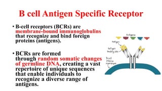
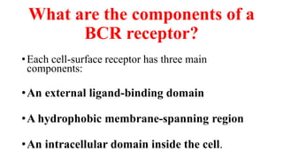
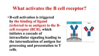
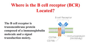
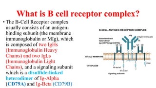
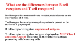
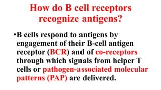
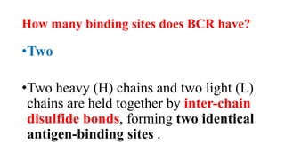
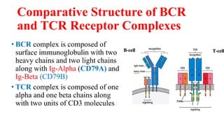
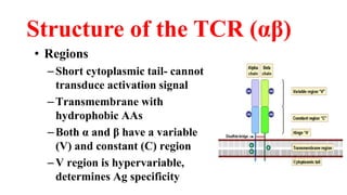
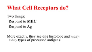
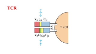
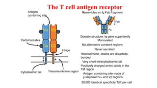
B Cell & T Cell Antigen
Specific Receptors
B cell Antigen Specific Receptor
• B-cell receptors (BCRs) are
membrane-bound immunoglobulins
that recognize and bind foreign
proteins (antigens).
• BCRs are formed
through random somatic changes
of germline DNA, creating a vast
repertoire of unique sequences
that enable individuals to
recognize a diverse range of
antigens.
What are the components of a
BCR receptor?
•Each cell-surface receptor has three main
components:
•An external ligand-binding domain
•A hydrophobic membrane-spanning region
•An intracellular domain inside the cell.
What activates the B cell receptor?
• B-cell activation is triggered
by the binding of ligand
(referred to as antigen) to the B-
cell receptor (BCR), which
initiates a cascade of
intracellular signaling leading to
the internalization of antigen for
processing and presentation to T
cells.
Where is the B cell receptor (BCR)
Located?
The B cell receptor is
transmembrane protein
composed of a immunoglobulin
molecule and a signal
transduction moiety.
What is B cell receptor complex?
• The B-Cell Receptor complex
usually consists of an antigen-
binding subunit (the membrane
immunoglobulin or MIg), which
is composed of two IgHs
(Immunoglobulin Heavy
Chains) and two IgLs
(Immunoglobulin Light
Chains), and a signaling subunit
which is a disulfide-linked
heterodimer of Ig-Alpha
(CD79A) and Ig-Beta (CD79B)
What are the differences between B cell
receptors and T cell receptors?
B cell receptor is a transmembrane receptor protein located on the
outer surface of B cells.
T cell receptor is an antigen recognizing molecule present on the
surface of T lymphocytes.
B cell receptor recognizes unprocessed antigens.
T cell receptor recognizes antigens displayed on MHC Class I
and MHC Class II molecules on the surface of antigen
presenting cells/accessary cells.
How do B cell receptors
recognize antigens?
•B cells respond to antigens by
engagement of their B-cell antigen
receptor (BCR) and of co-receptors
through which signals from helper T
cells or pathogen-associated molecular
patterns (PAP) are delivered.
Do B cells have MHC 1 or 2?
•Besides secreting antibodies, B cells
express MHC class II and serve as
antigen-presenting cells (APCs) for
CD4+ helper T cells.
What are antigen Presenting cells?
•Macrophages
•Dendritic cells
•B cells
How many binding sites does BCR have?
•Two
•Two heavy (H) chains and two light (L)
chains are held together by inter-chain
disulfide bonds, forming two identical
antigen-binding sites .
What is required for B cell activation?
•Naïve B cell activation requires antigen
recognition by the Ig receptor.
• The additional signals that can come
either from a CD4+ Helper T cell
(thymus-dependent)
•Or, in some cases, directly from microbial
components (T independent).
How does B cell activation begin?
•B cell activation is initiated by the binding
of antigen to the B cell receptor
(BCR) that triggers a number of signaling
cascades that ultimately lead to B cell
activation.
Do B cells bind to MHC?
T cells bind antigens that have been digested
and embedded in MHC molecules by APCs
B cells function as Antigen Presenting Cells
(APC) that bind intact antigens that have
not been processed.
How do B cells digest antigens?
•B Cells as Antigen-Presenting Cells
Professional antigen-presenting cells (APCs)
take up antigen through phagocytosis, fluid-
phase pinocytosis, or receptor-mediated
endocytosis for processing, loading of the
digestion products onto MHC Class II (MHC
II) and presentation to CD4 Helper T cells.
How many B cell receptors are
there?
•105
•Each B cell has approximately 105 such
receptors in its plasma membrane.
•Each of these receptors is stably associated
with a complex of transmembrane proteins
that activate intracellular signaling pathways
when antigen binds to the receptor.
Comparative Structure of BCR
and TCR Receptor Complexes
• BCR complex is composed of
surface immunoglobulin with two
heavy chains and two light chains
along with Ig-Alpha (CD79A) and
Ig-Beta (CD79B)
• TCR complex is composed of one
alpha and one beta chains along
with two units of CD3 molecules
T cell Receptor (TCR)
• Principal component TCR was a heterodimeric 90kDa
protein composed of a 40kDa and a 50kDa molecule (α
and β chains)
• Surface molecule on T cells
• Recognize Ag presented in MHC context
• Similar to Immunoglobulin
• Two types of TCR
• α β: predominant in lymphoid tissues
• γ δ: enriched at mucosal surfaces
T cell antigen receptor: Some
Fundamentals
One T-cell has one type of TCR
One T-cell has one TCR with a wholly unique
specificity
.
One T-cell has as many as 100,000 identical TCR’s.
There are two important types of T-cells: TH & TC.
Structure of the TCR (αβ)
• Regions
–Short cytoplasmic tail- cannot
transduce activation signal
–Transmembrane with
hydrophobic AAs
–Both α and β have a variable
(V) and constant (C) region
–V region is hypervariable,
determines Ag specificity
What Cell Receptors do?
Two things:
Respond to MHC
Respond to Ag
More exactly, they see one histotope and many,
many types of processed antigens.
TCR
Structure of the TCR (αβ)
• Heterodimer
•α and β chains
•approx equal length
Important Aspects of TCR
•Each T cell has TCR of only ONE specificity
•Allelic exclusion
•αβ TCR recognizes Ag only in the context of
cell-cell interaction and in correct MHC context
•γδ TCR recognizes Ag in MHC-independent
manner
•Response to certain viral and bacterial Ag
Genetic basis for receptor generation
•Accomplished by
recombination of V, D and J
gene segments
•TCR β chain genes have V,
D, and J
•TCR α chain genes have V
and J
TCR and CD3 complex
• TCR is closely associated with CD3
complex
• Group of 5 proteins
• Commonly called “invariant” chains of TCR
• Role of CD3 complex
• CD3 necessary for cell surface expression of
TCR
• transduces signal after Ag interaction with
TCR
Key steps in T cell activation
•APC must process and present peptides to T cells
•T cells must receive co-stimulatory signal
•Accessory adhesion molecules stabilize binding of
TCR and MHC
•Signal from cell surface is transmitted to nucleus
•Cytokines produced help drive cell proliferation
The T cell antigen receptor
Va Vb
Ca Cb
Carbohydrates
Hinge
Monovalent
Resembles an Ig Fab fragment
Fab
VH
VL
Fc
CL
CH
VL
VH
CH CL
CH CH
CH
CH
No alternative constant regions
Transmembrane region
Never secreted
Domain structure: Ig gene superfamily
Heterodimeric, chains are disuphide-
bonded
Cytoplasmic tail
Very short intracytoplasmic tail
+
+
+
Positively charged amino acids in the
TM region
Antigen
combining site
Antigen combining site made of
juxtaposed Va and Vb regions
30,000 identical specificity TcR per cell
 T cells
Distinct lineage of cells with unknown functions
1-5% of peripheral blood T cells
In the gut and epidermis of mice, most T cells
express  TcR
Ligands of  TcR are unknown
Possibly recognise:
Antigens without involvement of MHC antigens - CD1
Class IB genes
Role of MHC in immune response
•TCR recognizes Ag presented in MHC
•Context is important
•Binding of Ag peptides in non-covalent
•Two types of MHC (class I and class II) are
recognized by different subsets of T cells
•CTL recognizes Ag peptide in MHC class I
•T-helper recognizes Ag peptide in MHC class II
Structure of MHC class I
•Two polypeptide chains
•Long α chain and short β
Structure of MHC class I
• Four regions
• Cytoplasmic contains sites for
phosphorylation and binding to
cytoskeleton
• Transmembrane contains
hydrophobic AAs
• Highly conserved α3 domain binds
CD8
• Highly polymorphic peptide binding
region formed by α1 and α2
Structure of MHC class II
• Two polypeptide chains
• α and β
• approx equal length
Structure of MHC class II
• Four regions
• Cytoplasmic contains sites for
phosphorylation and binding
to cytoskeleton
• Transmembrane contains
hydrophobic AAs
• Highly conserved α2 and β2
domains binds CD4
• Highly polymorphic peptide
binding region formed by α1
and β1
Important aspects of MHC
• Individuals have a limited number of MHC alleles for each class
• High polymorphism in MHC for a species
• Alleles for MHC genes are co-dominant
• Each MHC gene product is expressed on surface of individual cell
Important aspects of MHC
• Each MHC has ONE peptide binding site
• But each MHC can bind many different peptides
• Only one at a time
• Peptide binding is “degenerate”
• MHC polymorphism is determined in germline
• NO recombination mechanisms for creating diversity in
MHC
• Peptide must bind with individual’s MHC to induce
immune response
B Cell & T Cell Receptors.pptx

B Cell & T Cell Receptors.pptx

  • 1.
    B Cell &T Cell Antigen Specific Receptors
  • 2.
    B cell AntigenSpecific Receptor • B-cell receptors (BCRs) are membrane-bound immunoglobulins that recognize and bind foreign proteins (antigens). • BCRs are formed through random somatic changes of germline DNA, creating a vast repertoire of unique sequences that enable individuals to recognize a diverse range of antigens.
  • 3.
    What are thecomponents of a BCR receptor? •Each cell-surface receptor has three main components: •An external ligand-binding domain •A hydrophobic membrane-spanning region •An intracellular domain inside the cell.
  • 4.
    What activates theB cell receptor? • B-cell activation is triggered by the binding of ligand (referred to as antigen) to the B- cell receptor (BCR), which initiates a cascade of intracellular signaling leading to the internalization of antigen for processing and presentation to T cells.
  • 5.
    Where is theB cell receptor (BCR) Located? The B cell receptor is transmembrane protein composed of a immunoglobulin molecule and a signal transduction moiety.
  • 6.
    What is Bcell receptor complex? • The B-Cell Receptor complex usually consists of an antigen- binding subunit (the membrane immunoglobulin or MIg), which is composed of two IgHs (Immunoglobulin Heavy Chains) and two IgLs (Immunoglobulin Light Chains), and a signaling subunit which is a disulfide-linked heterodimer of Ig-Alpha (CD79A) and Ig-Beta (CD79B)
  • 7.
    What are thedifferences between B cell receptors and T cell receptors? B cell receptor is a transmembrane receptor protein located on the outer surface of B cells. T cell receptor is an antigen recognizing molecule present on the surface of T lymphocytes. B cell receptor recognizes unprocessed antigens. T cell receptor recognizes antigens displayed on MHC Class I and MHC Class II molecules on the surface of antigen presenting cells/accessary cells.
  • 8.
    How do Bcell receptors recognize antigens? •B cells respond to antigens by engagement of their B-cell antigen receptor (BCR) and of co-receptors through which signals from helper T cells or pathogen-associated molecular patterns (PAP) are delivered.
  • 9.
    Do B cellshave MHC 1 or 2? •Besides secreting antibodies, B cells express MHC class II and serve as antigen-presenting cells (APCs) for CD4+ helper T cells.
  • 10.
    What are antigenPresenting cells? •Macrophages •Dendritic cells •B cells
  • 11.
    How many bindingsites does BCR have? •Two •Two heavy (H) chains and two light (L) chains are held together by inter-chain disulfide bonds, forming two identical antigen-binding sites .
  • 12.
    What is requiredfor B cell activation? •Naïve B cell activation requires antigen recognition by the Ig receptor. • The additional signals that can come either from a CD4+ Helper T cell (thymus-dependent) •Or, in some cases, directly from microbial components (T independent).
  • 13.
    How does Bcell activation begin? •B cell activation is initiated by the binding of antigen to the B cell receptor (BCR) that triggers a number of signaling cascades that ultimately lead to B cell activation.
  • 14.
    Do B cellsbind to MHC? T cells bind antigens that have been digested and embedded in MHC molecules by APCs B cells function as Antigen Presenting Cells (APC) that bind intact antigens that have not been processed.
  • 15.
    How do Bcells digest antigens? •B Cells as Antigen-Presenting Cells Professional antigen-presenting cells (APCs) take up antigen through phagocytosis, fluid- phase pinocytosis, or receptor-mediated endocytosis for processing, loading of the digestion products onto MHC Class II (MHC II) and presentation to CD4 Helper T cells.
  • 16.
    How many Bcell receptors are there? •105 •Each B cell has approximately 105 such receptors in its plasma membrane. •Each of these receptors is stably associated with a complex of transmembrane proteins that activate intracellular signaling pathways when antigen binds to the receptor.
  • 17.
    Comparative Structure ofBCR and TCR Receptor Complexes • BCR complex is composed of surface immunoglobulin with two heavy chains and two light chains along with Ig-Alpha (CD79A) and Ig-Beta (CD79B) • TCR complex is composed of one alpha and one beta chains along with two units of CD3 molecules
  • 18.
    T cell Receptor(TCR) • Principal component TCR was a heterodimeric 90kDa protein composed of a 40kDa and a 50kDa molecule (α and β chains) • Surface molecule on T cells • Recognize Ag presented in MHC context • Similar to Immunoglobulin • Two types of TCR • α β: predominant in lymphoid tissues • γ δ: enriched at mucosal surfaces
  • 19.
    T cell antigenreceptor: Some Fundamentals One T-cell has one type of TCR One T-cell has one TCR with a wholly unique specificity . One T-cell has as many as 100,000 identical TCR’s. There are two important types of T-cells: TH & TC.
  • 20.
    Structure of theTCR (αβ) • Regions –Short cytoplasmic tail- cannot transduce activation signal –Transmembrane with hydrophobic AAs –Both α and β have a variable (V) and constant (C) region –V region is hypervariable, determines Ag specificity
  • 21.
    What Cell Receptorsdo? Two things: Respond to MHC Respond to Ag More exactly, they see one histotope and many, many types of processed antigens.
  • 22.
  • 23.
    Structure of theTCR (αβ) • Heterodimer •α and β chains •approx equal length
  • 24.
    Important Aspects ofTCR •Each T cell has TCR of only ONE specificity •Allelic exclusion •αβ TCR recognizes Ag only in the context of cell-cell interaction and in correct MHC context •γδ TCR recognizes Ag in MHC-independent manner •Response to certain viral and bacterial Ag
  • 25.
    Genetic basis forreceptor generation •Accomplished by recombination of V, D and J gene segments •TCR β chain genes have V, D, and J •TCR α chain genes have V and J
  • 26.
    TCR and CD3complex • TCR is closely associated with CD3 complex • Group of 5 proteins • Commonly called “invariant” chains of TCR • Role of CD3 complex • CD3 necessary for cell surface expression of TCR • transduces signal after Ag interaction with TCR
  • 27.
    Key steps inT cell activation •APC must process and present peptides to T cells •T cells must receive co-stimulatory signal •Accessory adhesion molecules stabilize binding of TCR and MHC •Signal from cell surface is transmitted to nucleus •Cytokines produced help drive cell proliferation
  • 28.
    The T cellantigen receptor Va Vb Ca Cb Carbohydrates Hinge Monovalent Resembles an Ig Fab fragment Fab VH VL Fc CL CH VL VH CH CL CH CH CH CH No alternative constant regions Transmembrane region Never secreted Domain structure: Ig gene superfamily Heterodimeric, chains are disuphide- bonded Cytoplasmic tail Very short intracytoplasmic tail + + + Positively charged amino acids in the TM region Antigen combining site Antigen combining site made of juxtaposed Va and Vb regions 30,000 identical specificity TcR per cell
  • 29.
     T cells Distinctlineage of cells with unknown functions 1-5% of peripheral blood T cells In the gut and epidermis of mice, most T cells express  TcR Ligands of  TcR are unknown Possibly recognise: Antigens without involvement of MHC antigens - CD1 Class IB genes
  • 30.
    Role of MHCin immune response •TCR recognizes Ag presented in MHC •Context is important •Binding of Ag peptides in non-covalent •Two types of MHC (class I and class II) are recognized by different subsets of T cells •CTL recognizes Ag peptide in MHC class I •T-helper recognizes Ag peptide in MHC class II
  • 31.
    Structure of MHCclass I •Two polypeptide chains •Long α chain and short β
  • 32.
    Structure of MHCclass I • Four regions • Cytoplasmic contains sites for phosphorylation and binding to cytoskeleton • Transmembrane contains hydrophobic AAs • Highly conserved α3 domain binds CD8 • Highly polymorphic peptide binding region formed by α1 and α2
  • 33.
    Structure of MHCclass II • Two polypeptide chains • α and β • approx equal length
  • 34.
    Structure of MHCclass II • Four regions • Cytoplasmic contains sites for phosphorylation and binding to cytoskeleton • Transmembrane contains hydrophobic AAs • Highly conserved α2 and β2 domains binds CD4 • Highly polymorphic peptide binding region formed by α1 and β1
  • 35.
    Important aspects ofMHC • Individuals have a limited number of MHC alleles for each class • High polymorphism in MHC for a species • Alleles for MHC genes are co-dominant • Each MHC gene product is expressed on surface of individual cell
  • 36.
    Important aspects ofMHC • Each MHC has ONE peptide binding site • But each MHC can bind many different peptides • Only one at a time • Peptide binding is “degenerate” • MHC polymorphism is determined in germline • NO recombination mechanisms for creating diversity in MHC • Peptide must bind with individual’s MHC to induce immune response