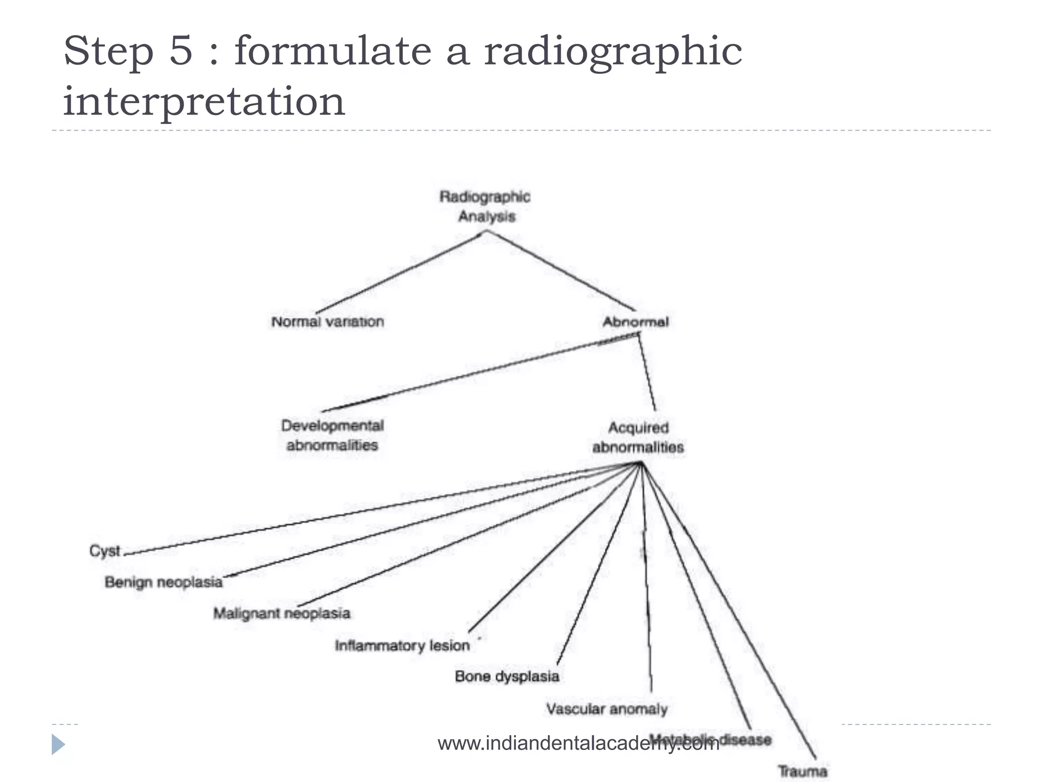
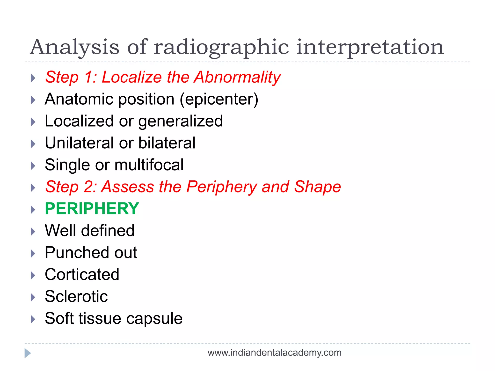
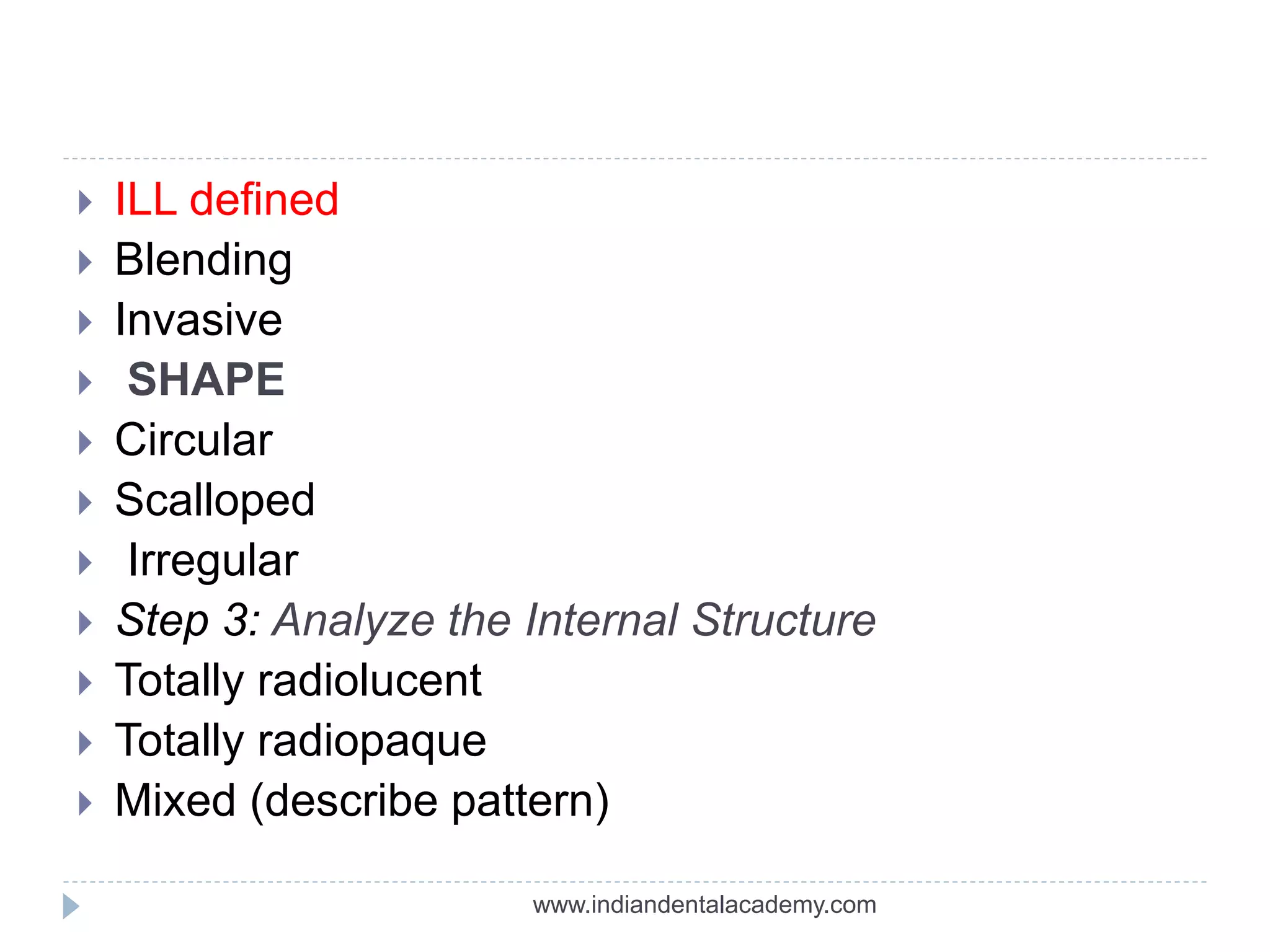
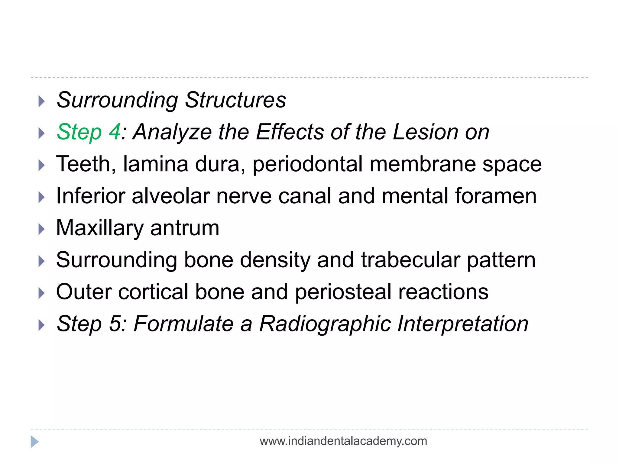
INDIAN DENTAL ACADEMY
Leader in continuing Dental Education
www.indiandentalacademy.com
Interpretation terminology
 Interpret: to offer an explanation
 Interpretation: an explanation
 Radiographic interpretation : an explanation of what
is viewed on dental radiograph.
 Diagnosis: the identification of a disease by
examination or analysis
www.indiandentalacademy.com
IMPORTANCE
 Dental radiographs are essential for diagnostic
purposes.
 All dental radiographs must be carefully reviewed
and interpreted.
 A great deal of information about the teeth and
supporting bone is obtained from radiographic
interpretation.
 It enables the dental professional to play vital role in
the detection of diseases , lesions and conditions of
the teeth and jaws that cannot be identified clinically.
www.indiandentalacademy.com
CLINICAL SITUATIONS NEED
RADIOGRAPHIC EXAMINATIONS
1-Dental caries
2-periodontal diseases
3-Dental anomalies
4-Growth&development and dental malocclusion
5-occult diseases
6-Tmj disorders
7-Implants
8-paranasal sinuses
9-Trauma
www.indiandentalacademy.com
Essential requirements for dental
radiographs
 Optimum viewing conditions
 Understanding the nature and limitations of the
black, white and grey radiographic image
 Knowledge of what the radiographs used in
dentistry should look like, so a critical assessment of
individual film quality can be made.
www.indiandentalacademy.com
 Detailed knowledge of the range of
radiographic appearances of normal
anatomical structures
 Detailed knowledge of the radiographic
appearances of the pathological conditions
affecting the head and neck
 A systematic approach to viewing the entire
radiograph and to viewing and describing
specific lesions
 Access to previous films for comparison.
www.indiandentalacademy.com
NEVER INTERPRET A FAULTY
RADIOGRAPH
Ideal radiograph:
 Visual : density & contrast
 Geometric : sharpness/detail, resolution/definition,
magnification, distortion
 Anatomical accuracy of radiographic images
 Adequate coverage of anatomical region of interest.
www.indiandentalacademy.com
RADIOGRAPHIC INTERPRETATION
 CLINICAL EXAMINATION
 QUALITY OF DIAGNOSTIC IMAGE
 NUMBER &TYPE OF AVAILABLE IMAGES
 VIEWING CONDITIONS
www.indiandentalacademy.com
Examples of how variations in radiographic technique
can alter the images produced of the same object. A
Correct projection. B Incorrect vertical angulation
producing an elongated image. C Incorrect vertical
angulation producing a foreshortened image. D and E
Incorrect horizontal angulations producing distorted
images.
www.indiandentalacademy.com
Examples of how variations in exposure
factors can alter the image quality of the
same object
. A Overexposed. B Slightly overexposed.
C Correctly exposed.D Underexposed.
www.indiandentalacademy.com
Viewing conditions
 This should be done in a quiet, darkened room
 At least two good, evenly-lit viewing boxes are required
 A bright light illuminator is required for relatively over-
exposed areas
 Mounted in holder
 Appropriate size of view box to accommodate film
 Magnifying glass-detailed examination of small regions
www.indiandentalacademy.com
A, ward ray viewing box incorporating an additional incorporating
bright light source for viewing overexposed dark films
B, SDI xray reader- an extraneous light excluding intra oral film
Viewer with built in magnification
www.indiandentalacademy.com
The effect of different viewing conditions on same periapical
Radiograph A, with a black surround B, with white surround
Note , increased details visible in A, particularly around molar teeth.
www.indiandentalacademy.com
Image analysis
 Systematic radiographic examination -Identify
normal anatomy and examinate the entire film
 Extra oral images - panoramic films , cephalometric
views & TMJ views
www.indiandentalacademy.com
First visual circuit: intraoral images
 Periapical before bitewing images
 Right maxilla to left; left mandible to right
 One anatomic structure at a time
 Eg: posterior maxilla-maxillary
sinus,tuberosity,zygomatic process
 Normal anatomy
bones, canals, foramina
www.indiandentalacademy.com
Second visual circuit
 Examination of bone:
 Height of alveolar bone
 Crest relative to teeth
 Loss of height-more than 1.5 mm-periodontal
disease
 Cortication
 Lamina dura + PDL space + tooth roots
 Carcinoma-erosion of alveolar crest+ ill defined
borders.
www.indiandentalacademy.com
Third visual circuit
 Examination of dentition & associated structures
 Number, Sequence, appearance, root structure
 Crowns –defective enamel, caries
 Intreproximal areas & restorations
 Pulp chambers-size, content
 Bone-radioluscent/radioopaque lesions
www.indiandentalacademy.com
Aunt minny approach
 Aunt Minny represents an abnormality which
looks like one that the evaluator has seen
before, or been told about.
 It would be difficult to recognise new findings
using this approach
www.indiandentalacademy.com
Analysis of intra osseous lesions
 STEP 1:LOCALIZE THE ABNORMALITY
LOCALIZED OR GENERALIZED
POSITION IN THE JAWS
SINGLE OR MULTIFOCAL
UNILATERAL OR BILATERAL
SIZE
www.indiandentalacademy.com
Characteristics of lesional tissue
 Described as
miltilocular,unilocular,circumscribed or not.
 Indicates
well circumscribed –benign or cystic
poorly circumscribed-malignant.
 Radiolucent lesions without septations have three
pattern of bone destruction
geographic,moth eaten and permeative.
www.indiandentalacademy.com
Geographic Pattern
 Single ,large area
 More than 1 cm
 Signifies
 large area of lysis
 Less aggressive form of
malignant lesion
 Monolocular or non septated
benign lesion
www.indiandentalacademy.com
Moth Eaten Pattern
 Smaller areas of bone
destruction
 Less well defined
 3 to 5 mm
 Signifies
 Can in both benign and
malignant
 Inflammatory conditions
like osteomyelitis and
osteonecrosis
 More destructive lesion
than with geographic
pattern.
www.indiandentalacademy.com
Permeative Pattern
 Much smaller and
poorly defined
 1-2 mm in size
 Signifies
 Aggressive,rapidly
destructive lesion.
 Cortex involvement
with this ,indicates
rapid destruction.
www.indiandentalacademy.com
Radiolucent Lesions with Septations
 True septations
vascular lesions
 False septations
 Erosion or scalloping of endosteal surface
.eg.ameloblastoma
 Filaments of remnant host boneform locules within
lesion.eg.aneurysmal bone cyst,central giant cell
granuloma
www.indiandentalacademy.com
Honeycomb Pattern
 Loculations are small
and numerous
 Represent earlier
change than soap
bubble pattern
 ameloblastoma
www.indiandentalacademy.com
Soap Bubble Pattern
 Larger and less
numerous loculations.
 Signifies
 Breakdown of
honeycomb pattern
 ameloblastoma
www.indiandentalacademy.com
Tennis Racket Pattern
 Septa intersect at
right angles.
 Odontogenic
myxoma
www.indiandentalacademy.com
Scalloped Pattern
 Incompete septation
gives a false
impression of
multilocularity.
 Odontogenic
keratocyst
www.indiandentalacademy.com
LOCATION AND EXTENT
 Location –helps in diagnosis
maxilla /mandible
unilateral/bilateral
incisor /premolar/molar
angle/ramus/body area
localized and generalized
www.indiandentalacademy.com
Position in jaws
 Epicenter -coronal to tooth- odontogenic epithelium
 Epicenter of the lesion is above the mandibular
canal-odontogenic in origin
 Epicenter -below IAC-non odontogenic (likely)
 Cartilaginous lesions, osteochondromas –condylar
region.
 If the epicenter of the lesion is in the sinus, not
odontogenic in origin-alveolar process of maxilla
www.indiandentalacademy.com
A ,cropped panoramic image of a lesion where epicenter
Is coronal to the mandibular first molar b,an occlusal view of
Same lesion.
www.indiandentalacademy.com
A cropped panoramic image displaying a lesion
(developmental salivary gland defect) below the inferior
alveolar canal and thus unlikely to be of odontogenic
origin.
www.indiandentalacademy.com
A lateral oblique view of the mandible revealing
a lesion within the inferior alveolar canal. The smooth
fusiform expansion of the canal indicates a neural lesion
www.indiandentalacademy.com
The lack of a peripheral cortex on this benign cyst
indicates that it originated in the sinus and not in the alveolar
process. It therefore is unlikely to be of odontogenic origin.
www.indiandentalacademy.com
STEP 2:ASSESS THE PERIPHERY &SHAPE
WELL DEFINED OR ILL DEFINED?
 Sharp margins
 Corticated margins
 Sclerotic margins
 Radiolucent band
 Blends into adjacent area
 Irregular margins
www.indiandentalacademy.com
 WELL OR POORLY DEMARCATED
 Punched out-sharp- (no bony reaction)-
multiple myeloma
 Corticated-uniform-periphery- (thin opaque border)
cyst
 Sclerotic (wide, uneven opaque border)
Periapical cemental dysplasia
 Radiolucent(periphery)+ corticated
Odontoma , cementoblastoma
www.indiandentalacademy.com
A cropped lateral skull view shows several punched out
Lesion of multiple myeloma
www.indiandentalacademy.com
Lateral periapical cyst in mandibular premolar region
With well defined corticated periphery
www.indiandentalacademy.com
Late lesions of periapical cemental dysplasia shows more
Radiopaque interior surrounded by more radiolucent margin
www.indiandentalacademy.com
Note, the thin radiolucent positioned between the internal
radiopaque structure of this odontoma and radiopaque outer cortical
border.
www.indiandentalacademy.com
ILL DEFINED BORDERS
 Blending border - gradual transition-normal
appearing bone & abnormal appearing trabeculae -
sclerosing osteitis
 Invasive border-bone destruction-malignancy
www.indiandentalacademy.com
Periapical (A) and occlusal (B) films revealing a squamous cell carcinoma
in the anterior maxilla. Note the invasive margin that extends beyond the
lateral incisor (arrow) and the bone destruction immediately behind this
margin
www.indiandentalacademy.com
Shape
 Circular
 Oval
 Scalloped
 Multilocular
www.indiandentalacademy.com
Periapical film of lateral periapical cyst ,
circular or oval In shape
www.indiandentalacademy.com
STEP3:ANALYZE THE INTERNAL
STRUCTURE
 Radiolucent
 Mixed
 Radiopaque
 Trabeculation
 Septa
 Calcifications
 Tooth or similar entities
www.indiandentalacademy.com
lateral periapical cyst in between two
premolars shows radiolucency
www.indiandentalacademy.com
Periapical cemental dysplasia showing
mixed lesion and mature radiopaque lesion
www.indiandentalacademy.com
A lateral oblique view of mandibular lesion
showing an internal Septa that divides the lesion
into several compartments
www.indiandentalacademy.com
A periapical film showing soft tissue mass of
shadow of polyp emanting from edentulous ridge
www.indiandentalacademy.com
STEP4:ANALYSE THE EFFECTS OF THE
LESION ON SURROUNDING STrUCTURES
 Teeth , lamina dura , periodontal membrane space
 Inferior alveolar canal & mental foramen
 Maxillary antrum
 Surrounding bone density & trabecular pattern
 Outer cortical bone & periosteal reaction
www.indiandentalacademy.com
A and B periapical films reveals malignant lymphoma invading
mandible.,irregular widening of pdl space is seen.
www.indiandentalacademy.com
Step 5 : formulate a radiographic
interpretation
 Decision 1: Normal V/S Abnormal
 Decision2: Developmental V/S Acquired
 Decision 3: Classification
 Decision 4: Ways To Proceed
www.indiandentalacademy.com
Step 5 : formulate a radiographic
interpretation
www.indiandentalacademy.com
Analysis of radiographic interpretation
 Step 1: Localize the Abnormality
 Anatomic position (epicenter)
 Localized or generalized
 Unilateral or bilateral
 Single or multifocal
 Step 2: Assess the Periphery and Shape
 PERIPHERY
 Well defined
 Punched out
 Corticated
 Sclerotic
 Soft tissue capsule
www.indiandentalacademy.com
 ILL defined
 Blending
 Invasive
 SHAPE
 Circular
 Scalloped
 Irregular
 Step 3: Analyze the Internal Structure
 Totally radiolucent
 Totally radiopaque
 Mixed (describe pattern)
www.indiandentalacademy.com
 Surrounding Structures
 Step 4: Analyze the Effects of the Lesion on
 Teeth, lamina dura, periodontal membrane space
 Inferior alveolar nerve canal and mental foramen
 Maxillary antrum
 Surrounding bone density and trabecular pattern
 Outer cortical bone and periosteal reactions
 Step 5: Formulate a Radiographic Interpretation
www.indiandentalacademy.com
A cropped panoramic image of a lesion related to the unerupted
mandibular first molar. B, An occlusal
projection providing a right-angled view of the same lesion
www.indiandentalacademy.com
 Location. The abnormality is singular and
unilateral,and the epicenter lies coronal to the
mandibular firstmolar.
 Periphery and shape. The lesion has a well-
defined cortical boundary and a spherical or round
shape.
 The periphery also attaches to the cemento enamel
junction.
 Internal structure. The internal structure is totally
radiolucent.
www.indiandentalacademy.com
 Effects. This lesion has displaced the first molar
in an apical direction, which reinforces the decision
that the origin was coronal to this tooth. Also, the
lesion has displaced the second molar distally and
the second premolar in an anterior direction. Apical
resorption distal root of the second deciduous molar
has occurred.
 The occlusal radiograph reveals that the buccal
cortical plate has expanded in a smooth, curved
shape, and a thin cortical boundary still exists.
 RADIOGRAPHIC DIAGNOSIS: follicular cyst
www.indiandentalacademy.com
Radiographic report
 Patient & general information
 Imaging procedure
 Clinical information
 Findings
 Radiographic interpretation
www.indiandentalacademy.com
Systematic viewing
 This approach ensures that all areas of the film are
observed and that the important features of the
tooth apex are examined.
www.indiandentalacademy.com
 GENERAL OVERVIEW OF ENTIRE RADIOGRAPH
 1. Note the chronological and development age of
the patient
 2. Note the position, outline and density of all the
normal I superimposed anatomical shadows
including any developing teeth
www.indiandentalacademy.com
 EXAMINE EACH TOOTH ON THE RADIOGRAPH
AND ASSESS
 3. THE CROWN
 Note particularly:
The presence of caries
The state of existing restorations
www.indiandentalacademy.com
 THE ROOT(S)
 Note particularly:
 • The length of the root
 • The number(s)
 • The morphology
 • The size and shape of canals
 • The presence of:
a. Pulp stones
b. Root fillings
c. Internal resorption
d. External resorption
e. Root fractures www.indiandentalacademy.com
THE APICAL TISSUES
 Note particularly:
 • The integrity, continuity and thickness of:
a. The radiolucent line of the periodontal
ligament space
b. The radiopaque line of the lamina dura
 • Any associated radiolucent areas
 • Any associated radiopaque areas
 • The pattern of the trabecular bone
www.indiandentalacademy.com
 Diagrams showing the
various radiographic
appearances of infection
and inflammation in the
apical tissues.
 A Normal.
 B Early apical change —
widening of the
radiolucent periodontal
ligament space (acute
apical periodontitis)
(arrowed).
 C Early apical change —
loss of the radiopaque
lamina dura (early
periapical abscess}
(arrowed).
www.indiandentalacademy.com
 D Extensive destructive
acute inflammation —
diffuse, ill-defined area of
radiolucency at the apex
(periapical abscess).
 E Low grade chronic
inflammation — diffuse
radiopaque area at the
apex (sclerosing osteitis).
 F Longstanding chronic
inflammation — well-
defined area of
radiolucency surrounded
by dense sclerotic bone
(periapical granuloma or
radicular cyst).
www.indiandentalacademy.com
 THE PERIODONTAL TISSUES
 Note particularly:
 • The width of the periodontal ligament
 • The level and quality of the crestal bone
 • Any vertical or horizontal bone loss
 • Any calculus deposits
 • Any furcation involvements
www.indiandentalacademy.com
REFERENCES
1. Oral Radiology Principles and Interpretations –
White and Pharoah edition 5.
2. Dental radiology principles and techniques-
Joen Iannucci Haring edition 2.
3. Essentials of dental radiography and radiology
– Eric Whaites – 3rd edition.
www.indiandentalacademy.com

Principles of radiographic interpretation/ dental courses

  • 1.
    INDIAN DENTAL ACADEMY Leaderin continuing Dental Education www.indiandentalacademy.com
  • 2.
    Interpretation terminology  Interpret:to offer an explanation  Interpretation: an explanation  Radiographic interpretation : an explanation of what is viewed on dental radiograph.  Diagnosis: the identification of a disease by examination or analysis www.indiandentalacademy.com
  • 3.
    IMPORTANCE  Dental radiographsare essential for diagnostic purposes.  All dental radiographs must be carefully reviewed and interpreted.  A great deal of information about the teeth and supporting bone is obtained from radiographic interpretation.  It enables the dental professional to play vital role in the detection of diseases , lesions and conditions of the teeth and jaws that cannot be identified clinically. www.indiandentalacademy.com
  • 4.
    CLINICAL SITUATIONS NEED RADIOGRAPHICEXAMINATIONS 1-Dental caries 2-periodontal diseases 3-Dental anomalies 4-Growth&development and dental malocclusion 5-occult diseases 6-Tmj disorders 7-Implants 8-paranasal sinuses 9-Trauma www.indiandentalacademy.com
  • 5.
    Essential requirements fordental radiographs  Optimum viewing conditions  Understanding the nature and limitations of the black, white and grey radiographic image  Knowledge of what the radiographs used in dentistry should look like, so a critical assessment of individual film quality can be made. www.indiandentalacademy.com
  • 6.
     Detailed knowledgeof the range of radiographic appearances of normal anatomical structures  Detailed knowledge of the radiographic appearances of the pathological conditions affecting the head and neck  A systematic approach to viewing the entire radiograph and to viewing and describing specific lesions  Access to previous films for comparison. www.indiandentalacademy.com
  • 7.
    NEVER INTERPRET AFAULTY RADIOGRAPH Ideal radiograph:  Visual : density & contrast  Geometric : sharpness/detail, resolution/definition, magnification, distortion  Anatomical accuracy of radiographic images  Adequate coverage of anatomical region of interest. www.indiandentalacademy.com
  • 8.
    RADIOGRAPHIC INTERPRETATION  CLINICALEXAMINATION  QUALITY OF DIAGNOSTIC IMAGE  NUMBER &TYPE OF AVAILABLE IMAGES  VIEWING CONDITIONS www.indiandentalacademy.com
  • 9.
    Examples of howvariations in radiographic technique can alter the images produced of the same object. A Correct projection. B Incorrect vertical angulation producing an elongated image. C Incorrect vertical angulation producing a foreshortened image. D and E Incorrect horizontal angulations producing distorted images. www.indiandentalacademy.com
  • 10.
    Examples of howvariations in exposure factors can alter the image quality of the same object . A Overexposed. B Slightly overexposed. C Correctly exposed.D Underexposed. www.indiandentalacademy.com
  • 11.
    Viewing conditions  Thisshould be done in a quiet, darkened room  At least two good, evenly-lit viewing boxes are required  A bright light illuminator is required for relatively over- exposed areas  Mounted in holder  Appropriate size of view box to accommodate film  Magnifying glass-detailed examination of small regions www.indiandentalacademy.com
  • 12.
    A, ward rayviewing box incorporating an additional incorporating bright light source for viewing overexposed dark films B, SDI xray reader- an extraneous light excluding intra oral film Viewer with built in magnification www.indiandentalacademy.com
  • 13.
    The effect ofdifferent viewing conditions on same periapical Radiograph A, with a black surround B, with white surround Note , increased details visible in A, particularly around molar teeth. www.indiandentalacademy.com
  • 14.
    Image analysis  Systematicradiographic examination -Identify normal anatomy and examinate the entire film  Extra oral images - panoramic films , cephalometric views & TMJ views www.indiandentalacademy.com
  • 15.
    First visual circuit:intraoral images  Periapical before bitewing images  Right maxilla to left; left mandible to right  One anatomic structure at a time  Eg: posterior maxilla-maxillary sinus,tuberosity,zygomatic process  Normal anatomy bones, canals, foramina www.indiandentalacademy.com
  • 16.
    Second visual circuit Examination of bone:  Height of alveolar bone  Crest relative to teeth  Loss of height-more than 1.5 mm-periodontal disease  Cortication  Lamina dura + PDL space + tooth roots  Carcinoma-erosion of alveolar crest+ ill defined borders. www.indiandentalacademy.com
  • 17.
    Third visual circuit Examination of dentition & associated structures  Number, Sequence, appearance, root structure  Crowns –defective enamel, caries  Intreproximal areas & restorations  Pulp chambers-size, content  Bone-radioluscent/radioopaque lesions www.indiandentalacademy.com
  • 18.
    Aunt minny approach Aunt Minny represents an abnormality which looks like one that the evaluator has seen before, or been told about.  It would be difficult to recognise new findings using this approach www.indiandentalacademy.com
  • 19.
    Analysis of intraosseous lesions  STEP 1:LOCALIZE THE ABNORMALITY LOCALIZED OR GENERALIZED POSITION IN THE JAWS SINGLE OR MULTIFOCAL UNILATERAL OR BILATERAL SIZE www.indiandentalacademy.com
  • 20.
    Characteristics of lesionaltissue  Described as miltilocular,unilocular,circumscribed or not.  Indicates well circumscribed –benign or cystic poorly circumscribed-malignant.  Radiolucent lesions without septations have three pattern of bone destruction geographic,moth eaten and permeative. www.indiandentalacademy.com
  • 21.
    Geographic Pattern  Single,large area  More than 1 cm  Signifies  large area of lysis  Less aggressive form of malignant lesion  Monolocular or non septated benign lesion www.indiandentalacademy.com
  • 22.
    Moth Eaten Pattern Smaller areas of bone destruction  Less well defined  3 to 5 mm  Signifies  Can in both benign and malignant  Inflammatory conditions like osteomyelitis and osteonecrosis  More destructive lesion than with geographic pattern. www.indiandentalacademy.com
  • 23.
    Permeative Pattern  Muchsmaller and poorly defined  1-2 mm in size  Signifies  Aggressive,rapidly destructive lesion.  Cortex involvement with this ,indicates rapid destruction. www.indiandentalacademy.com
  • 24.
    Radiolucent Lesions withSeptations  True septations vascular lesions  False septations  Erosion or scalloping of endosteal surface .eg.ameloblastoma  Filaments of remnant host boneform locules within lesion.eg.aneurysmal bone cyst,central giant cell granuloma www.indiandentalacademy.com
  • 25.
    Honeycomb Pattern  Loculationsare small and numerous  Represent earlier change than soap bubble pattern  ameloblastoma www.indiandentalacademy.com
  • 26.
    Soap Bubble Pattern Larger and less numerous loculations.  Signifies  Breakdown of honeycomb pattern  ameloblastoma www.indiandentalacademy.com
  • 27.
    Tennis Racket Pattern Septa intersect at right angles.  Odontogenic myxoma www.indiandentalacademy.com
  • 28.
    Scalloped Pattern  Incompeteseptation gives a false impression of multilocularity.  Odontogenic keratocyst www.indiandentalacademy.com
  • 29.
    LOCATION AND EXTENT Location –helps in diagnosis maxilla /mandible unilateral/bilateral incisor /premolar/molar angle/ramus/body area localized and generalized www.indiandentalacademy.com
  • 30.
  • 31.
    Position in jaws Epicenter -coronal to tooth- odontogenic epithelium  Epicenter of the lesion is above the mandibular canal-odontogenic in origin  Epicenter -below IAC-non odontogenic (likely)  Cartilaginous lesions, osteochondromas –condylar region.  If the epicenter of the lesion is in the sinus, not odontogenic in origin-alveolar process of maxilla www.indiandentalacademy.com
  • 32.
    A ,cropped panoramicimage of a lesion where epicenter Is coronal to the mandibular first molar b,an occlusal view of Same lesion. www.indiandentalacademy.com
  • 33.
  • 34.
    A cropped panoramicimage displaying a lesion (developmental salivary gland defect) below the inferior alveolar canal and thus unlikely to be of odontogenic origin. www.indiandentalacademy.com
  • 35.
    A lateral obliqueview of the mandible revealing a lesion within the inferior alveolar canal. The smooth fusiform expansion of the canal indicates a neural lesion www.indiandentalacademy.com
  • 36.
    The lack ofa peripheral cortex on this benign cyst indicates that it originated in the sinus and not in the alveolar process. It therefore is unlikely to be of odontogenic origin. www.indiandentalacademy.com
  • 37.
    STEP 2:ASSESS THEPERIPHERY &SHAPE WELL DEFINED OR ILL DEFINED?  Sharp margins  Corticated margins  Sclerotic margins  Radiolucent band  Blends into adjacent area  Irregular margins www.indiandentalacademy.com
  • 38.
     WELL ORPOORLY DEMARCATED  Punched out-sharp- (no bony reaction)- multiple myeloma  Corticated-uniform-periphery- (thin opaque border) cyst  Sclerotic (wide, uneven opaque border) Periapical cemental dysplasia  Radiolucent(periphery)+ corticated Odontoma , cementoblastoma www.indiandentalacademy.com
  • 39.
    A cropped lateralskull view shows several punched out Lesion of multiple myeloma www.indiandentalacademy.com
  • 40.
    Lateral periapical cystin mandibular premolar region With well defined corticated periphery www.indiandentalacademy.com
  • 41.
    Late lesions ofperiapical cemental dysplasia shows more Radiopaque interior surrounded by more radiolucent margin www.indiandentalacademy.com
  • 42.
  • 43.
    Note, the thinradiolucent positioned between the internal radiopaque structure of this odontoma and radiopaque outer cortical border. www.indiandentalacademy.com
  • 44.
    ILL DEFINED BORDERS Blending border - gradual transition-normal appearing bone & abnormal appearing trabeculae - sclerosing osteitis  Invasive border-bone destruction-malignancy www.indiandentalacademy.com
  • 45.
  • 46.
    Periapical (A) andocclusal (B) films revealing a squamous cell carcinoma in the anterior maxilla. Note the invasive margin that extends beyond the lateral incisor (arrow) and the bone destruction immediately behind this margin www.indiandentalacademy.com
  • 47.
  • 48.
    Shape  Circular  Oval Scalloped  Multilocular www.indiandentalacademy.com
  • 49.
    Periapical film oflateral periapical cyst , circular or oval In shape www.indiandentalacademy.com
  • 50.
  • 51.
  • 52.
    STEP3:ANALYZE THE INTERNAL STRUCTURE Radiolucent  Mixed  Radiopaque  Trabeculation  Septa  Calcifications  Tooth or similar entities www.indiandentalacademy.com
  • 53.
    lateral periapical cystin between two premolars shows radiolucency www.indiandentalacademy.com
  • 54.
    Periapical cemental dysplasiashowing mixed lesion and mature radiopaque lesion www.indiandentalacademy.com
  • 55.
  • 56.
    A lateral obliqueview of mandibular lesion showing an internal Septa that divides the lesion into several compartments www.indiandentalacademy.com
  • 57.
    A periapical filmshowing soft tissue mass of shadow of polyp emanting from edentulous ridge www.indiandentalacademy.com
  • 58.
    STEP4:ANALYSE THE EFFECTSOF THE LESION ON SURROUNDING STrUCTURES  Teeth , lamina dura , periodontal membrane space  Inferior alveolar canal & mental foramen  Maxillary antrum  Surrounding bone density & trabecular pattern  Outer cortical bone & periosteal reaction www.indiandentalacademy.com
  • 59.
  • 60.
    A and Bperiapical films reveals malignant lymphoma invading mandible.,irregular widening of pdl space is seen. www.indiandentalacademy.com
  • 61.
  • 62.
    Step 5 :formulate a radiographic interpretation  Decision 1: Normal V/S Abnormal  Decision2: Developmental V/S Acquired  Decision 3: Classification  Decision 4: Ways To Proceed www.indiandentalacademy.com
  • 63.
    Step 5 :formulate a radiographic interpretation www.indiandentalacademy.com
  • 64.
    Analysis of radiographicinterpretation  Step 1: Localize the Abnormality  Anatomic position (epicenter)  Localized or generalized  Unilateral or bilateral  Single or multifocal  Step 2: Assess the Periphery and Shape  PERIPHERY  Well defined  Punched out  Corticated  Sclerotic  Soft tissue capsule www.indiandentalacademy.com
  • 65.
     ILL defined Blending  Invasive  SHAPE  Circular  Scalloped  Irregular  Step 3: Analyze the Internal Structure  Totally radiolucent  Totally radiopaque  Mixed (describe pattern) www.indiandentalacademy.com
  • 66.
     Surrounding Structures Step 4: Analyze the Effects of the Lesion on  Teeth, lamina dura, periodontal membrane space  Inferior alveolar nerve canal and mental foramen  Maxillary antrum  Surrounding bone density and trabecular pattern  Outer cortical bone and periosteal reactions  Step 5: Formulate a Radiographic Interpretation www.indiandentalacademy.com
  • 67.
    A cropped panoramicimage of a lesion related to the unerupted mandibular first molar. B, An occlusal projection providing a right-angled view of the same lesion www.indiandentalacademy.com
  • 68.
     Location. Theabnormality is singular and unilateral,and the epicenter lies coronal to the mandibular firstmolar.  Periphery and shape. The lesion has a well- defined cortical boundary and a spherical or round shape.  The periphery also attaches to the cemento enamel junction.  Internal structure. The internal structure is totally radiolucent. www.indiandentalacademy.com
  • 69.
     Effects. Thislesion has displaced the first molar in an apical direction, which reinforces the decision that the origin was coronal to this tooth. Also, the lesion has displaced the second molar distally and the second premolar in an anterior direction. Apical resorption distal root of the second deciduous molar has occurred.  The occlusal radiograph reveals that the buccal cortical plate has expanded in a smooth, curved shape, and a thin cortical boundary still exists.  RADIOGRAPHIC DIAGNOSIS: follicular cyst www.indiandentalacademy.com
  • 70.
    Radiographic report  Patient& general information  Imaging procedure  Clinical information  Findings  Radiographic interpretation www.indiandentalacademy.com
  • 71.
    Systematic viewing  Thisapproach ensures that all areas of the film are observed and that the important features of the tooth apex are examined. www.indiandentalacademy.com
  • 72.
     GENERAL OVERVIEWOF ENTIRE RADIOGRAPH  1. Note the chronological and development age of the patient  2. Note the position, outline and density of all the normal I superimposed anatomical shadows including any developing teeth www.indiandentalacademy.com
  • 73.
     EXAMINE EACHTOOTH ON THE RADIOGRAPH AND ASSESS  3. THE CROWN  Note particularly: The presence of caries The state of existing restorations www.indiandentalacademy.com
  • 74.
     THE ROOT(S) Note particularly:  • The length of the root  • The number(s)  • The morphology  • The size and shape of canals  • The presence of: a. Pulp stones b. Root fillings c. Internal resorption d. External resorption e. Root fractures www.indiandentalacademy.com
  • 75.
    THE APICAL TISSUES Note particularly:  • The integrity, continuity and thickness of: a. The radiolucent line of the periodontal ligament space b. The radiopaque line of the lamina dura  • Any associated radiolucent areas  • Any associated radiopaque areas  • The pattern of the trabecular bone www.indiandentalacademy.com
  • 76.
     Diagrams showingthe various radiographic appearances of infection and inflammation in the apical tissues.  A Normal.  B Early apical change — widening of the radiolucent periodontal ligament space (acute apical periodontitis) (arrowed).  C Early apical change — loss of the radiopaque lamina dura (early periapical abscess} (arrowed). www.indiandentalacademy.com
  • 77.
     D Extensivedestructive acute inflammation — diffuse, ill-defined area of radiolucency at the apex (periapical abscess).  E Low grade chronic inflammation — diffuse radiopaque area at the apex (sclerosing osteitis).  F Longstanding chronic inflammation — well- defined area of radiolucency surrounded by dense sclerotic bone (periapical granuloma or radicular cyst). www.indiandentalacademy.com
  • 78.
     THE PERIODONTALTISSUES  Note particularly:  • The width of the periodontal ligament  • The level and quality of the crestal bone  • Any vertical or horizontal bone loss  • Any calculus deposits  • Any furcation involvements www.indiandentalacademy.com
  • 79.
    REFERENCES 1. Oral RadiologyPrinciples and Interpretations – White and Pharoah edition 5. 2. Dental radiology principles and techniques- Joen Iannucci Haring edition 2. 3. Essentials of dental radiography and radiology – Eric Whaites – 3rd edition. www.indiandentalacademy.com