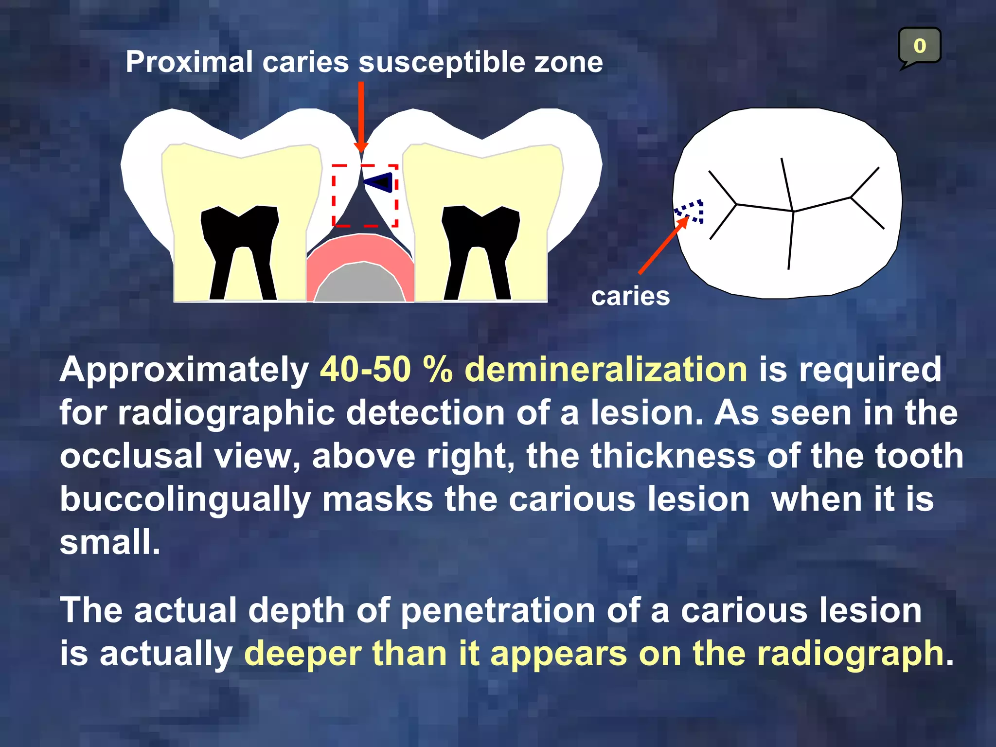
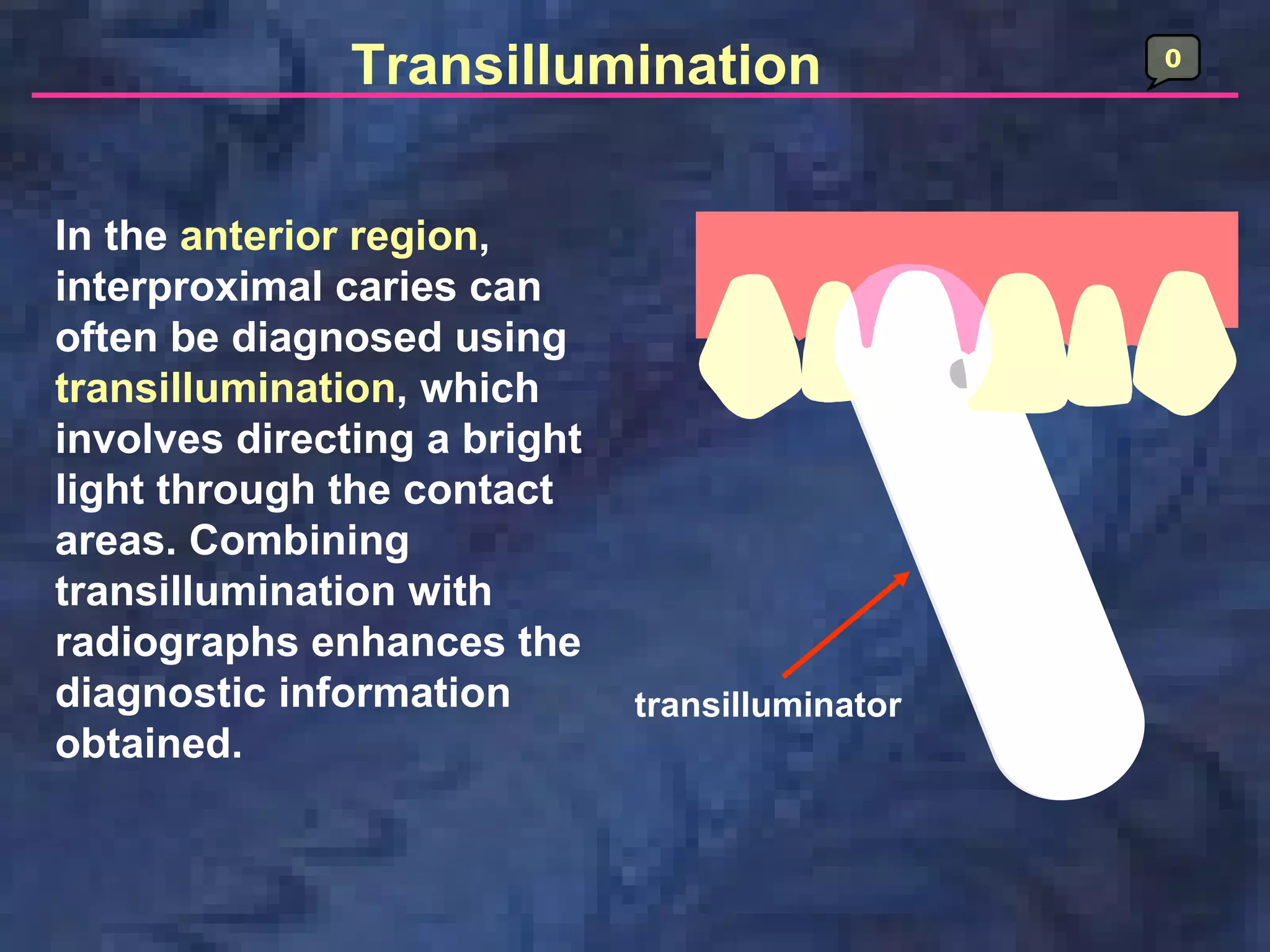
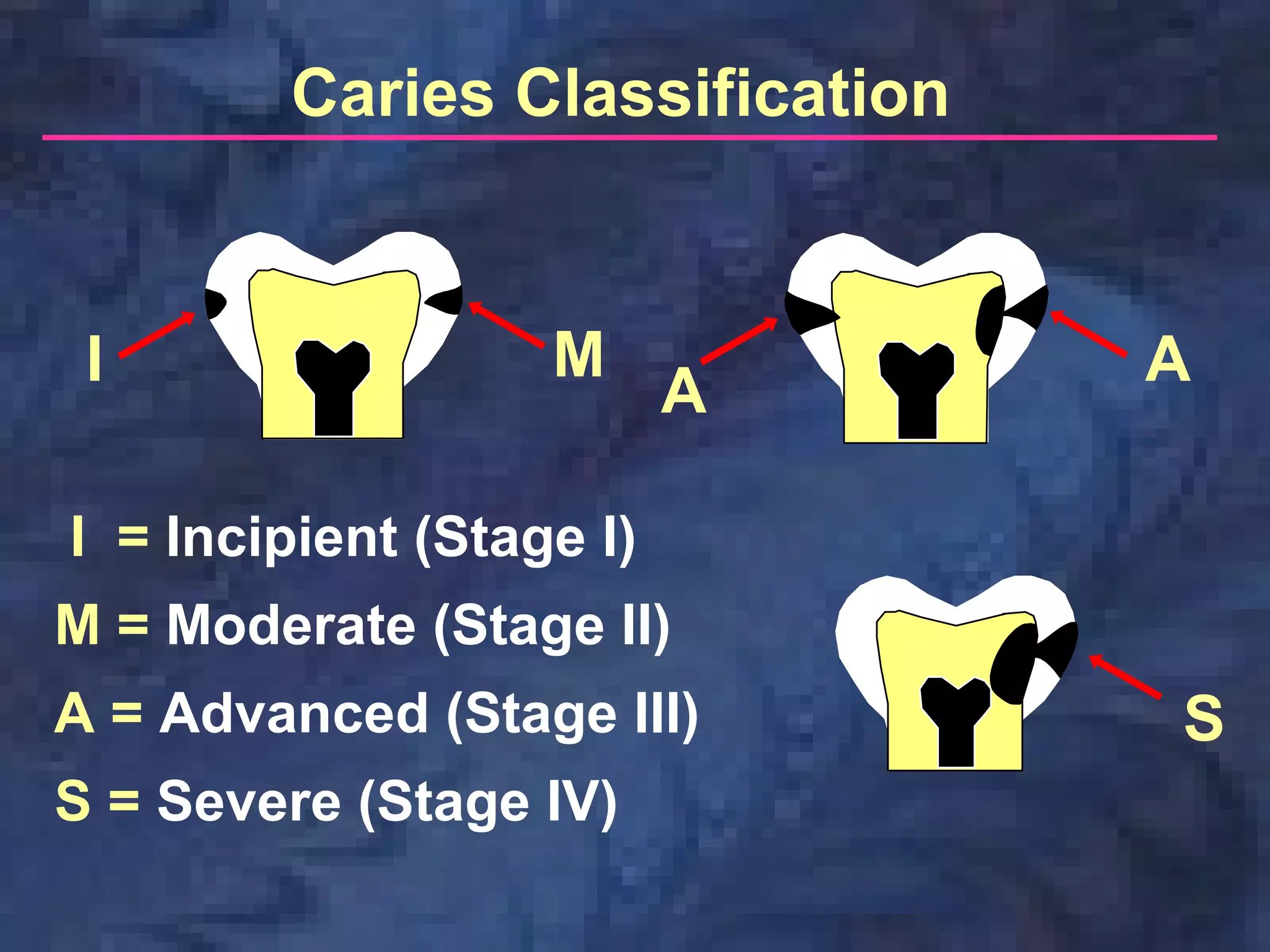
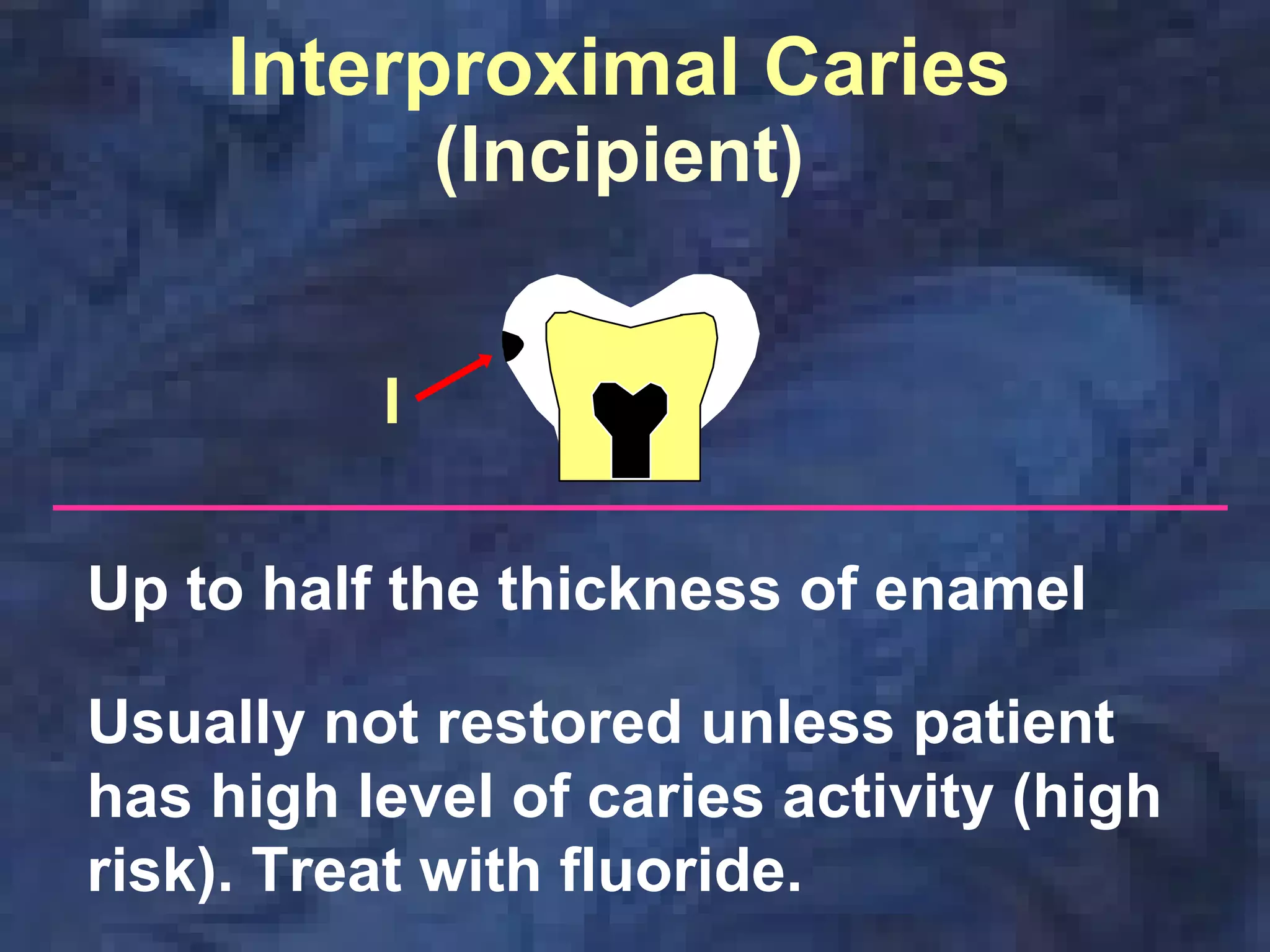
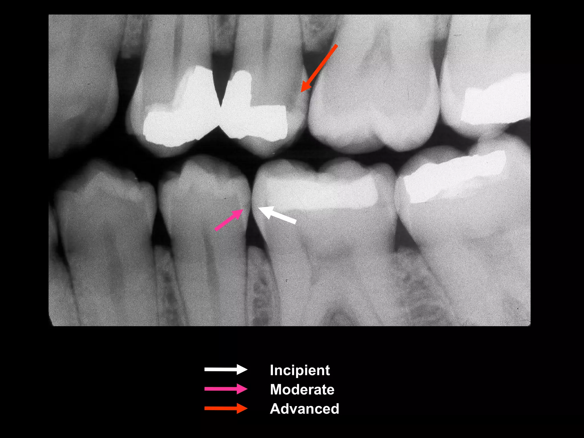
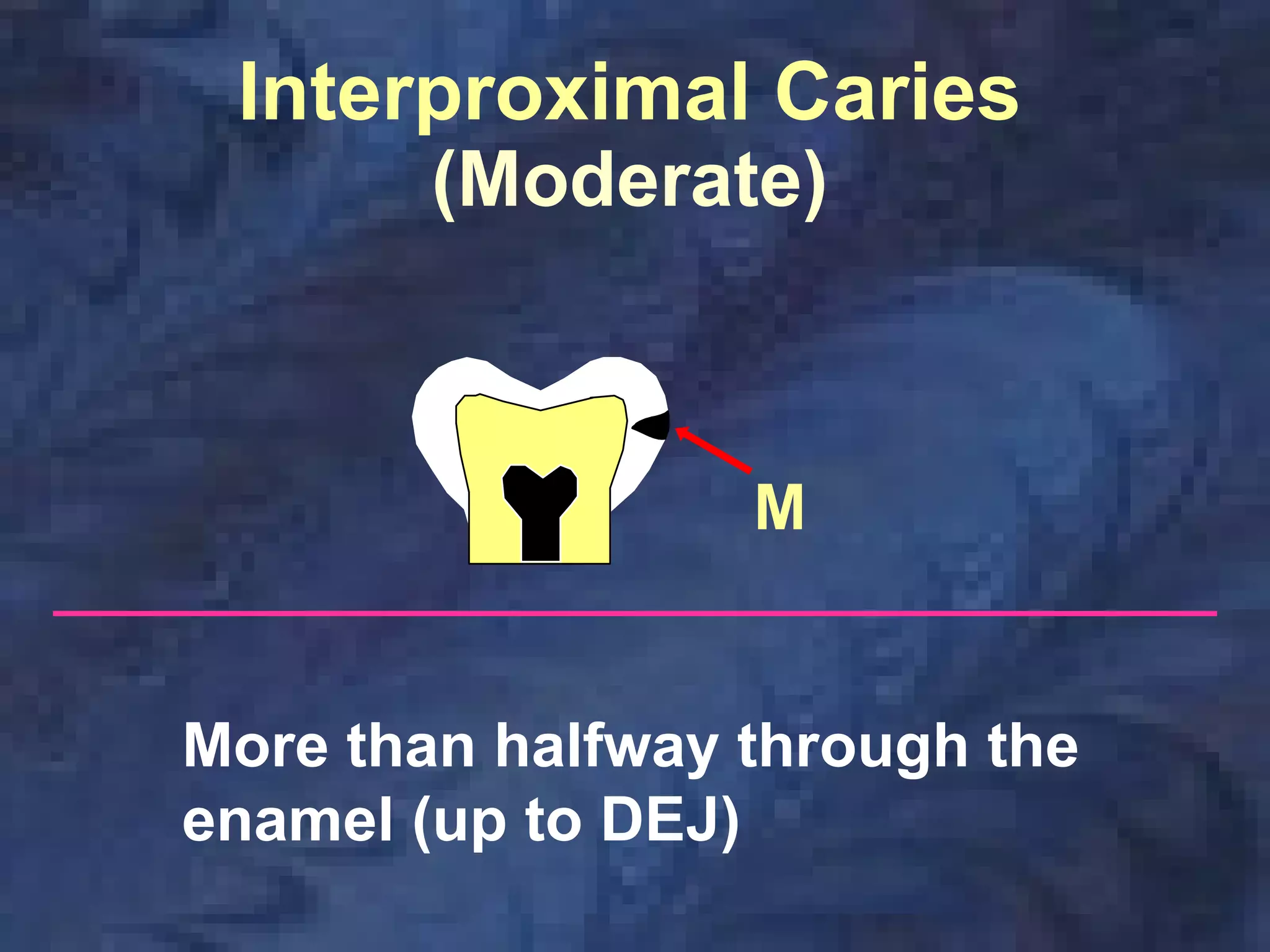
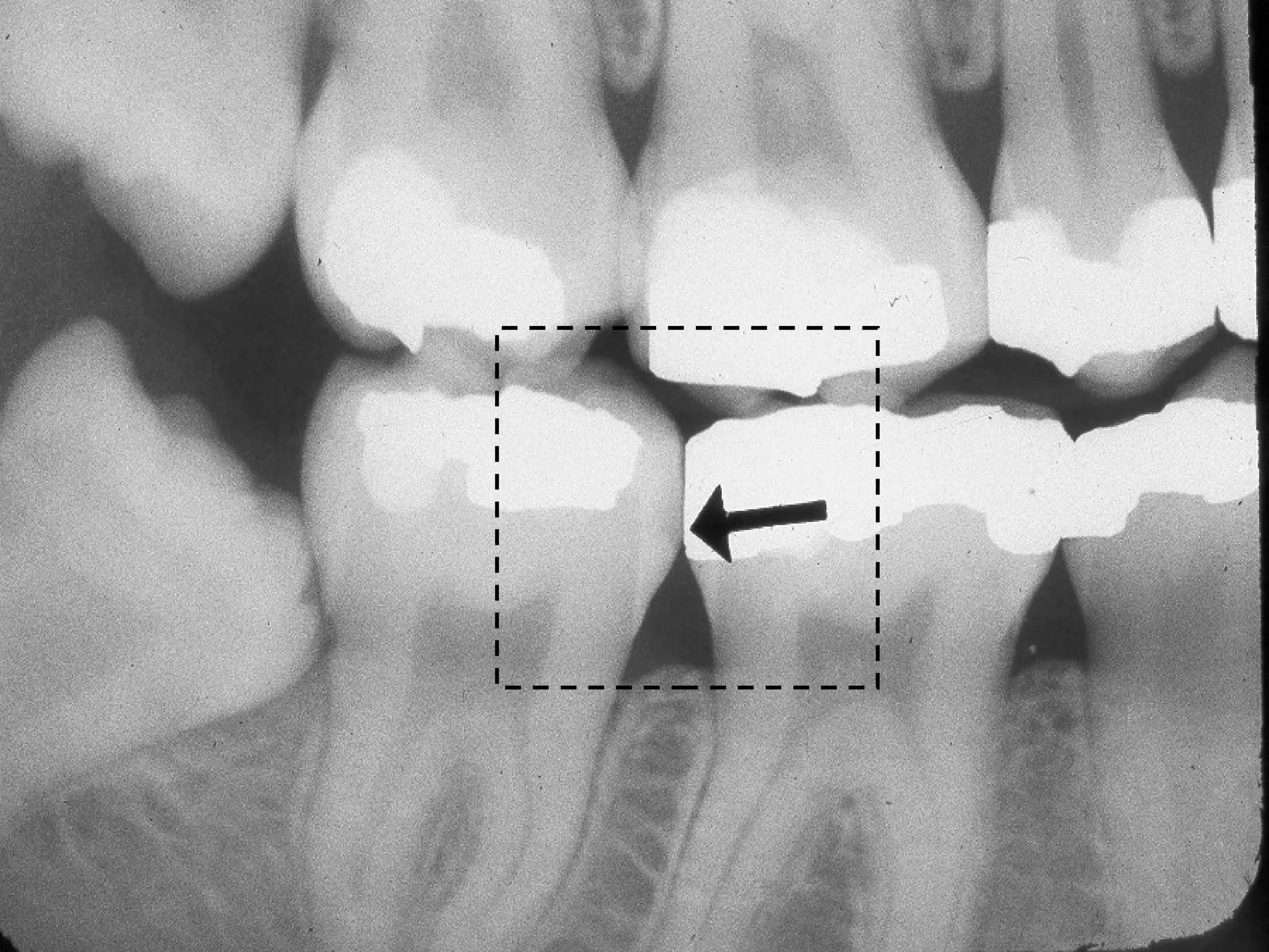
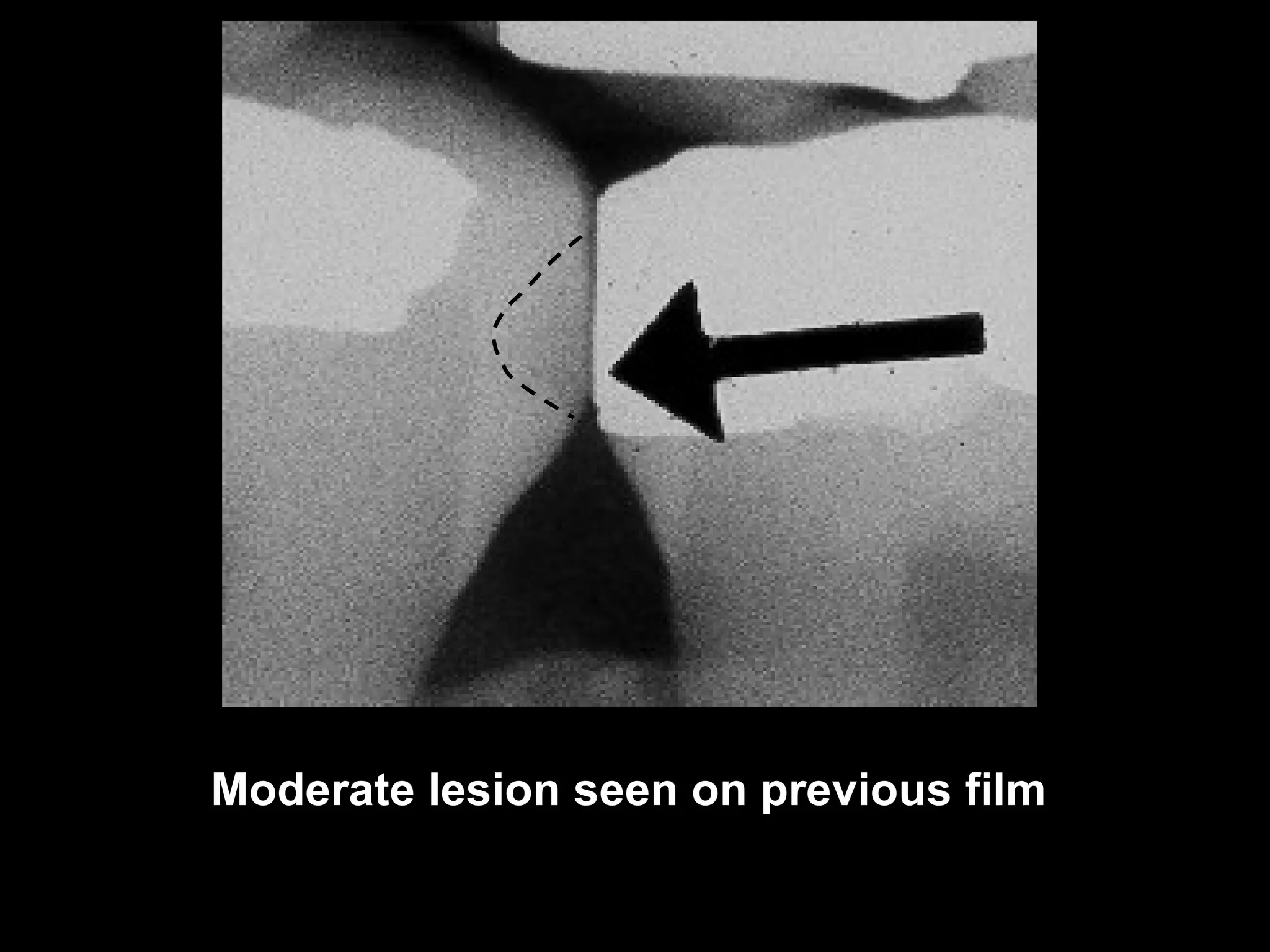
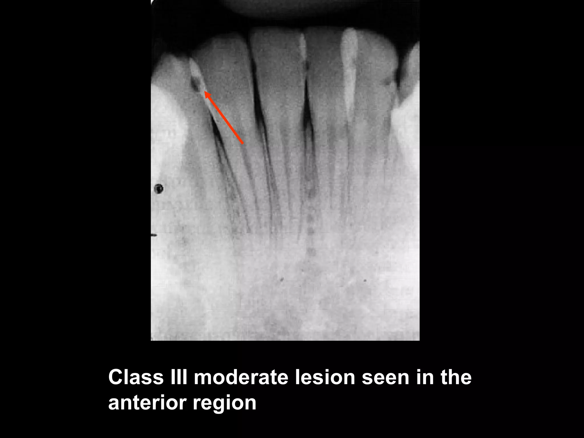
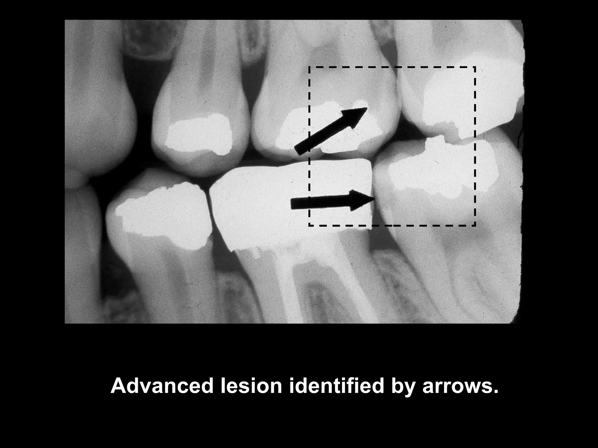
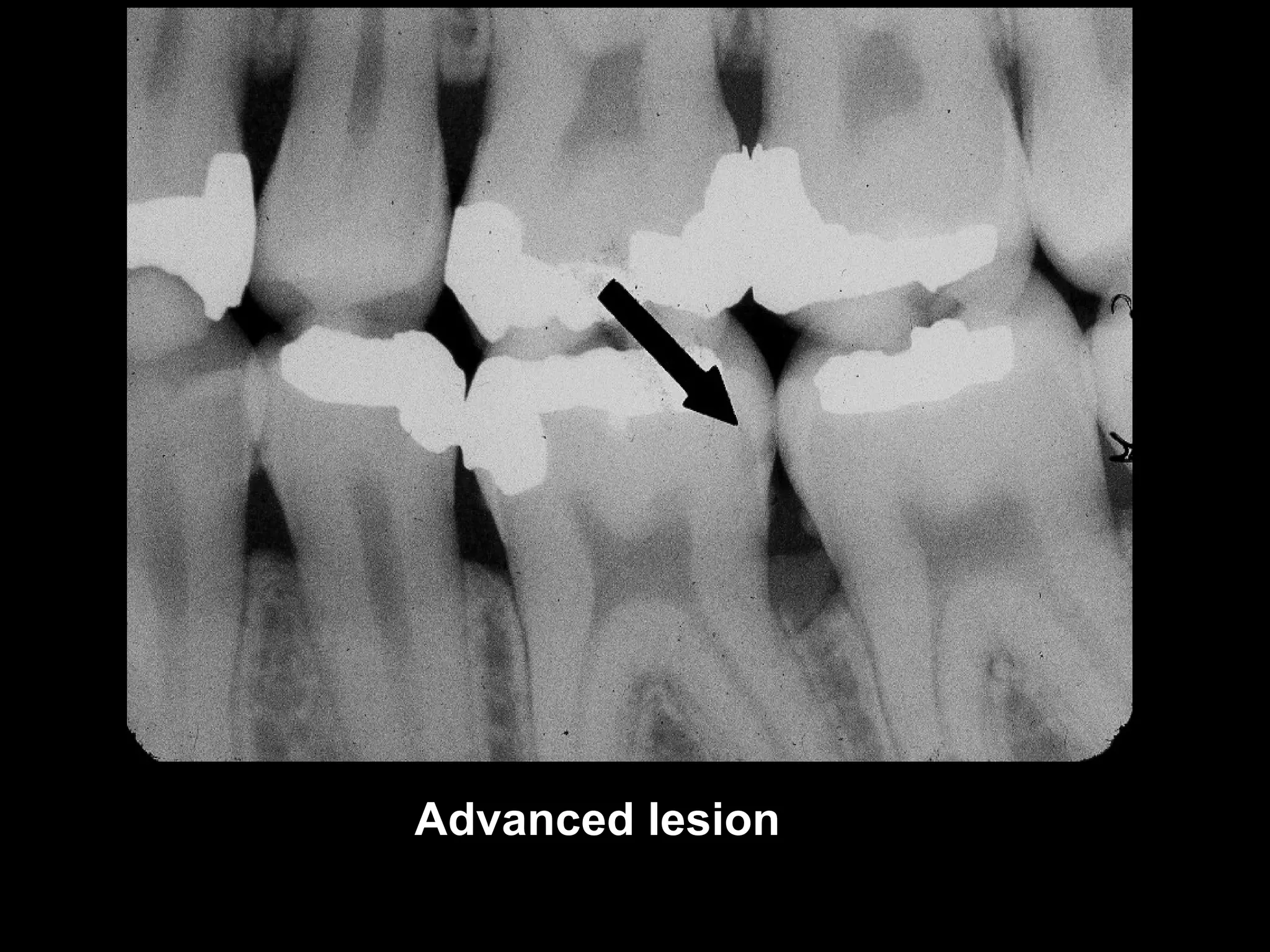
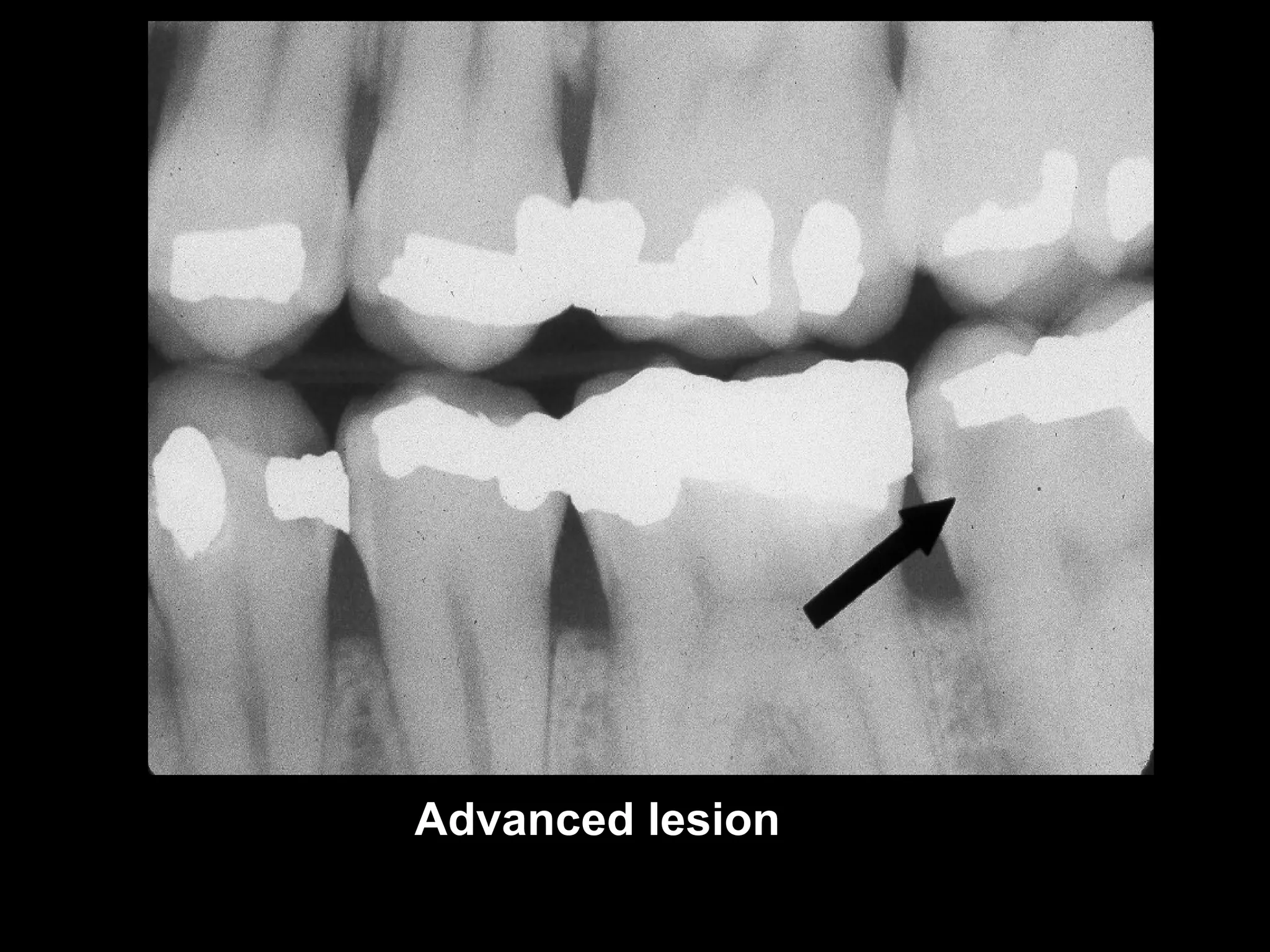
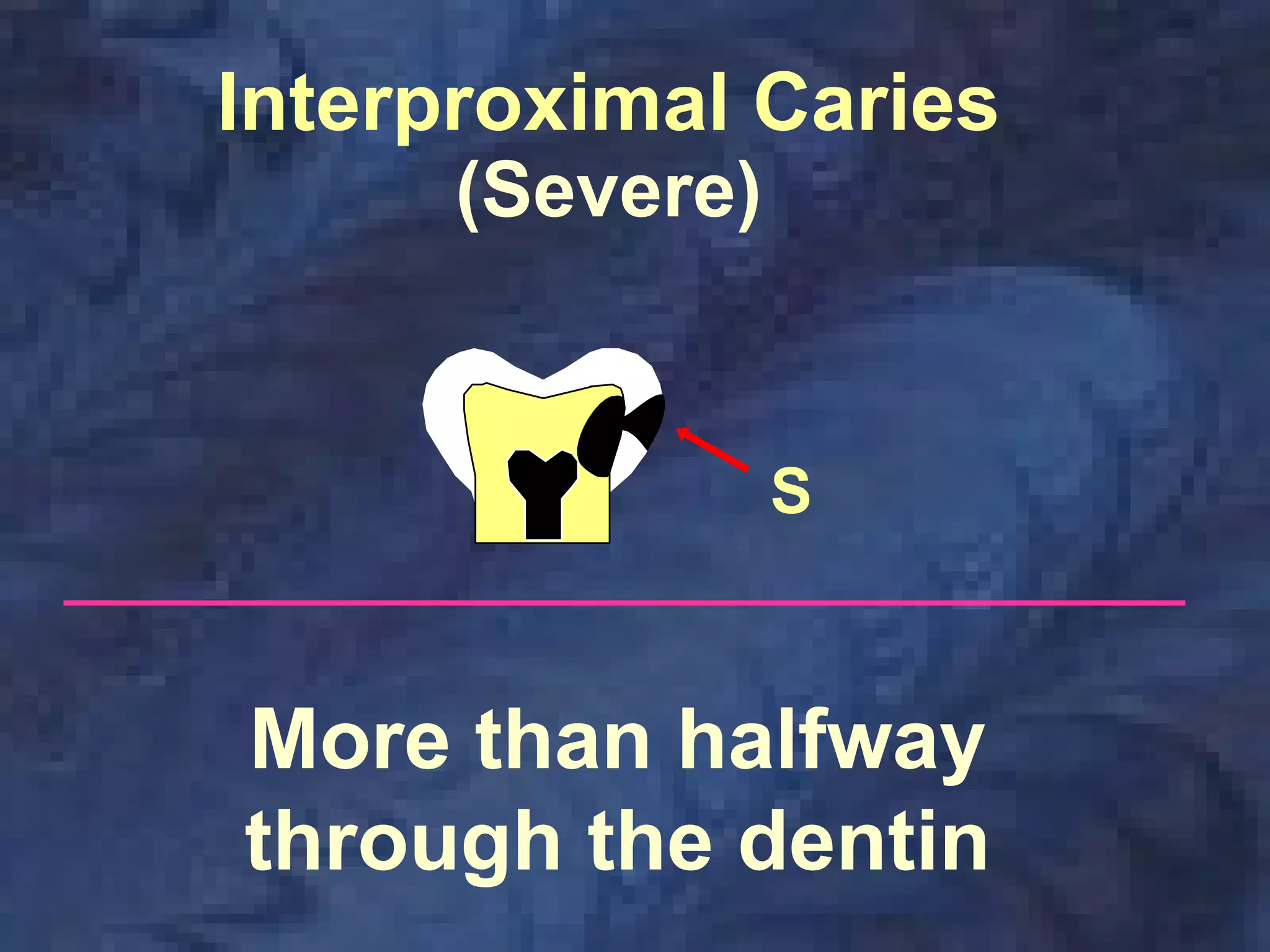
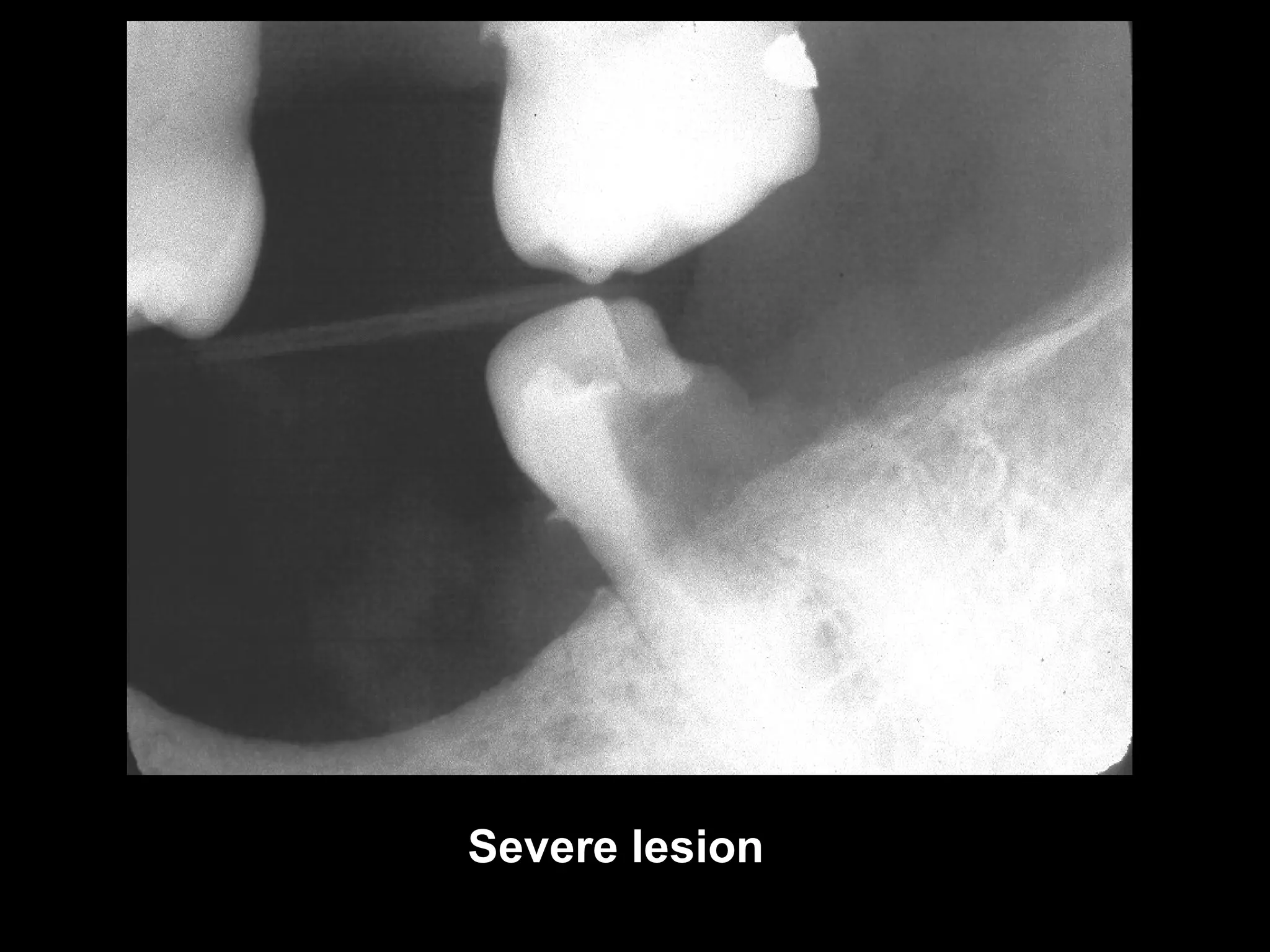
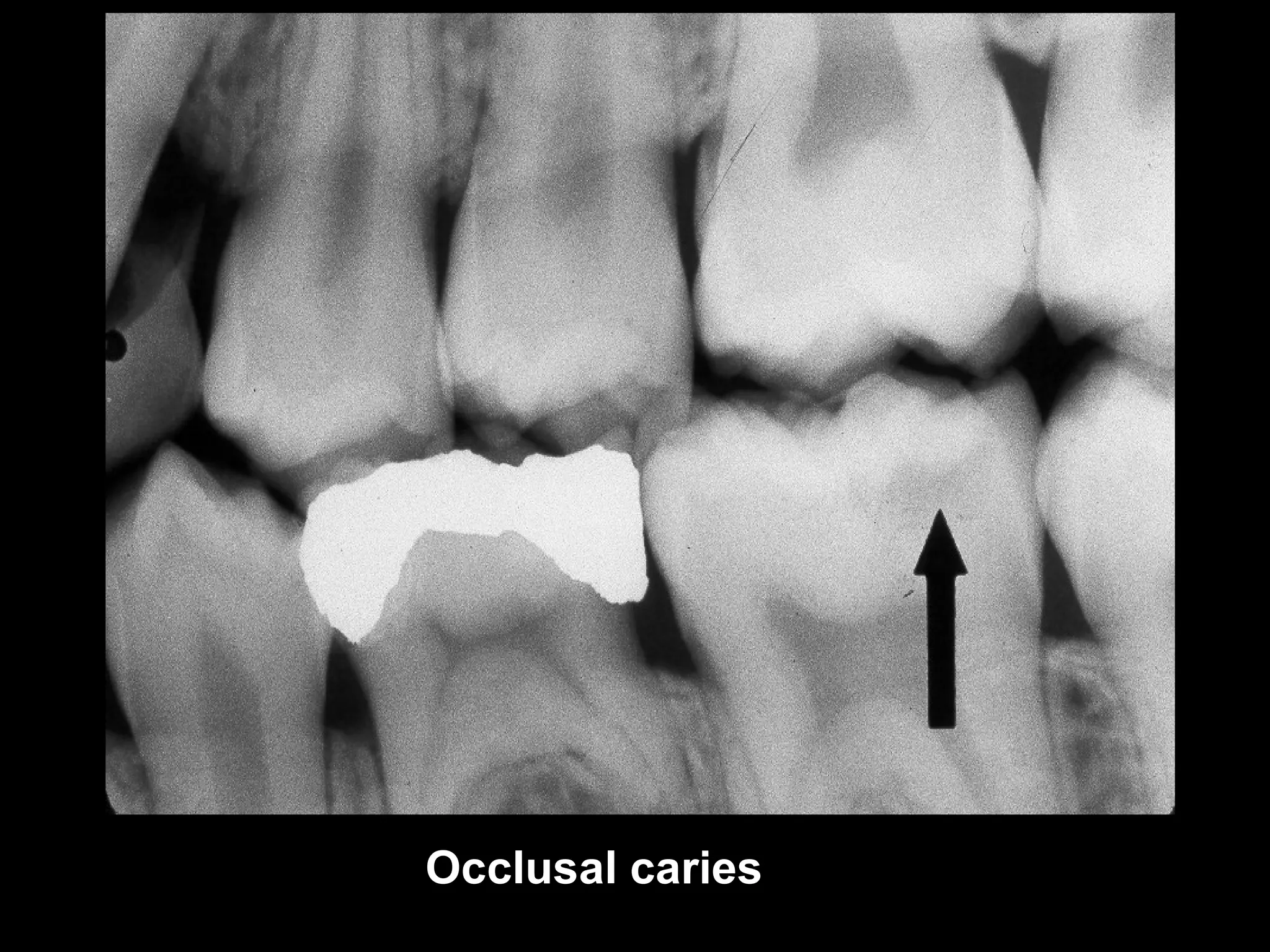
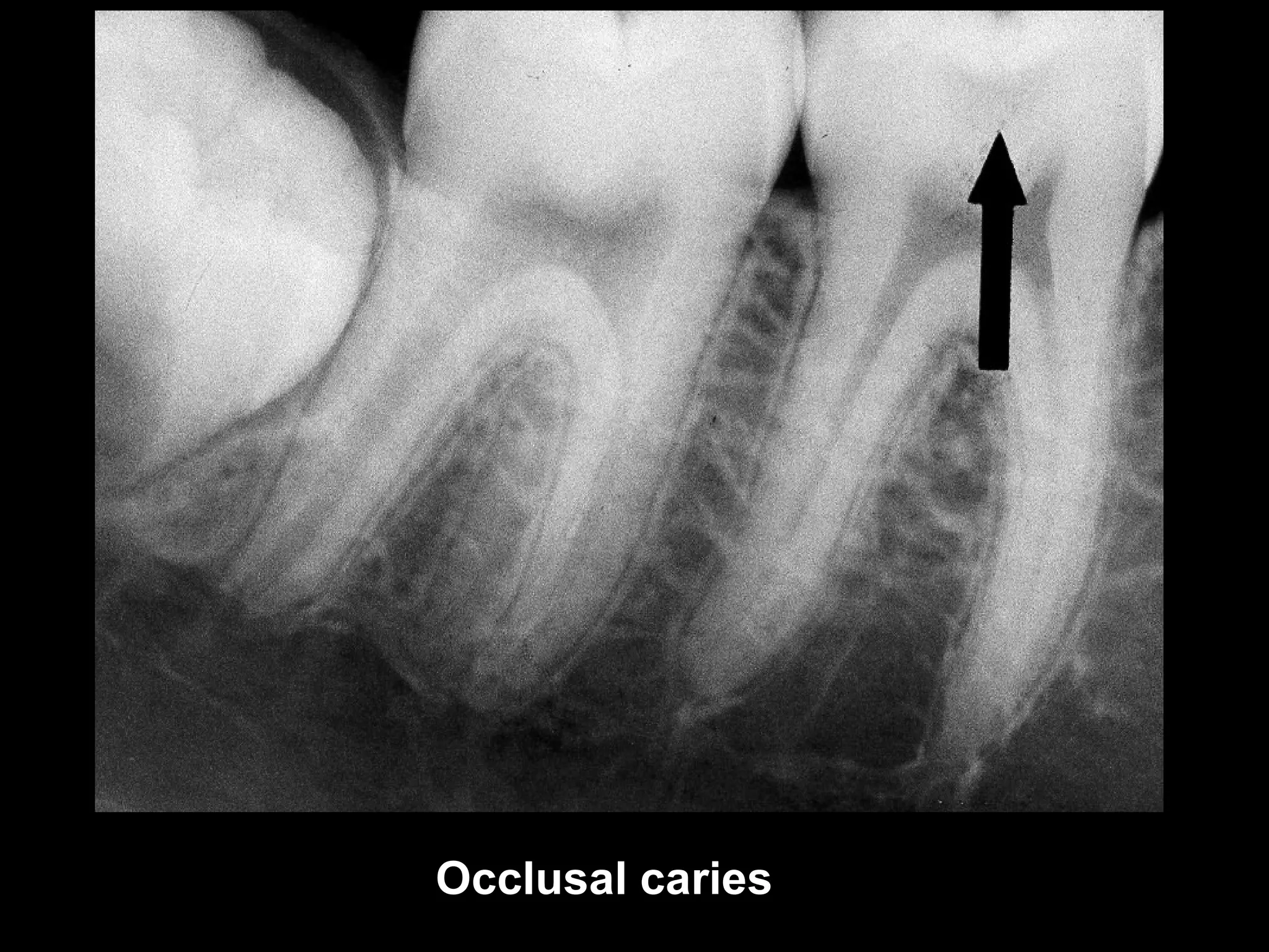
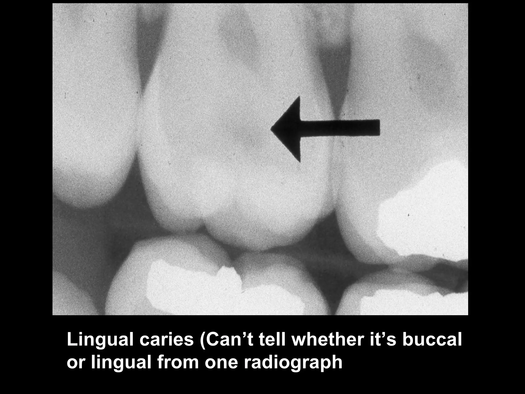
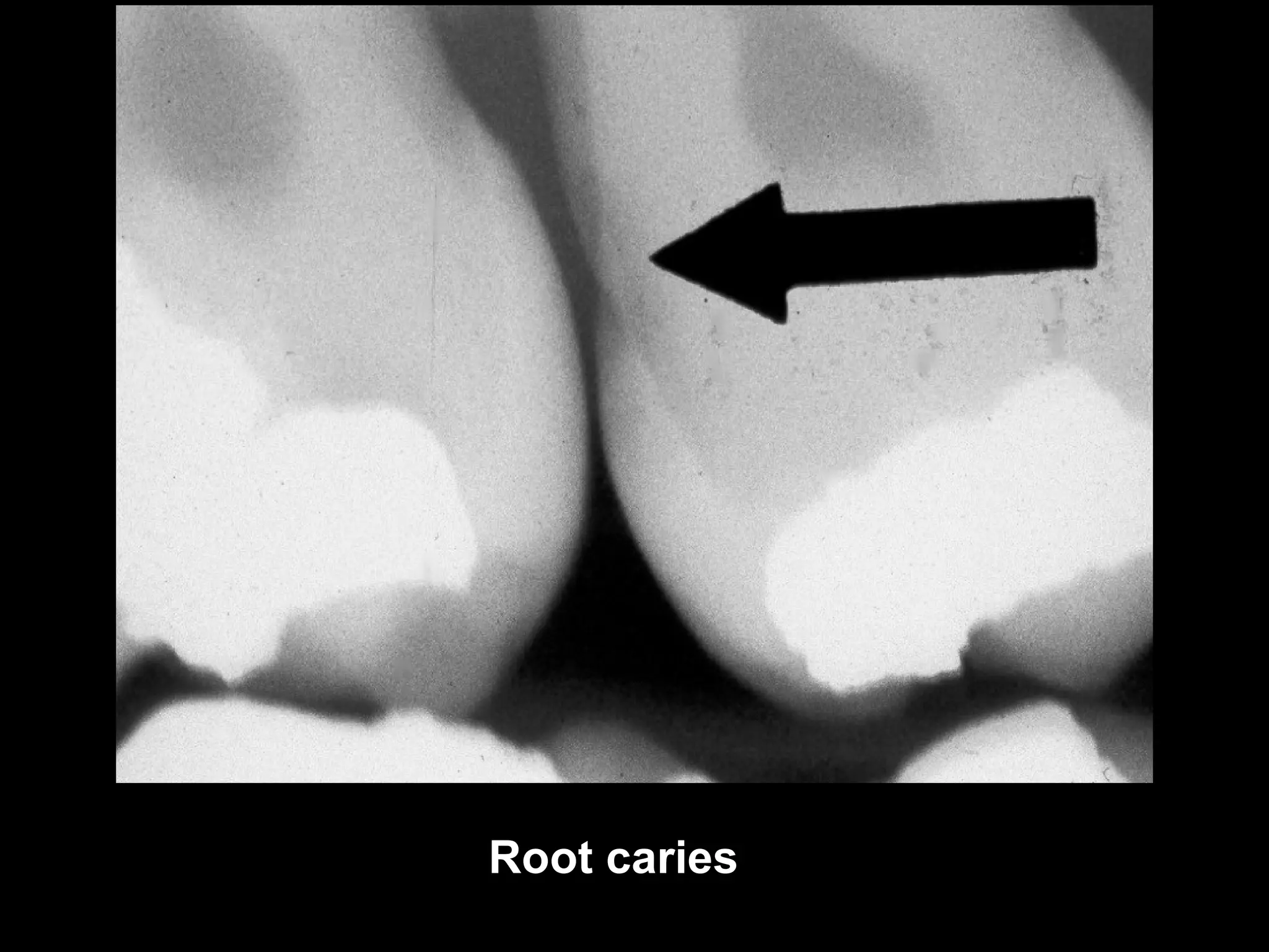
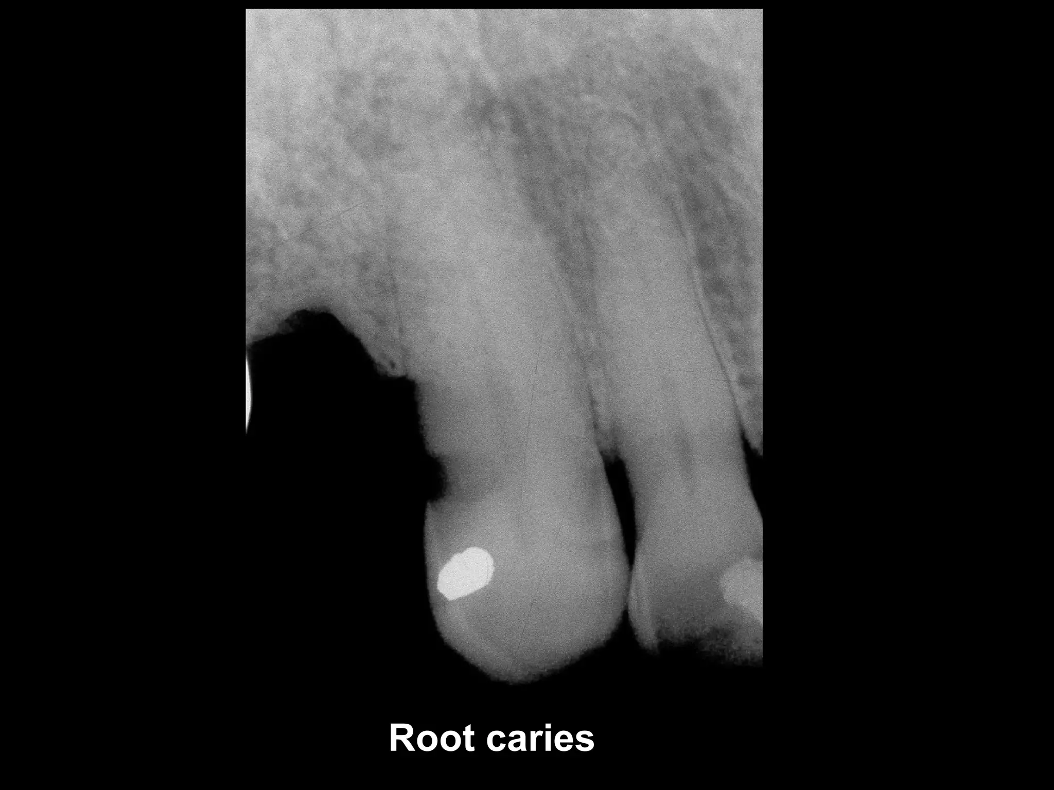
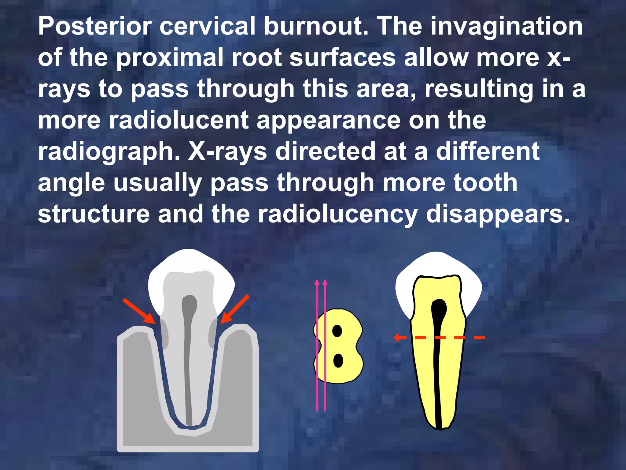
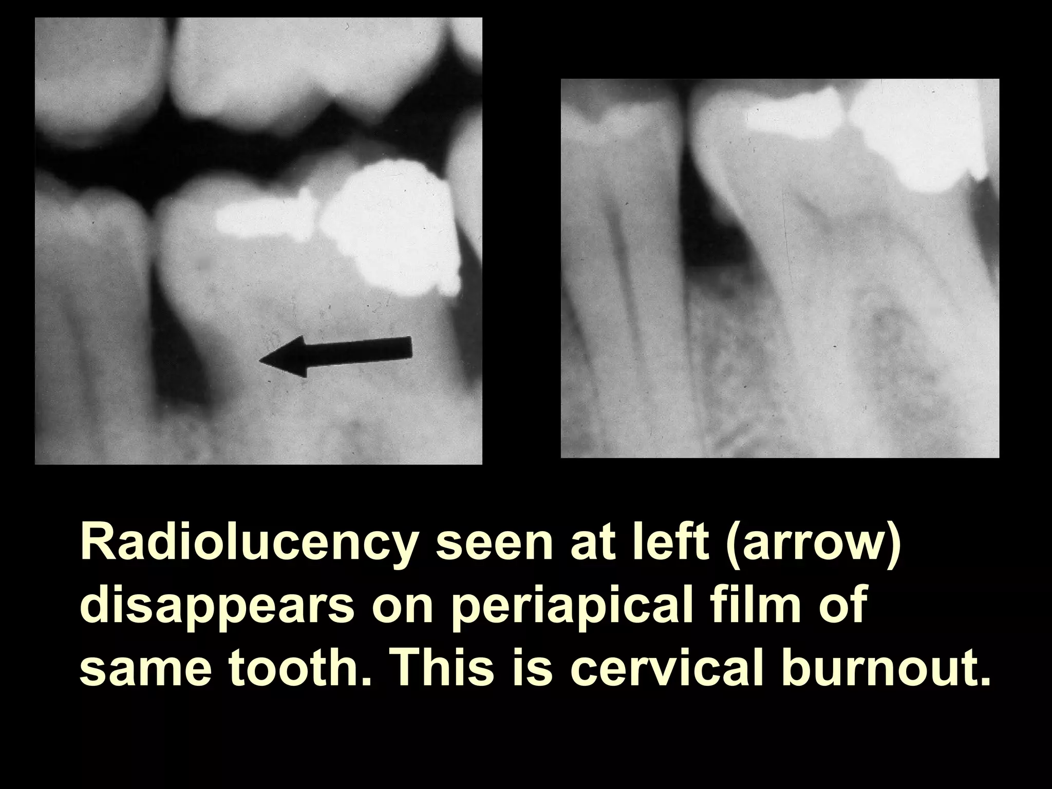
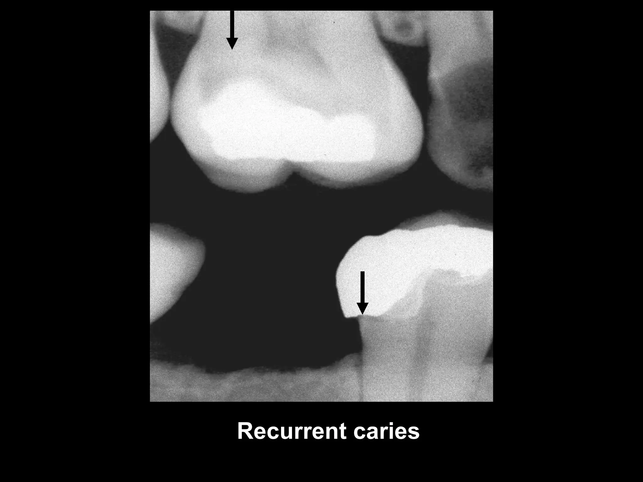
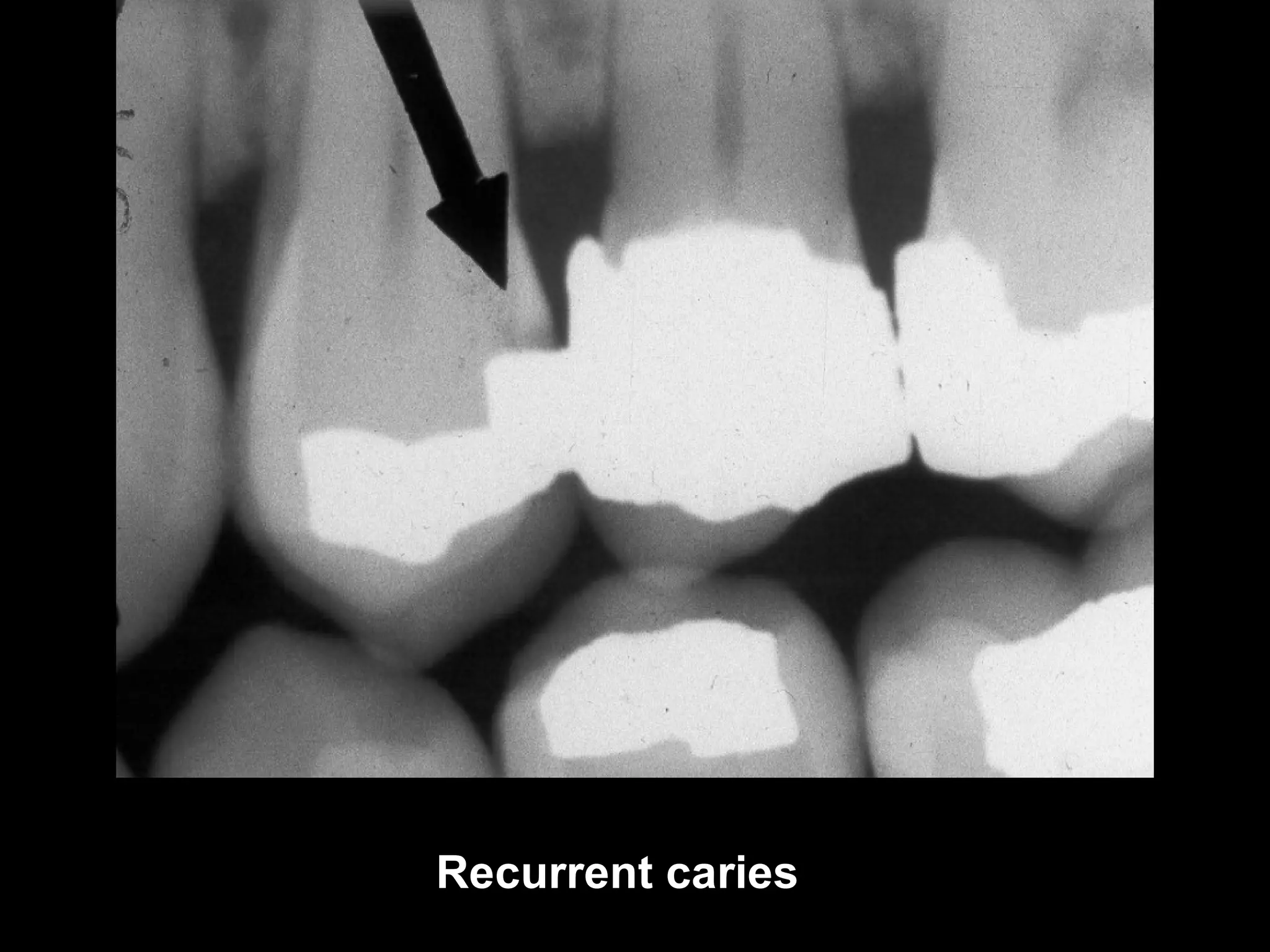
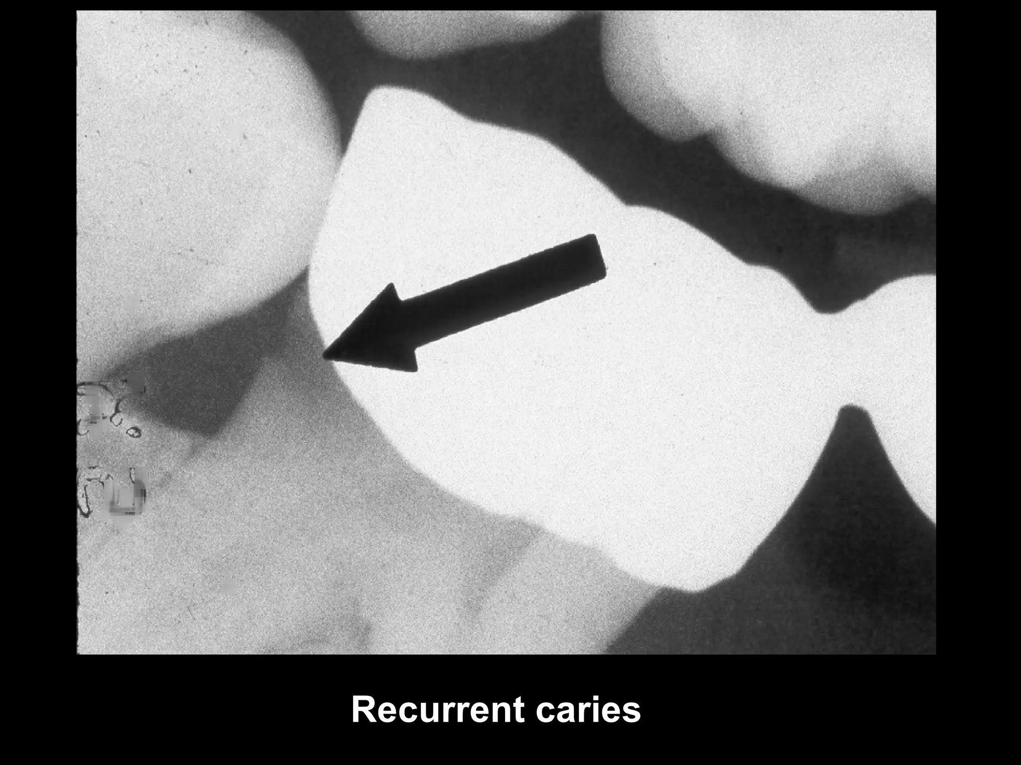
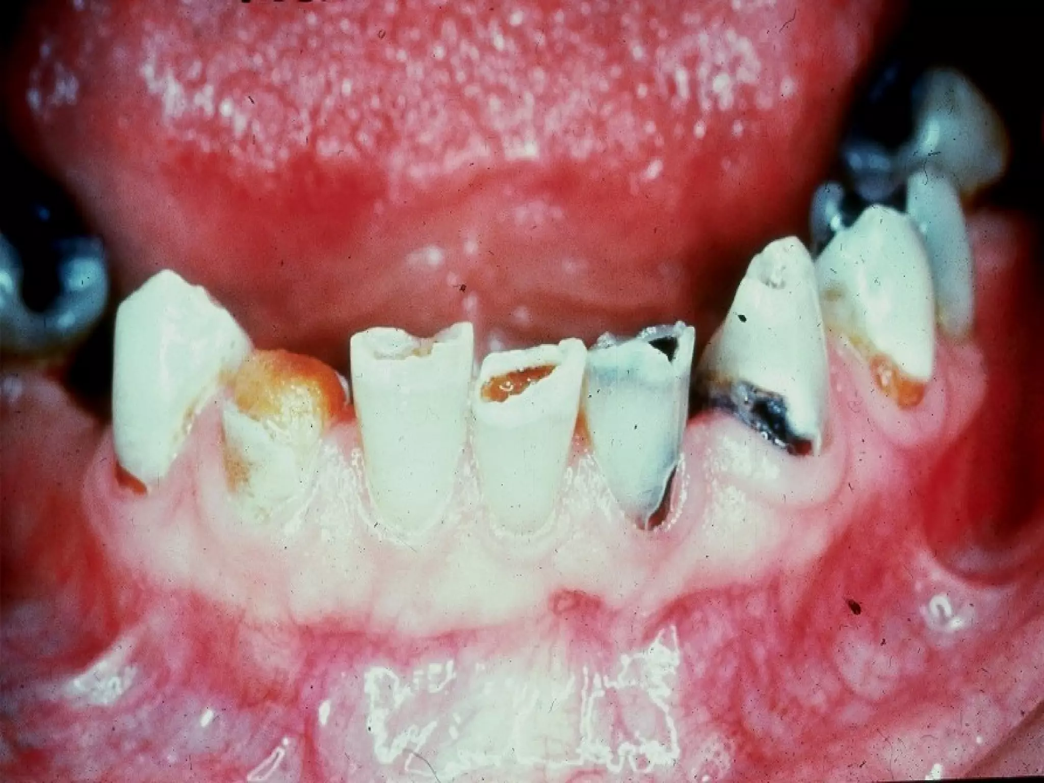
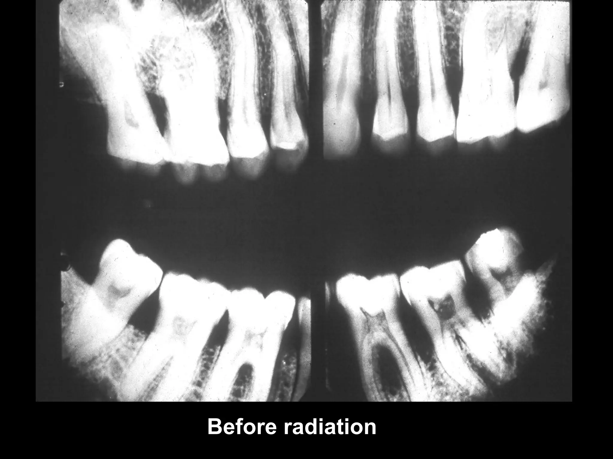
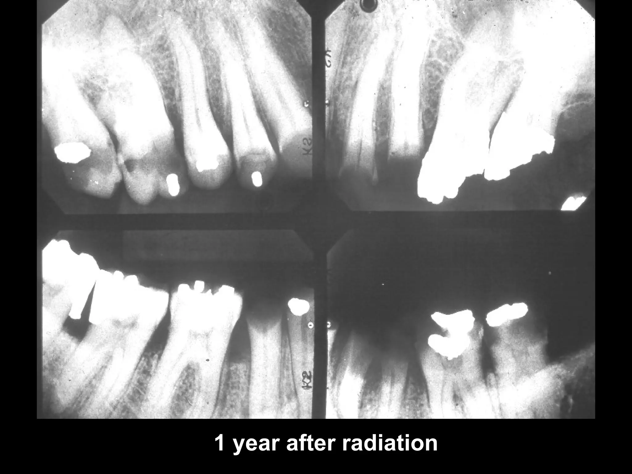
0


          Caries Diagnosis

The following slides describe the radiographic
diagnosis of caries.

In navigating through the slides, you should click
on the left mouse button when you see the
mouse holding an x-ray tubehead or you are
done reading a slide. Hitting “Enter” or “Page
Down” will also work. To go back to the previous
slide, hit “backspace” or “page up”.
Caries
Caries is the breakdown of tooth structure
caused by acid-producing bacteria in the mouth.
These bacteria are found in the white or pale
yellow plaque that builds up on the teeth if they
are not cleaned properly on a regular basis. The
bacteria break down carbohydrates (sugars) to
form the acid that demineralizes tooth structure,
leading to caries.
The diagnosis of caries is made through a
combination of the clinical examination and
radiographs.
Unless fairly large, interproximal caries in the
posterior region usually requires radiographs to
make a diagnosis.
Radiographs
The bitewing film is primarily used for caries
identification, but the periapical film is also helpful.
The difference in angulation between the two films
gives two different perspectives and can be especially
helpful in diagnosing recurrent caries around existing
restorations.
There is a lot of discussion on which film speed (D or
F) should be used. Many dentists use D-speed film
because they feel it provides sharper images as a
result of the smaller grain size. Most educators, on the
other hand, recommend the F-speed film (Insight)
because of the significant reduction in x-ray exposure
to the patient (approximately 60% less than when using
D-speed film).
0
   Proximal caries susceptible zone


                                  caries

Approximately 40-50 % demineralization is required
for radiographic detection of a lesion. As seen in the
occlusal view, above right, the thickness of the tooth
buccolingually masks the carious lesion when it is
small.
The actual depth of penetration of a carious lesion
is actually deeper than it appears on the radiograph.
Factors affecting appearance of caries
on radiographs:

Buccolingual thickness of tooth. The thicker the
tooth, the more difficult it is to see the extent of
the caries.

Limitations of two-dimensional film. The extent of
carious involvement can not be seen in a
buccolingual (cheek to tongue) direction.
Factors affecting appearance of caries
on radiographs (continued):
X-ray beam angle (horizontal or vertical). This is
especially important when trying to identify
recurrent caries, since changes in angulation may
cause the superimposition of the existing
restoration with the carious lesion. Overlap due to
improper horizontal angulation makes it very
difficult to diagnose early interproximal caries.

Exposure factors. Caries detection is improved
with a lower kVp setting, which provides a higher
contrast. If the overall density of the film is too
light or too dark, the diagnostic potential of the
film is limited.
Transillumination                 0


In the anterior region,
interproximal caries can
often be diagnosed using
transillumination, which
involves directing a bright
light through the contact
areas. Combining
transillumination with
radiographs enhances the
diagnostic information        transilluminator
obtained.
Caries Classification


 I                 M            A
                          A

I = Incipient (Stage I)
M = Moderate (Stage II)
A = Advanced (Stage III)        S
S = Severe (Stage IV)
Interproximal Caries
           (Incipient)


          I

Up to half the thickness of enamel

Usually not restored unless patient
has high level of caries activity (high
risk). Treat with fluoride.
The arrow points to incipient lesions on the
mesial of # 19 and the distal of # 20.
Incipient
Moderate
Advanced
Interproximal Caries
      (Moderate)


                  M


More than halfway through the
enamel (up to DEJ)
The bottom arrow points to a moderate lesion
on the distal of # 20. The upper arrow points to
one of several incipient lesions on the molar
and premolars.
Moderate lesion seen on previous film
Class III moderate lesion seen in the
anterior region
Interproximal Caries
     (Advanced)


               A
   A
Advanced lesion identified by arrows.
Advanced lesions seen on previous film
Advanced lesion
Advanced lesion
Interproximal Caries
      (Severe)


              S


More than halfway
through the dentin
Severe lesion
Severe lesion
Occlusal Caries
Must have penetrated into dentin
Diagnosed from clinical exam
May be seen as thin radiolucent line or
cup-shaped zone underlying occlusal
enamel, but difficult to see on
radiographs unless lesion is large.

Some feel that a sharp explorer used too
forcefully may contribute to spread
of caries by opening up pit or fissure
Occlusal caries
Occlusal caries
Buccal/Lingual
     Caries
Should be identified from clinical
exam. Sometimes seen as well-
defined circular area in middle of
tooth, although it is not very
radiolucent. Depth can not be
determined radiographically.
Lingual caries (Can’t tell whether it’s buccal
or lingual from one radiograph
Buccal caries with severe interproximal
caries on # 12
Root Caries
Saucer-like cratering on the roots of the
teeth, involving the cementum. Usually
found on older individuals with
prominent recession and/or
periodontitis. May have xerostomia due
to medications. May be confused with
cervical burnout (discussed on later
slide).
Root caries
Root caries
Cervical Burnout
Cervical burnout is an apparent radiolucency
found just below the CE junction on the root
due to anatomical variation (concave root
formation posteriorly) or a gap between the
enamel and bone covering the root
(anteriorly). Mimica root caries. Posteriorly,
this radiolucency usually disappears when
another film of the region is examined. Caries
does not occur on the root of the tooth unless
there is loss of alveolar bone and gingival
tissue due to recession or periodontitis.
Posterior cervical burnout. The invagination
of the proximal root surfaces allow more x-
rays to pass through this area, resulting in a
more radiolucent appearance on the
radiograph. X-rays directed at a different
angle usually pass through more tooth
structure and the radiolucency disappears.
Radiolucency seen at left (arrow)
disappears on periapical film of
same tooth. This is cervical burnout.
Anterior cervical burnout. The space between
 the enamel and the bone overlying the tooth
 will appear more radiolucent than either the
 enamel or the bone-tooth combination.


bone level
Cervical burnout in the
anterior region due to
gap between enamel
(red arrows) and
alveolar bone over root
(blue arrows).
Recurrent Caries
Found around the margins of existing
restorations. May be due to unusual
susceptibility to caries, poor oral
hygiene, failure to remove all of the
caries during cavity preparation, a
defective restoration or a combination
of the above.
Recurrent caries
Recurrent caries
Recurrent caries
Rampant Caries

Extensive and rapidly progressing
caries usually found in children
and teens with poor diet and
inadequate oral hygiene
Radiation Caries


Found in head/neck radiation
therapy patients with xerostomia

Fluoride used for control
Before radiation
1 year after radiation
Mach Band
Optical illusion giving appearance of increased
radiolucency at the junction of differing tissue
densities, such as enamel and dentin. If you block
off the enamel with a fingernail, the radiolucency
will disappear if due to the mach band effect. If the
radiolucency persists, it may be caries.
0

This concludes the section on Caries.

Additional self-study modules are available
at: http://dent.osu.edu/radiology/resources.htm

If you have any questions, you may e-mail
me at: jaynes.1@osu.edu.

Robert M. Jaynes, DDS, MS
Director, Radiology Group
College of Dentistry
Ohio State University

radiographic-caries-diagnosis

  • 1.
    0 Caries Diagnosis The following slides describe the radiographic diagnosis of caries. In navigating through the slides, you should click on the left mouse button when you see the mouse holding an x-ray tubehead or you are done reading a slide. Hitting “Enter” or “Page Down” will also work. To go back to the previous slide, hit “backspace” or “page up”.
  • 2.
    Caries Caries is thebreakdown of tooth structure caused by acid-producing bacteria in the mouth. These bacteria are found in the white or pale yellow plaque that builds up on the teeth if they are not cleaned properly on a regular basis. The bacteria break down carbohydrates (sugars) to form the acid that demineralizes tooth structure, leading to caries. The diagnosis of caries is made through a combination of the clinical examination and radiographs. Unless fairly large, interproximal caries in the posterior region usually requires radiographs to make a diagnosis.
  • 3.
    Radiographs The bitewing filmis primarily used for caries identification, but the periapical film is also helpful. The difference in angulation between the two films gives two different perspectives and can be especially helpful in diagnosing recurrent caries around existing restorations. There is a lot of discussion on which film speed (D or F) should be used. Many dentists use D-speed film because they feel it provides sharper images as a result of the smaller grain size. Most educators, on the other hand, recommend the F-speed film (Insight) because of the significant reduction in x-ray exposure to the patient (approximately 60% less than when using D-speed film).
  • 4.
    0 Proximal caries susceptible zone caries Approximately 40-50 % demineralization is required for radiographic detection of a lesion. As seen in the occlusal view, above right, the thickness of the tooth buccolingually masks the carious lesion when it is small. The actual depth of penetration of a carious lesion is actually deeper than it appears on the radiograph.
  • 5.
    Factors affecting appearanceof caries on radiographs: Buccolingual thickness of tooth. The thicker the tooth, the more difficult it is to see the extent of the caries. Limitations of two-dimensional film. The extent of carious involvement can not be seen in a buccolingual (cheek to tongue) direction.
  • 6.
    Factors affecting appearanceof caries on radiographs (continued): X-ray beam angle (horizontal or vertical). This is especially important when trying to identify recurrent caries, since changes in angulation may cause the superimposition of the existing restoration with the carious lesion. Overlap due to improper horizontal angulation makes it very difficult to diagnose early interproximal caries. Exposure factors. Caries detection is improved with a lower kVp setting, which provides a higher contrast. If the overall density of the film is too light or too dark, the diagnostic potential of the film is limited.
  • 7.
    Transillumination 0 In the anterior region, interproximal caries can often be diagnosed using transillumination, which involves directing a bright light through the contact areas. Combining transillumination with radiographs enhances the diagnostic information transilluminator obtained.
  • 8.
    Caries Classification I M A A I = Incipient (Stage I) M = Moderate (Stage II) A = Advanced (Stage III) S S = Severe (Stage IV)
  • 9.
    Interproximal Caries (Incipient) I Up to half the thickness of enamel Usually not restored unless patient has high level of caries activity (high risk). Treat with fluoride.
  • 10.
    The arrow pointsto incipient lesions on the mesial of # 19 and the distal of # 20.
  • 11.
  • 12.
    Interproximal Caries (Moderate) M More than halfway through the enamel (up to DEJ)
  • 13.
    The bottom arrowpoints to a moderate lesion on the distal of # 20. The upper arrow points to one of several incipient lesions on the molar and premolars.
  • 15.
    Moderate lesion seenon previous film
  • 16.
    Class III moderatelesion seen in the anterior region
  • 17.
    Interproximal Caries (Advanced) A A
  • 18.
  • 19.
    Advanced lesions seenon previous film
  • 20.
  • 21.
  • 22.
    Interproximal Caries (Severe) S More than halfway through the dentin
  • 23.
  • 24.
  • 25.
    Occlusal Caries Must havepenetrated into dentin Diagnosed from clinical exam May be seen as thin radiolucent line or cup-shaped zone underlying occlusal enamel, but difficult to see on radiographs unless lesion is large. Some feel that a sharp explorer used too forcefully may contribute to spread of caries by opening up pit or fissure
  • 26.
  • 27.
  • 28.
    Buccal/Lingual Caries Should be identified from clinical exam. Sometimes seen as well- defined circular area in middle of tooth, although it is not very radiolucent. Depth can not be determined radiographically.
  • 29.
    Lingual caries (Can’ttell whether it’s buccal or lingual from one radiograph
  • 30.
    Buccal caries withsevere interproximal caries on # 12
  • 31.
    Root Caries Saucer-like crateringon the roots of the teeth, involving the cementum. Usually found on older individuals with prominent recession and/or periodontitis. May have xerostomia due to medications. May be confused with cervical burnout (discussed on later slide).
  • 32.
  • 33.
  • 34.
    Cervical Burnout Cervical burnoutis an apparent radiolucency found just below the CE junction on the root due to anatomical variation (concave root formation posteriorly) or a gap between the enamel and bone covering the root (anteriorly). Mimica root caries. Posteriorly, this radiolucency usually disappears when another film of the region is examined. Caries does not occur on the root of the tooth unless there is loss of alveolar bone and gingival tissue due to recession or periodontitis.
  • 35.
    Posterior cervical burnout.The invagination of the proximal root surfaces allow more x- rays to pass through this area, resulting in a more radiolucent appearance on the radiograph. X-rays directed at a different angle usually pass through more tooth structure and the radiolucency disappears.
  • 36.
    Radiolucency seen atleft (arrow) disappears on periapical film of same tooth. This is cervical burnout.
  • 37.
    Anterior cervical burnout.The space between the enamel and the bone overlying the tooth will appear more radiolucent than either the enamel or the bone-tooth combination. bone level
  • 38.
    Cervical burnout inthe anterior region due to gap between enamel (red arrows) and alveolar bone over root (blue arrows).
  • 39.
    Recurrent Caries Found aroundthe margins of existing restorations. May be due to unusual susceptibility to caries, poor oral hygiene, failure to remove all of the caries during cavity preparation, a defective restoration or a combination of the above.
  • 40.
  • 41.
  • 42.
  • 43.
    Rampant Caries Extensive andrapidly progressing caries usually found in children and teens with poor diet and inadequate oral hygiene
  • 45.
    Radiation Caries Found inhead/neck radiation therapy patients with xerostomia Fluoride used for control
  • 47.
  • 48.
    1 year afterradiation
  • 49.
    Mach Band Optical illusiongiving appearance of increased radiolucency at the junction of differing tissue densities, such as enamel and dentin. If you block off the enamel with a fingernail, the radiolucency will disappear if due to the mach band effect. If the radiolucency persists, it may be caries.
  • 50.
    0 This concludes thesection on Caries. Additional self-study modules are available at: http://dent.osu.edu/radiology/resources.htm If you have any questions, you may e-mail me at: jaynes.1@osu.edu. Robert M. Jaynes, DDS, MS Director, Radiology Group College of Dentistry Ohio State University