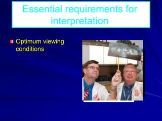
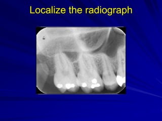
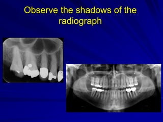
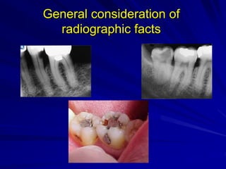
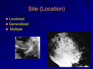
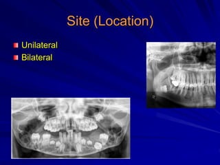
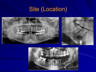
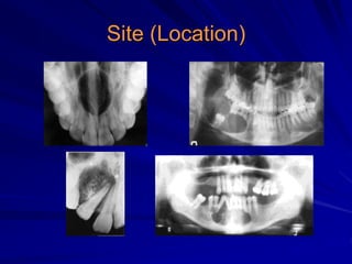
Principles of
Radiographic
Interpretation
Interpretation
An explanation of what is viewed on dental
radiograph
The ability to read what is revealed in
dental radiograph
Diagnosis
The identification of disease by examination
or analysis
Objectives of radiographic
interpretation
3D
Objectives of radiographic
interpretation
Detection: presence or absence of
disease
Description: providing information on the
disease according to systematic approach
Differentiation: diseases of similar
radiographic appearance
Essential requirements for
interpretation
Optimum viewing
conditions
Essential requirements for
interpretation
Knowledge of what the radiograph should
look like
Essential requirements for
interpretation
Knowledge of appearance of normal
anatomical landmarks as well as
variations of normal.
Essential requirements for
interpretation
Knowledge of radiographic appearance of
pathological conditions affecting the
maxillofacial region.
Essential requirements for
interpretation
How to interpret a
radiographic image?
L O G I C
LOGIC
Localize the radiograph
Observe the shadows of the radiograph
General consideration of radiographic
facts
Interpretation formulation
Correlation of radiographic interpretation,
history, and clinical findings
Localize the radiograph
Observe the shadows of the
radiograph
General consideration of
radiographic facts
Interpretation formulation
Systematic description and analysis of
findings revealed in the radiograph
Interpretation formulation
Site (Location)
Shape and Periphery
Internal structure
Effects on surrounding structures
Size
Number
Site (Location)
Localized
Site (Location)
Localized
Generalized
Site (Location)
Localized
Generalized
Multiple
Site (Location)
Unilateral
Bilateral
Site (Location)
Site (Location)
Shape and Periphery
1. Border
2. Margin
3. Shape
Shape and Periphery
1. Border
Well defined
Ill defined
Shape and Periphery
1. Border
Well defined
Shape and Periphery
1. Border
Ill defined
Shape and Periphery
2. Margin
Corticated
Non corticated
Sclerotic
Radiolucent rim
Shape and Periphery
3. Shape
Round
Oval
Scalloped
Irregular
Inverted pear
Heart shape
Internal structure
Radiodensity
Radiolucent Radiopaque Mixed (RL-RO)
Radiolucent
Radiopaque
Mixed (RL-RO)
Radiolucent
Unilocular Multilocular
Soap bubble Honey comb Tennis Racket
Internal structure
Unilocular Multilocular
Internal structure
Soap bubble
Honey comb
Tennis Racket
Radiopaque or Mixed
Sunray
Orange peal
Ground glass
Cotton wool
Driven snow
Internal structure
Associated with
impacted tooth
Effects on surrounding structures
Teeth
Bone
Effects on surrounding structures
Teeth
Widening of Periodontal
membrane space
Loss of lamina dura
Root resorption
Tooth displacement
Root divergence
Hindering of tooth
eruption
Effects on surrounding structures
Bone
Expansion
Thinning
Erosion &
perforation
Displacement of
cortices
Obliteration of max.
sinus
Size
Small – medium –
large – Huge
Measured in
centimeters
Number
Single
Multiple:
Systemic (blood or
endocrinal diseases)
Syndromes
Again
Interpretation does not equal diagnosis
Diagnosis
History
Clinical Examination
Special Investigations
Conventional radiographs (periapical-
occlusal- panoramic-lateral oblique-etc…)
Advanced imaging modalities (CT-MRI-
US-etc….)
Biopsy (aspiration-smear-incisional-
excisional)
Blood analysis
Urine analysis
Diagnosis
Non differential diagnosis
Differential diagnosis
Diagnosis
Non differential
diagnosis
Diagnosis
Differential diagnosis
How to differentiate between different
lesions which have similar radiographic
appearance
Wood and Goaz Classification
Radiolucencies
Periapical
Pericoronal
Interradicular
Solitary cyst-like
lesions
Multilocular
Ill defined
Multiple separate
Generalized
rarefaction
Mixed (RL-RO)
Periapical
Pericoronal
Not necessarily
contacting the
teeth
Radiopacities
• Periapical
• Not necessarily
contacting the
teeth
• Multiple
• Generalized

Principles Of Radiographic Interpretation