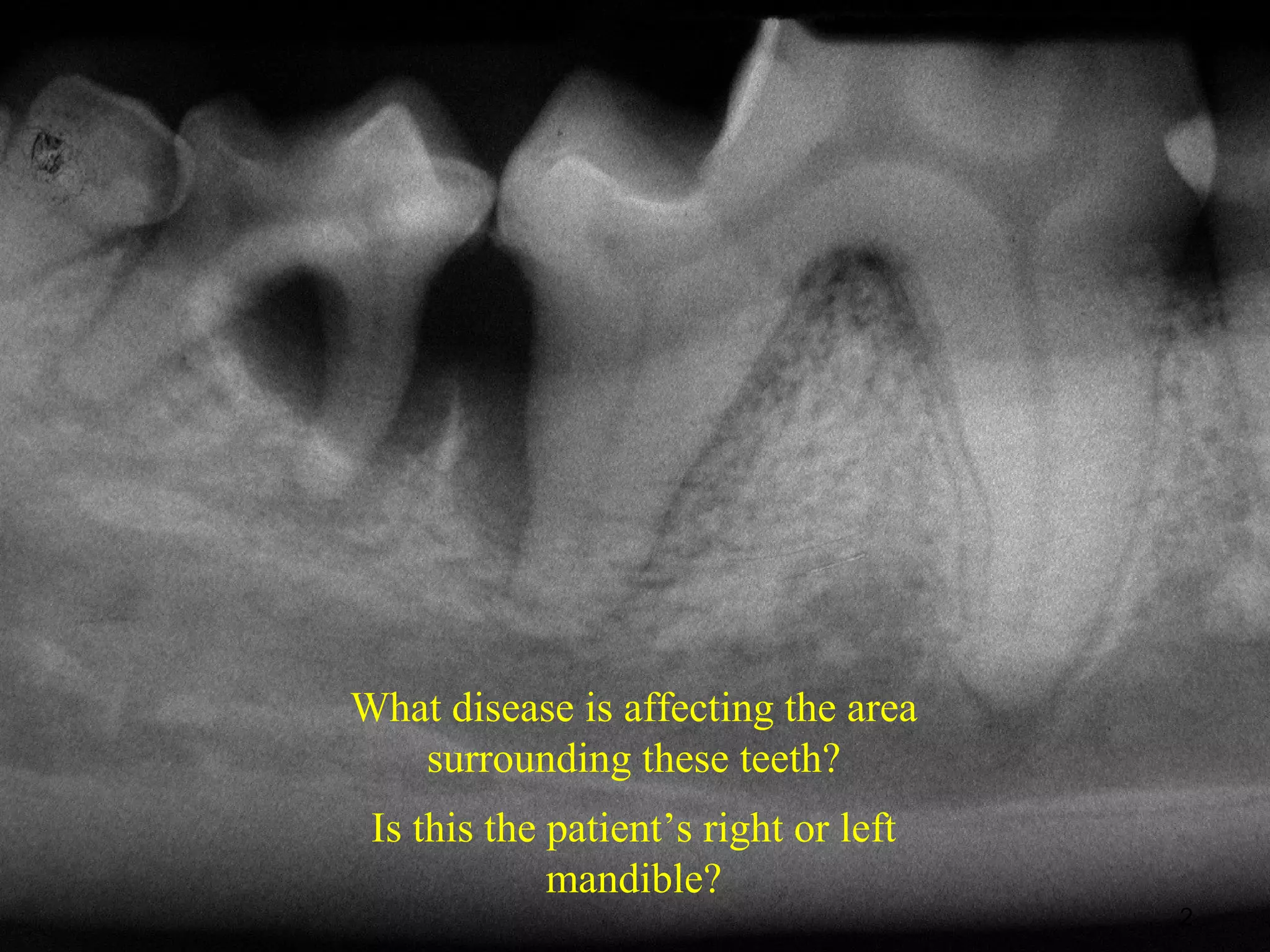
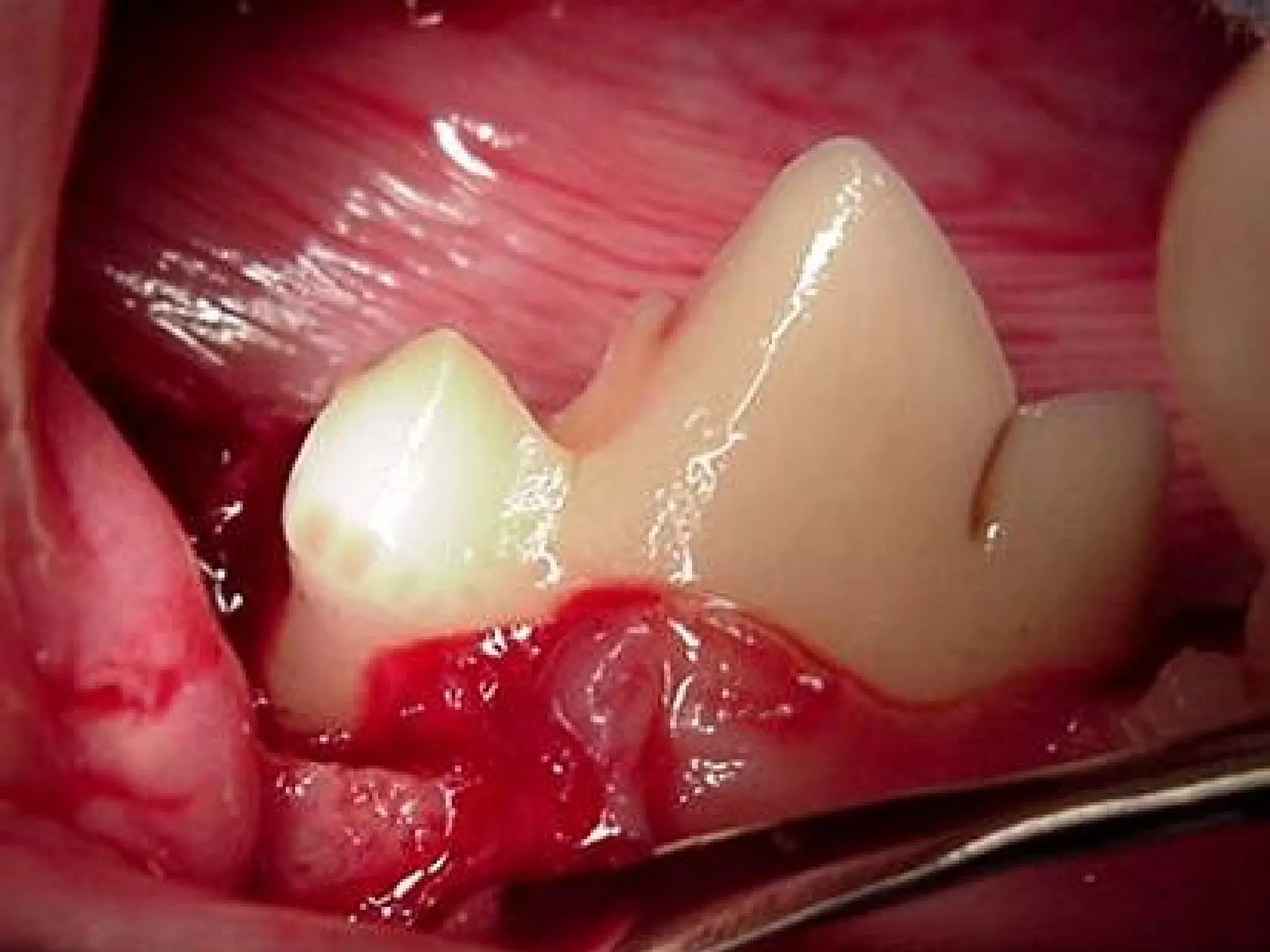
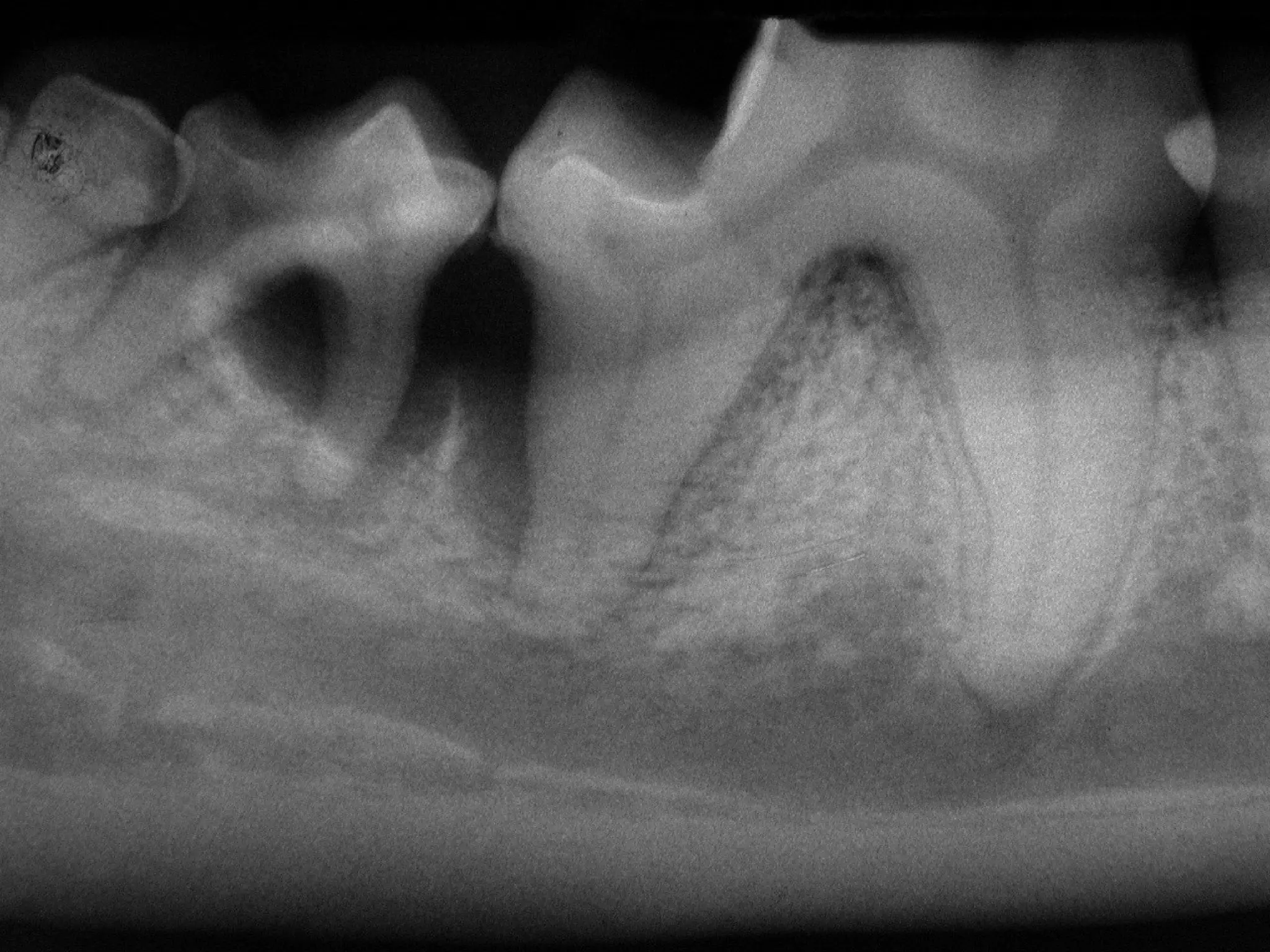
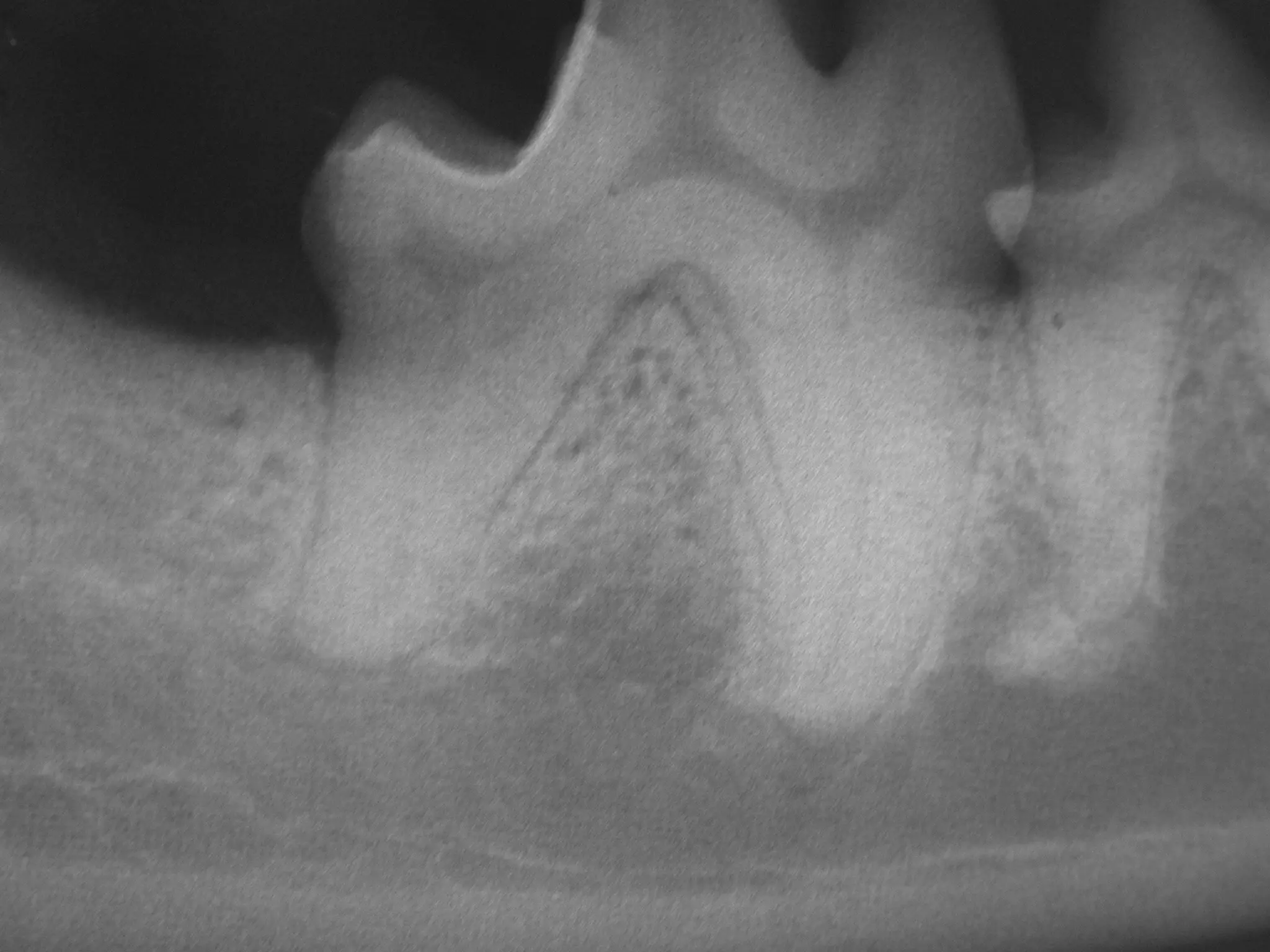
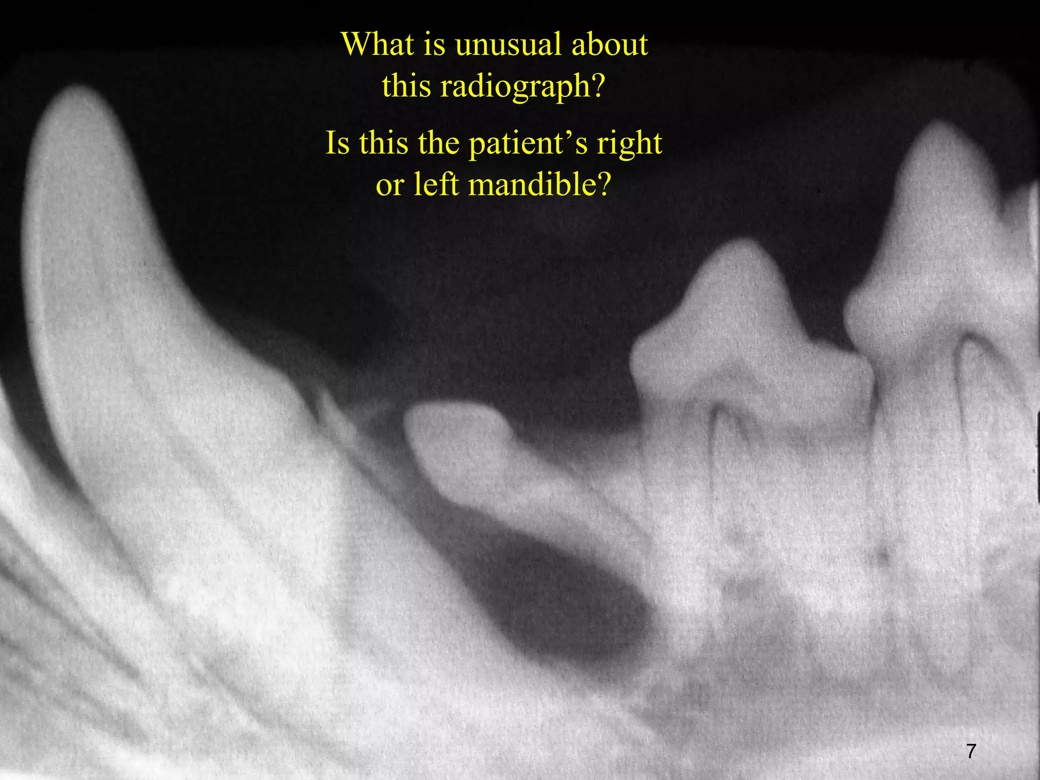
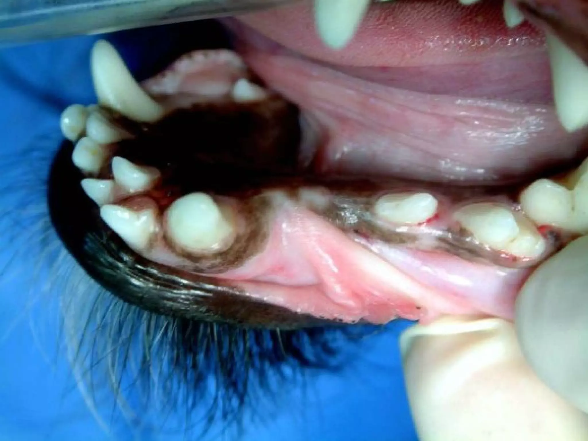
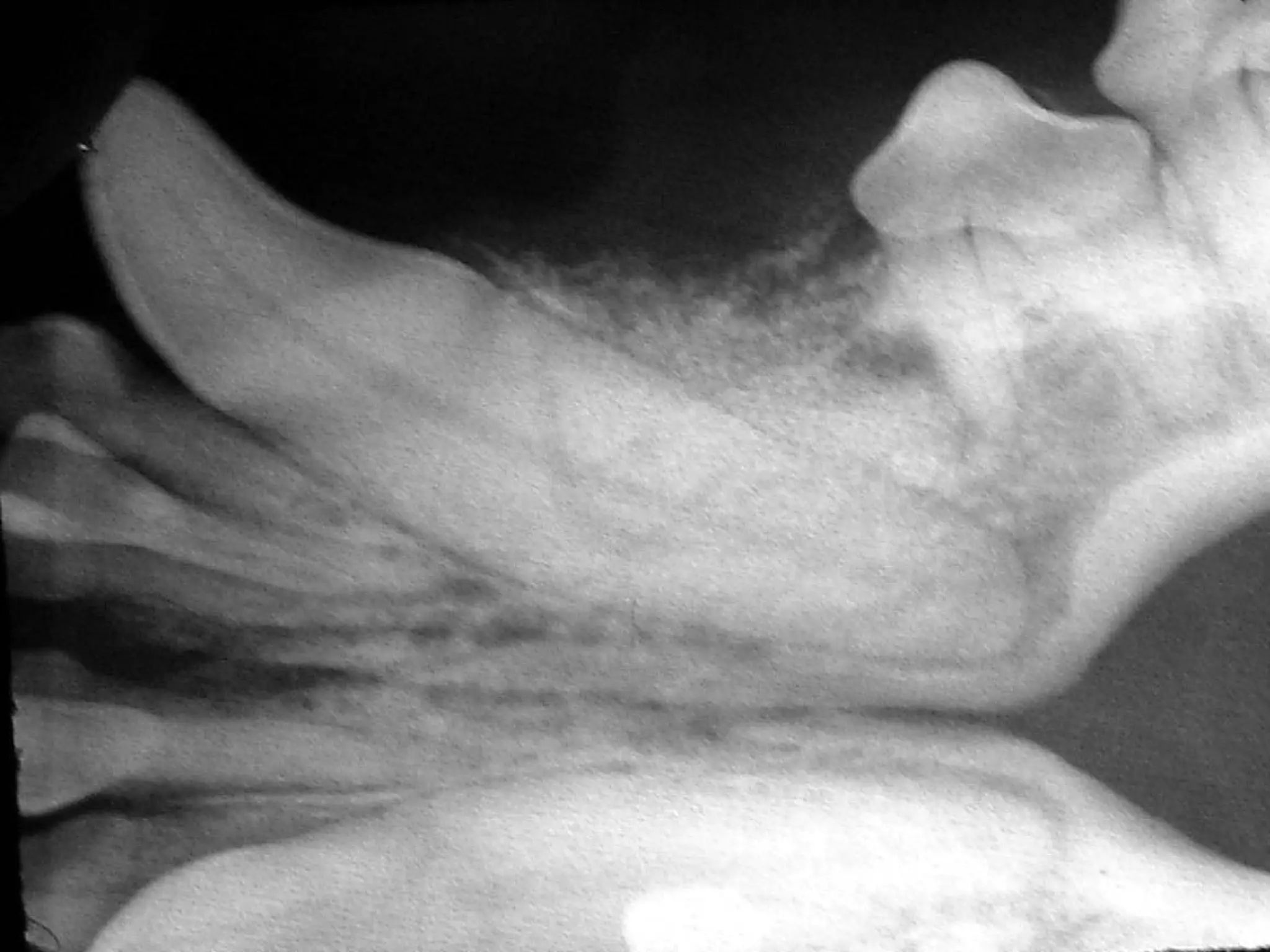
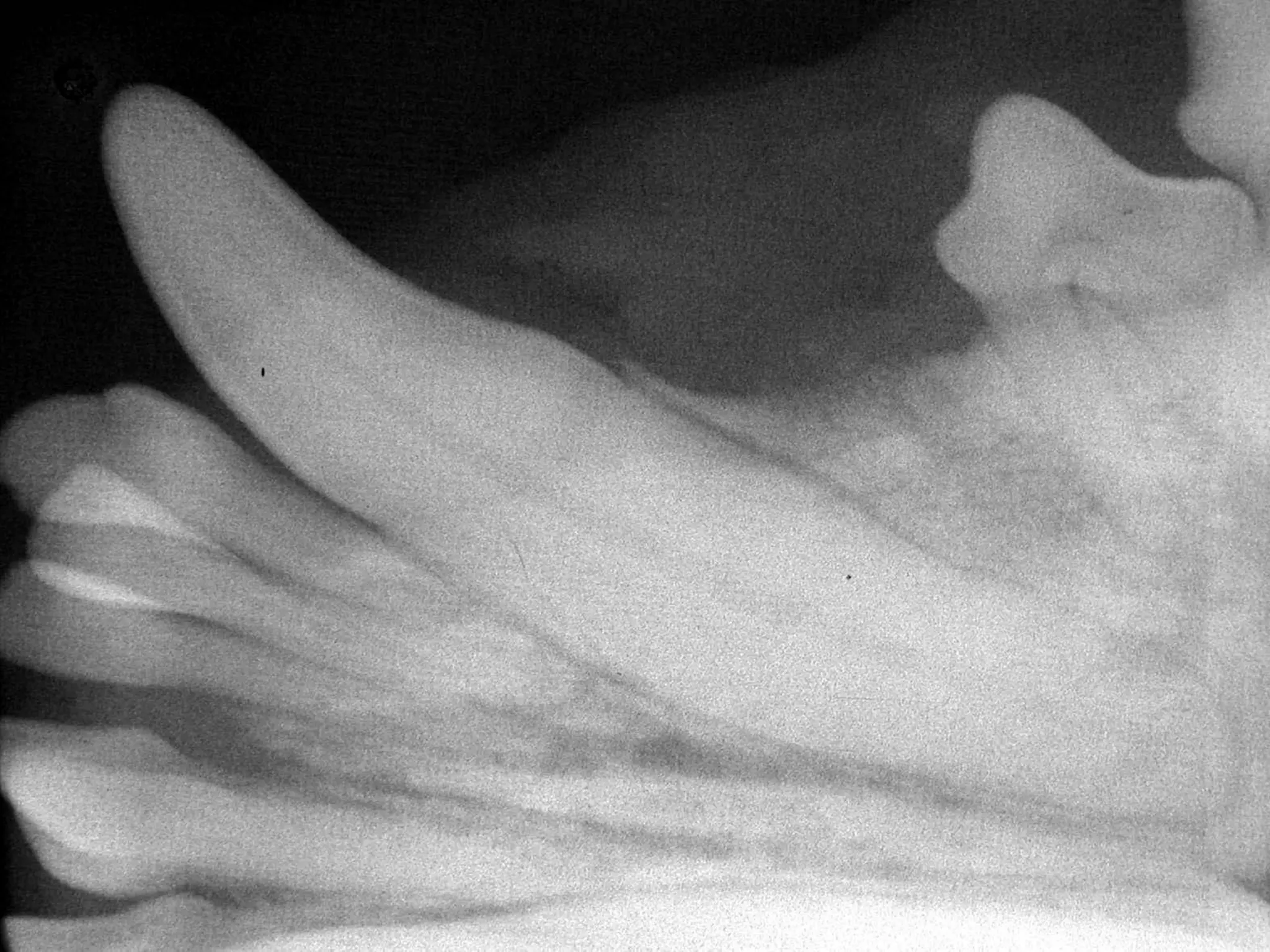
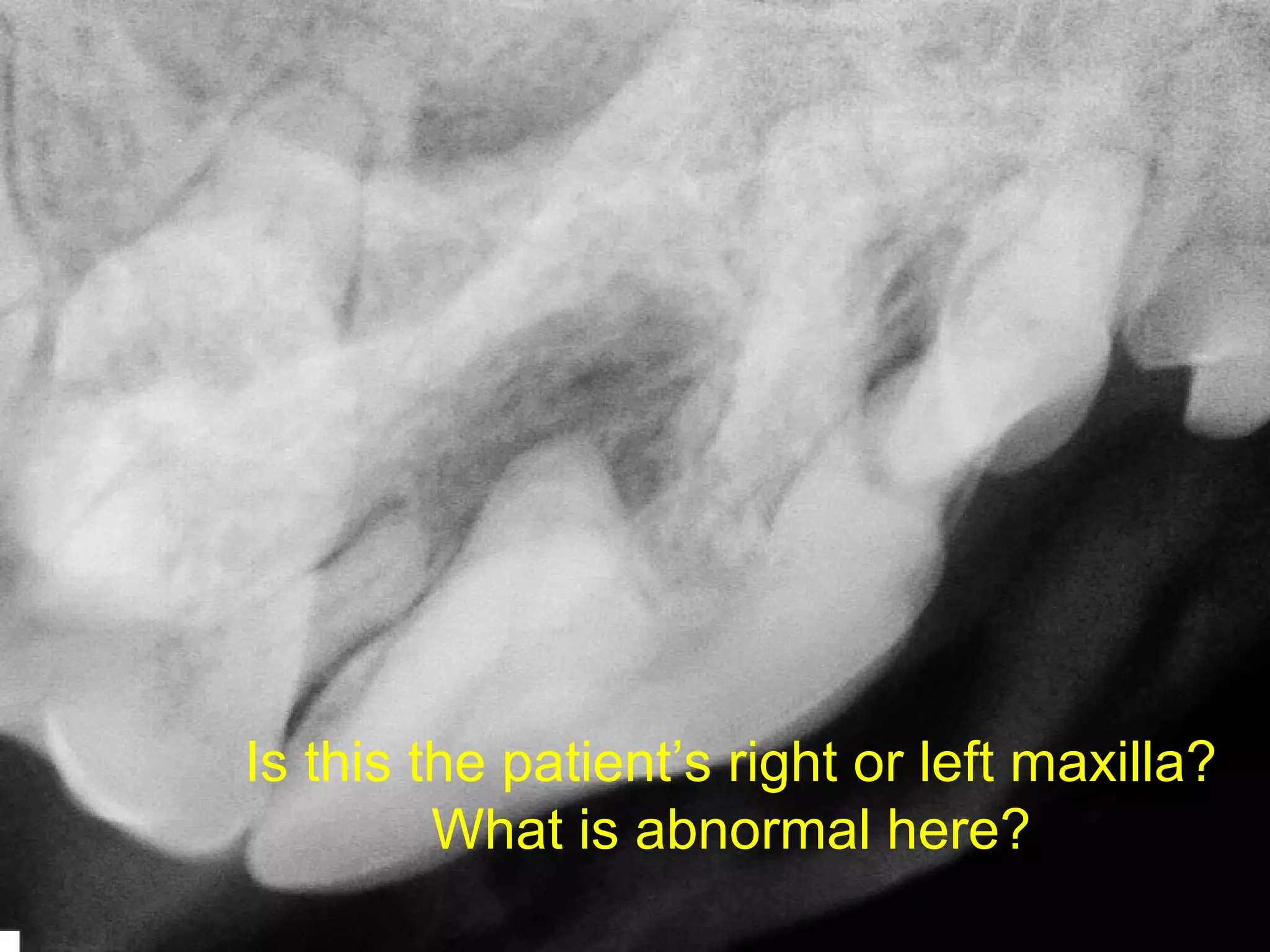
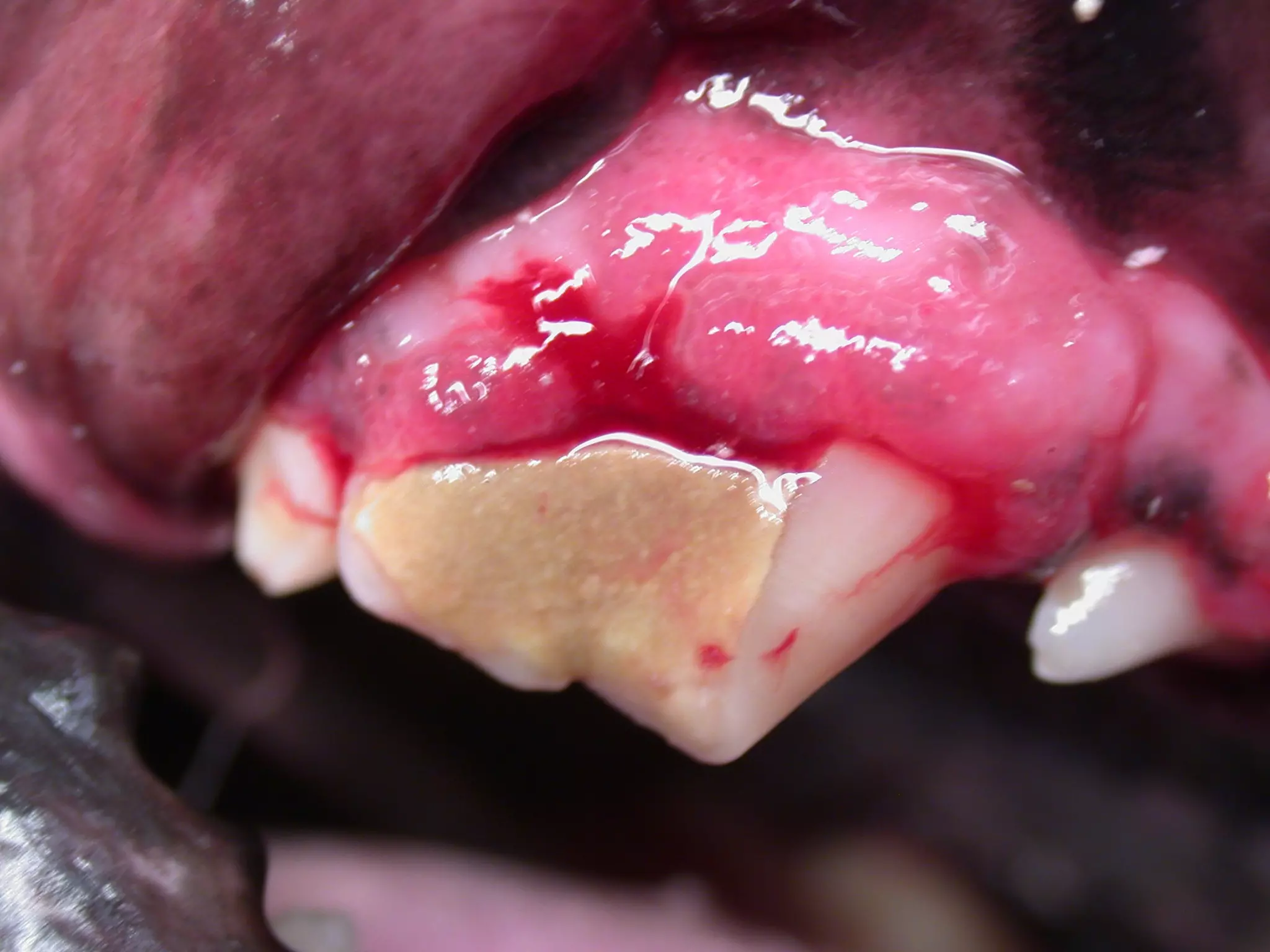
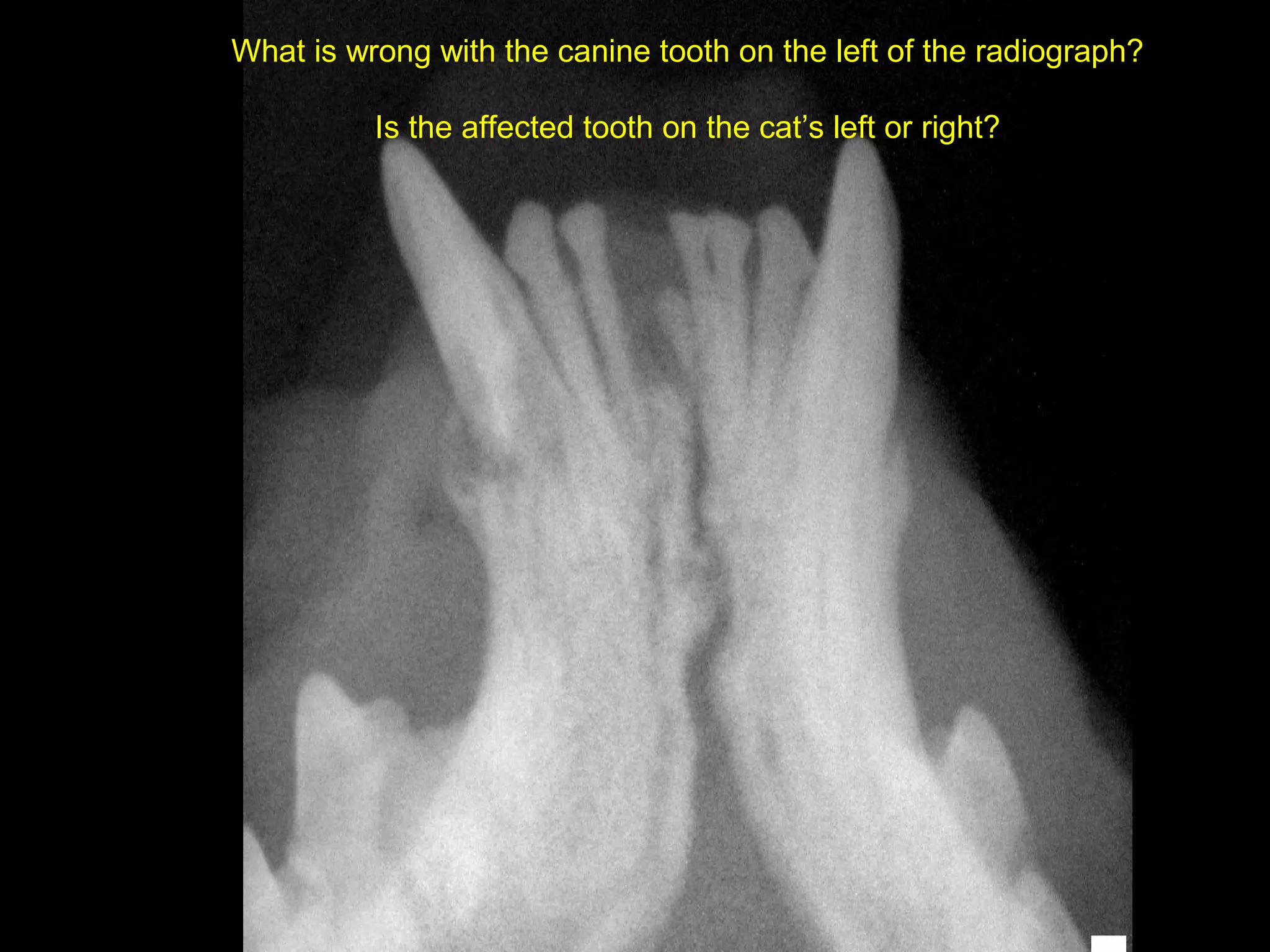
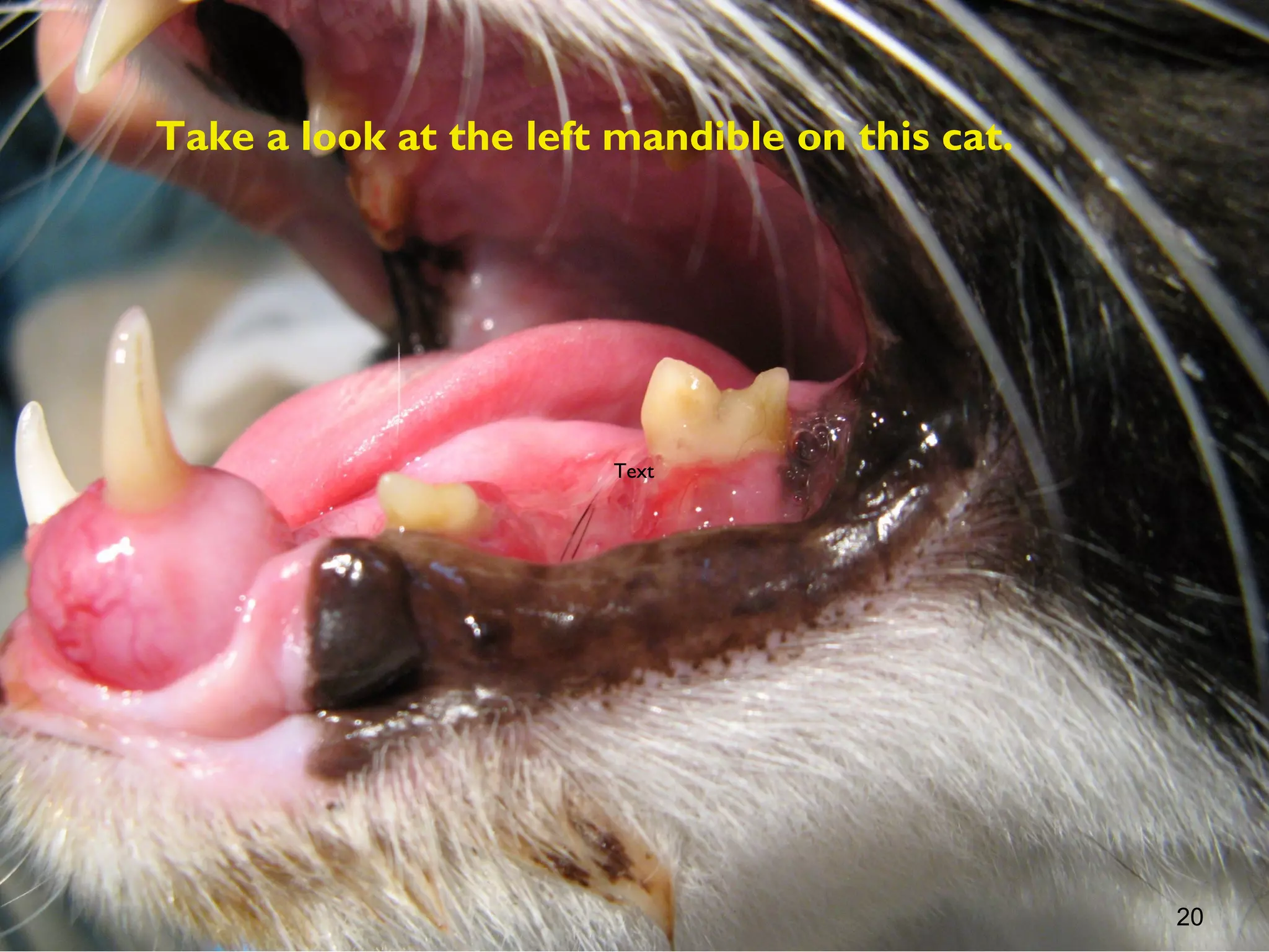
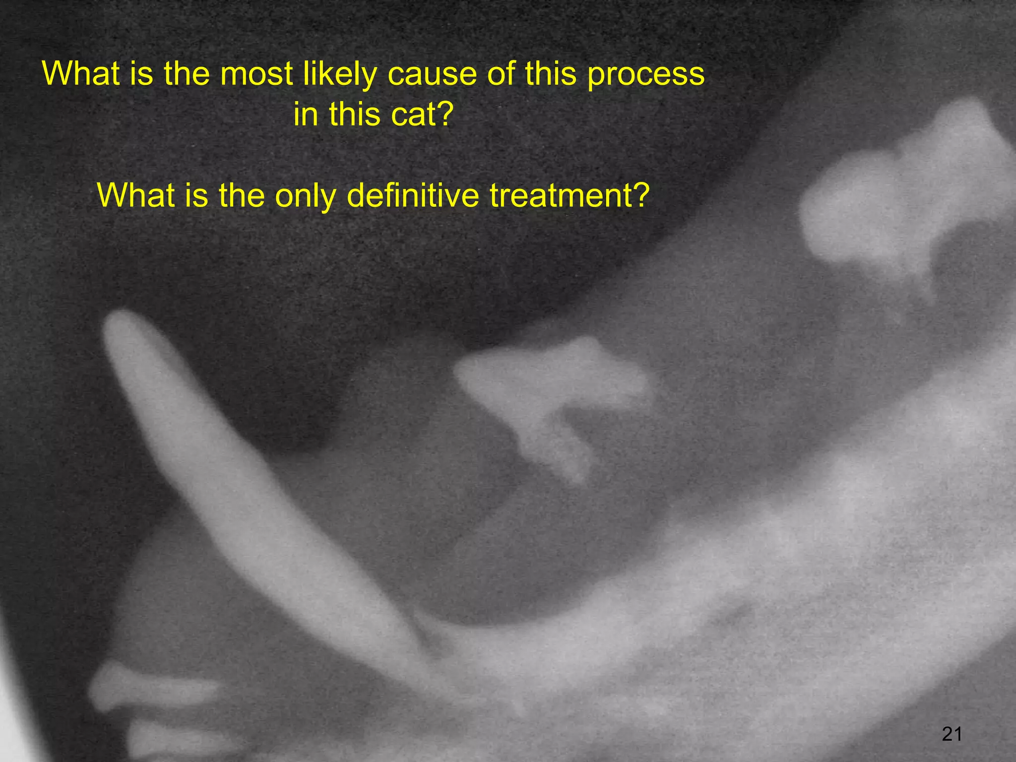
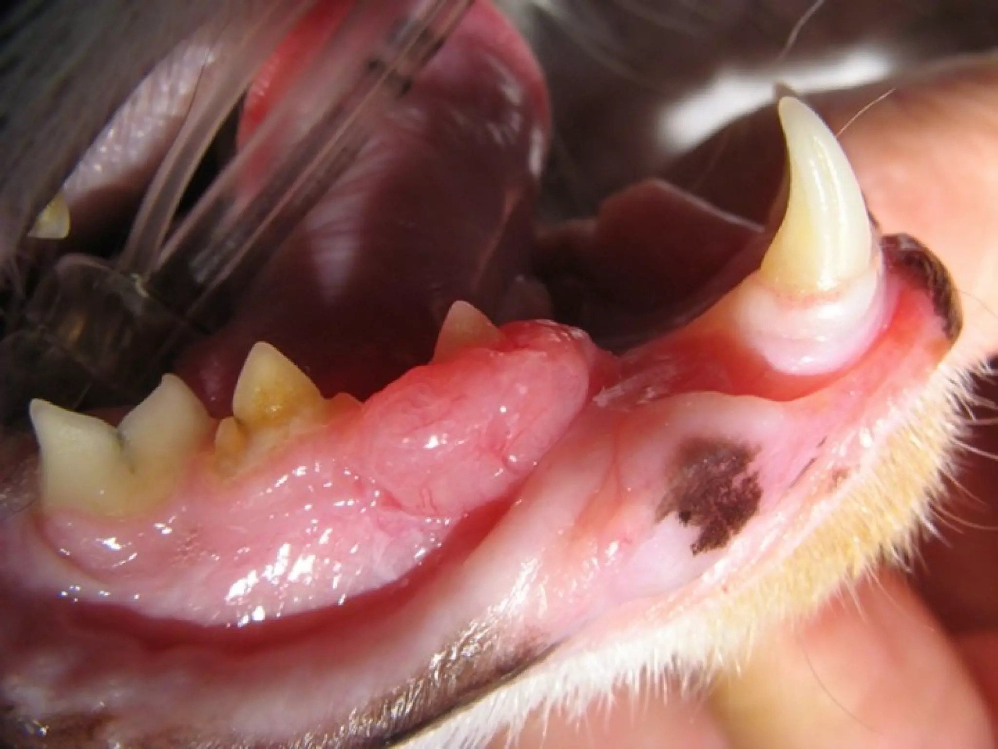
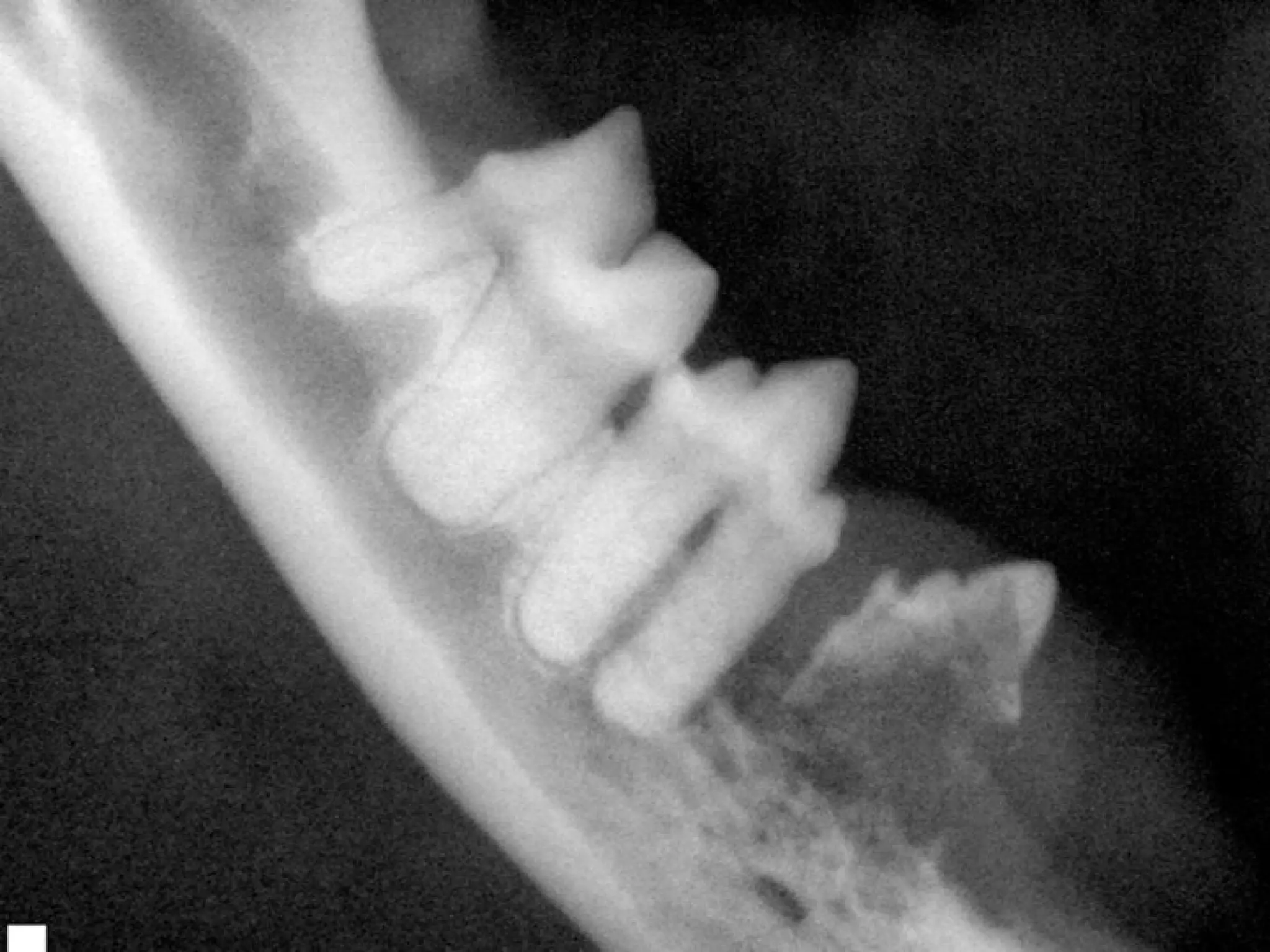
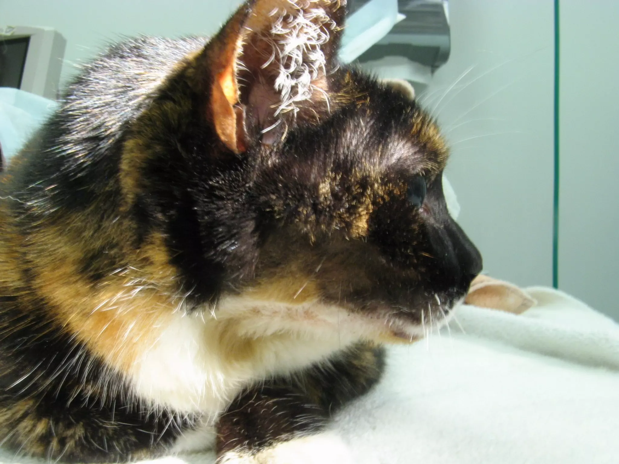
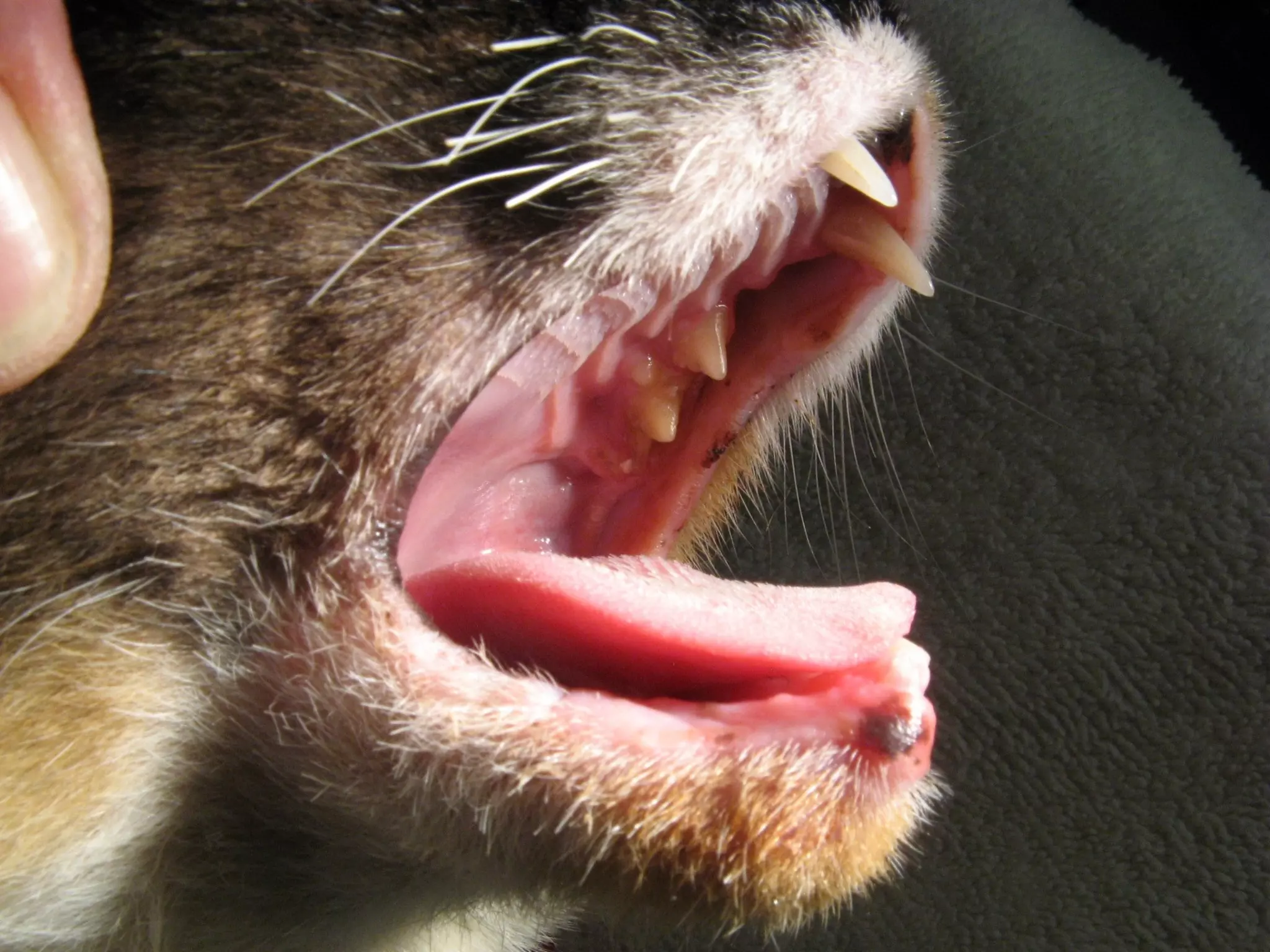
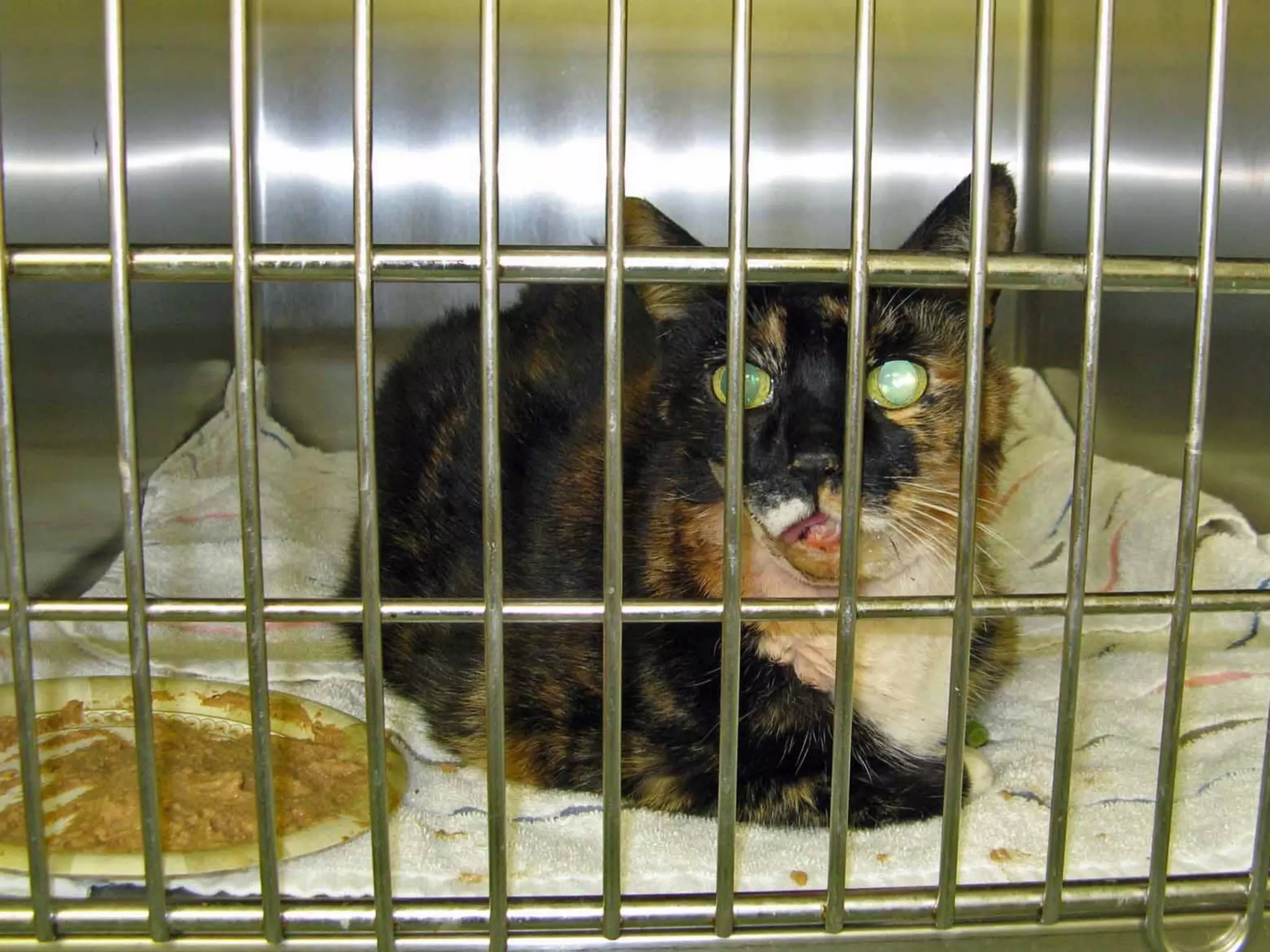
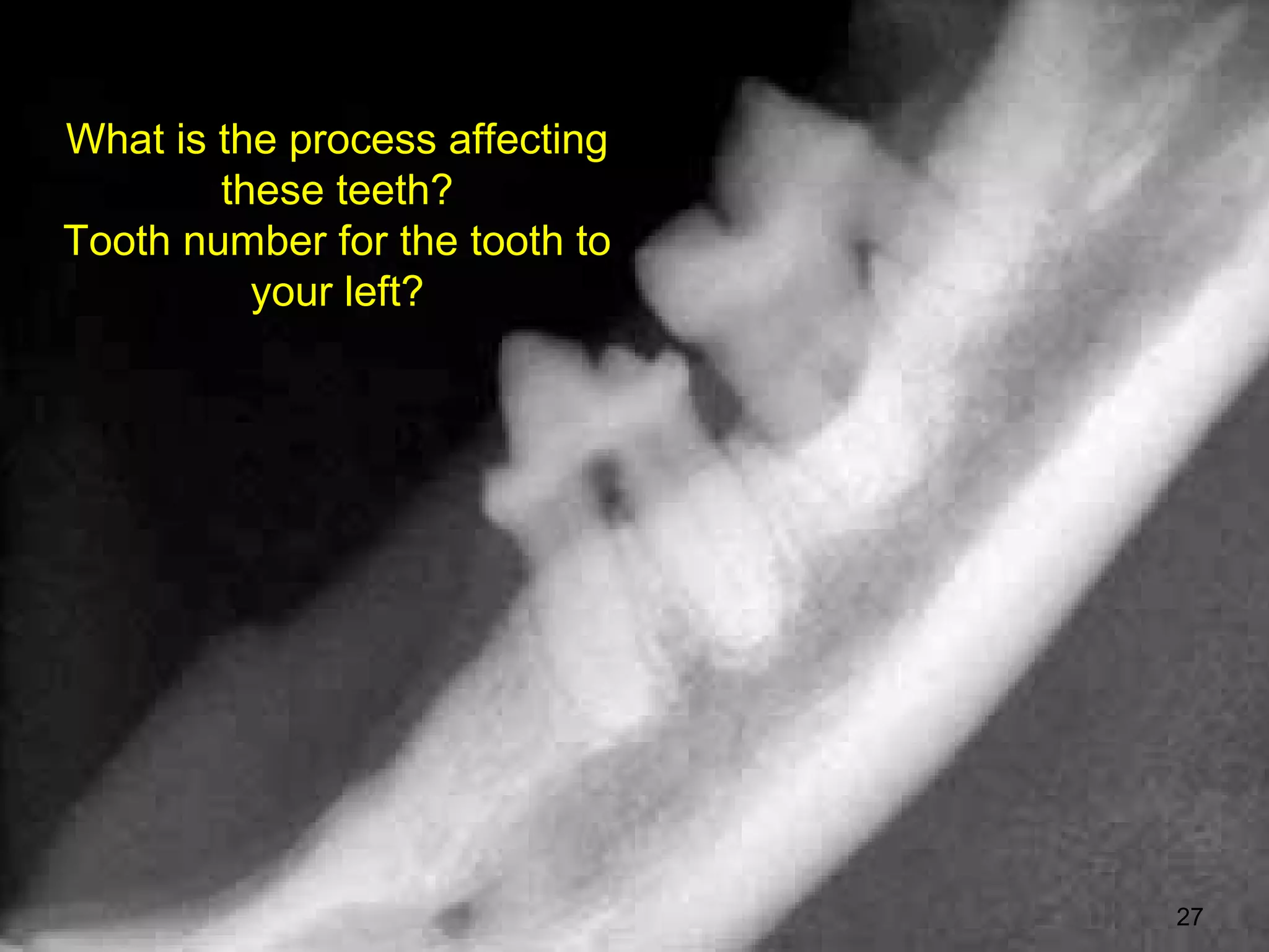
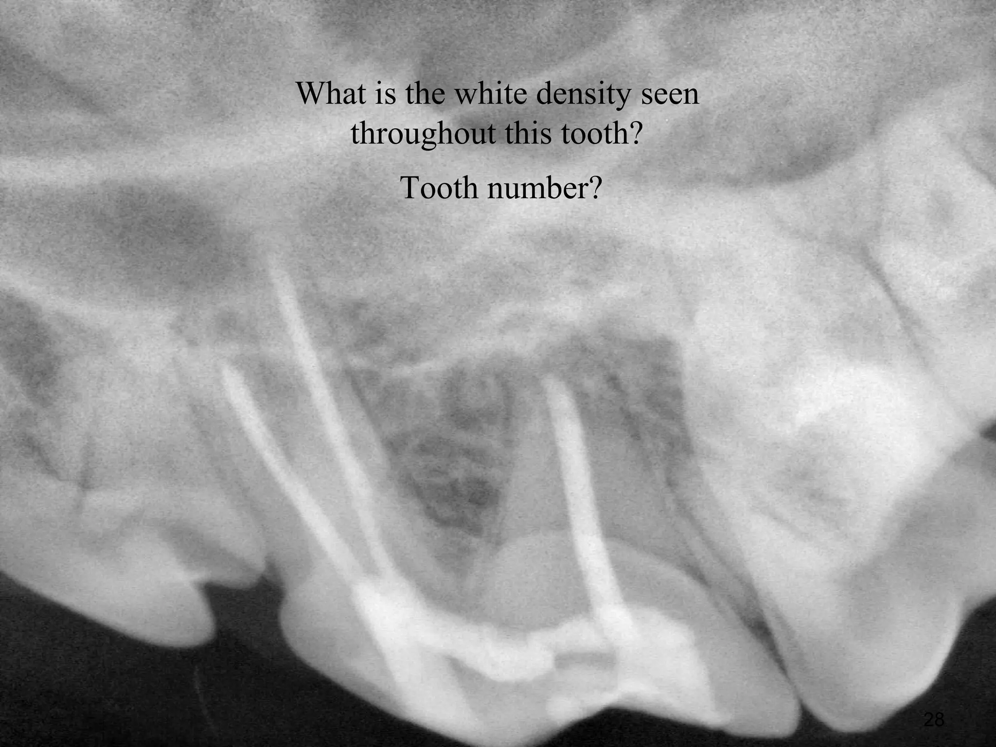
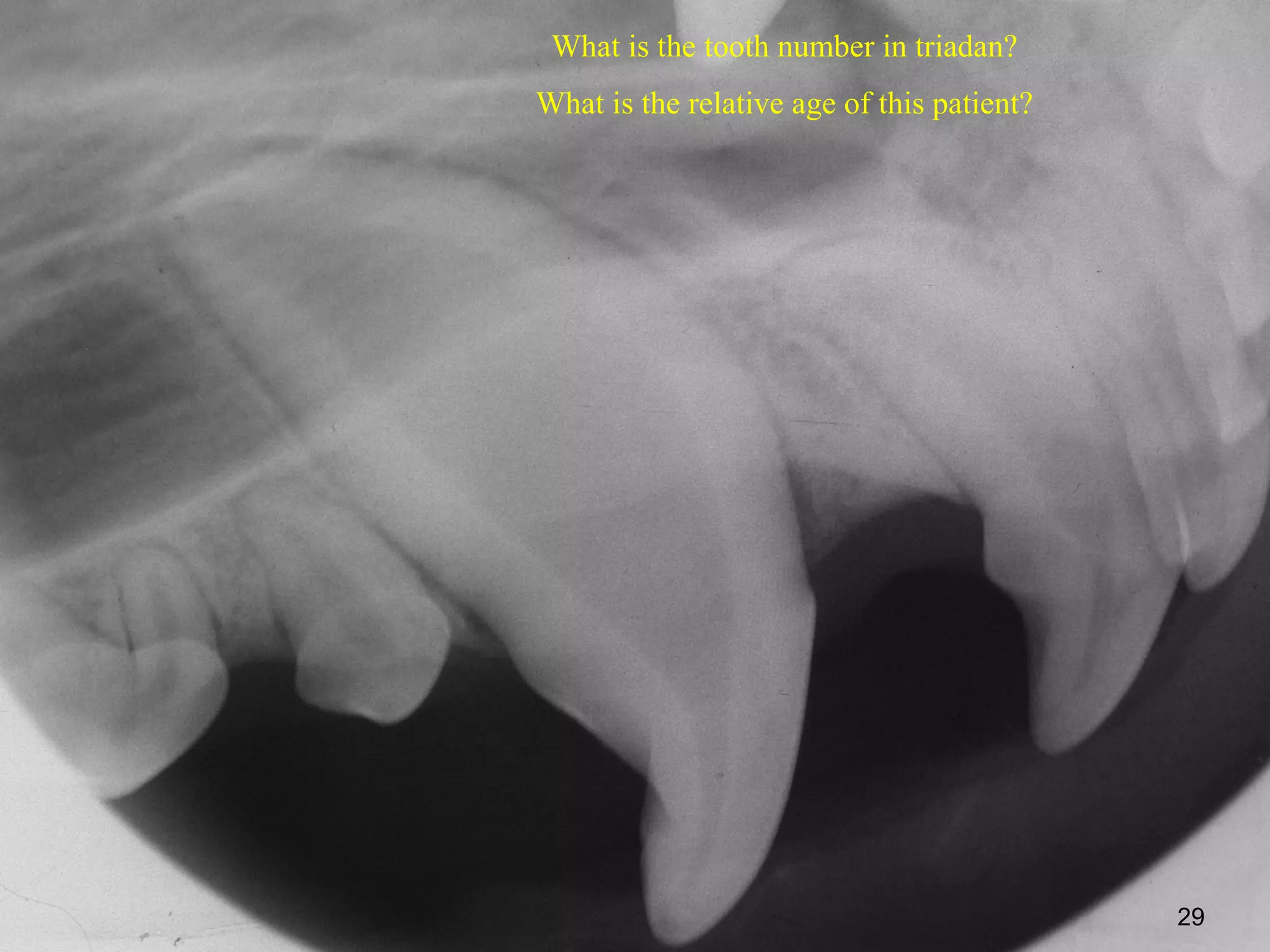
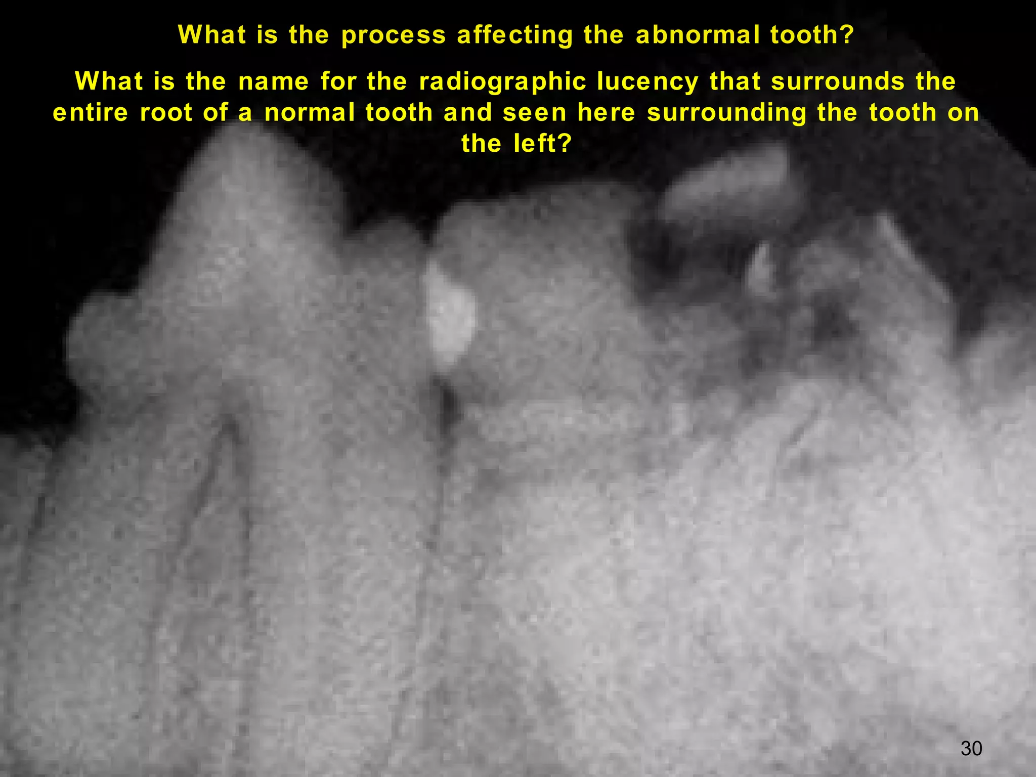
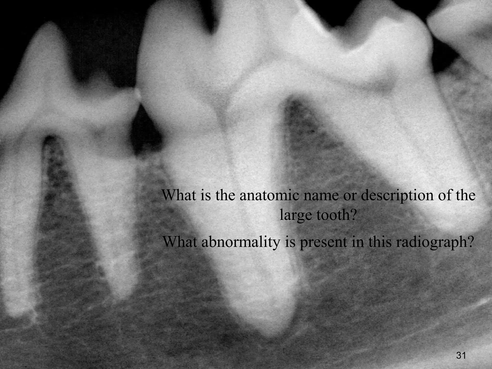
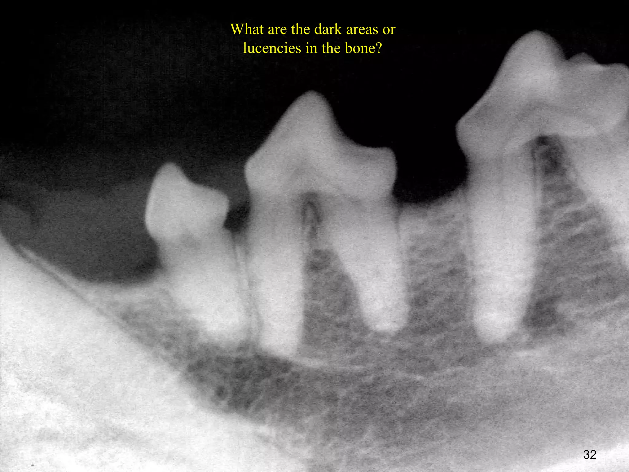
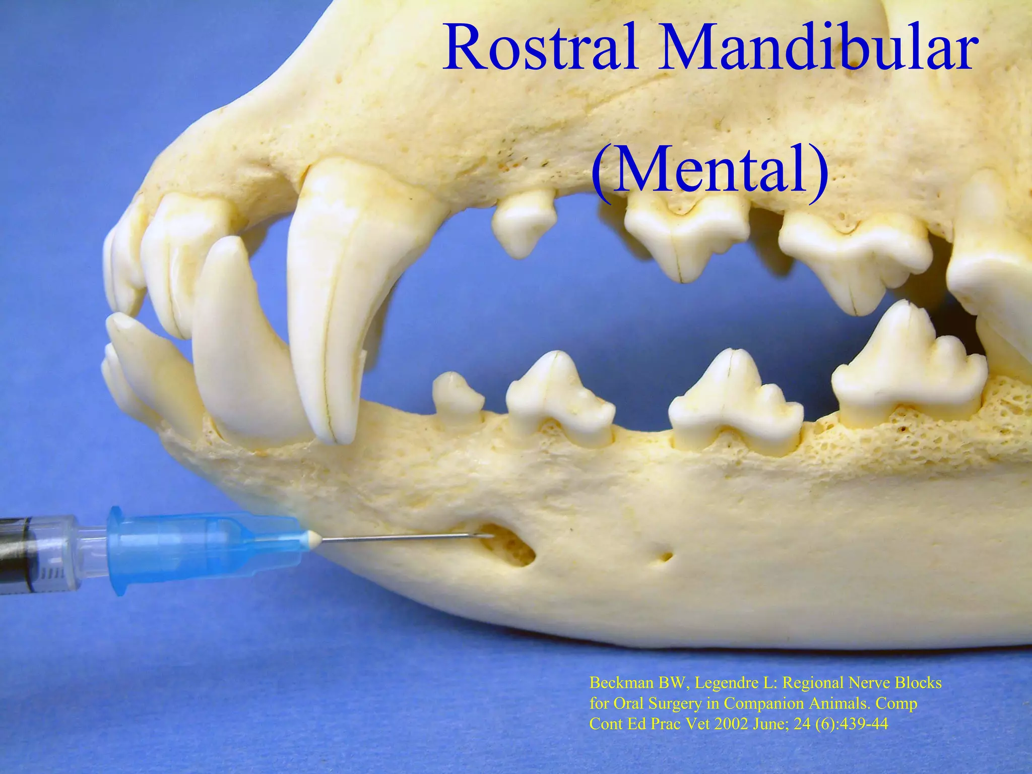
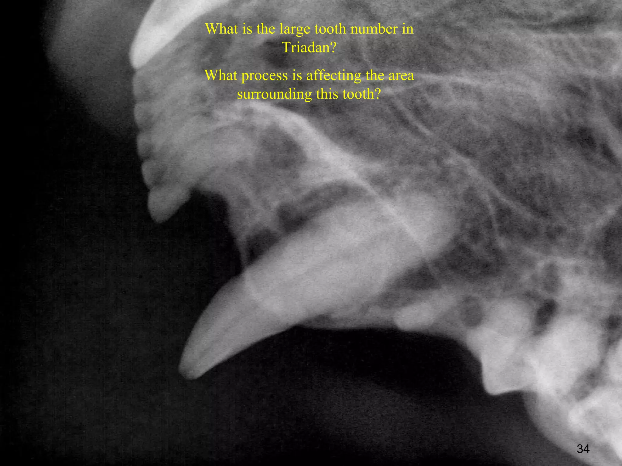
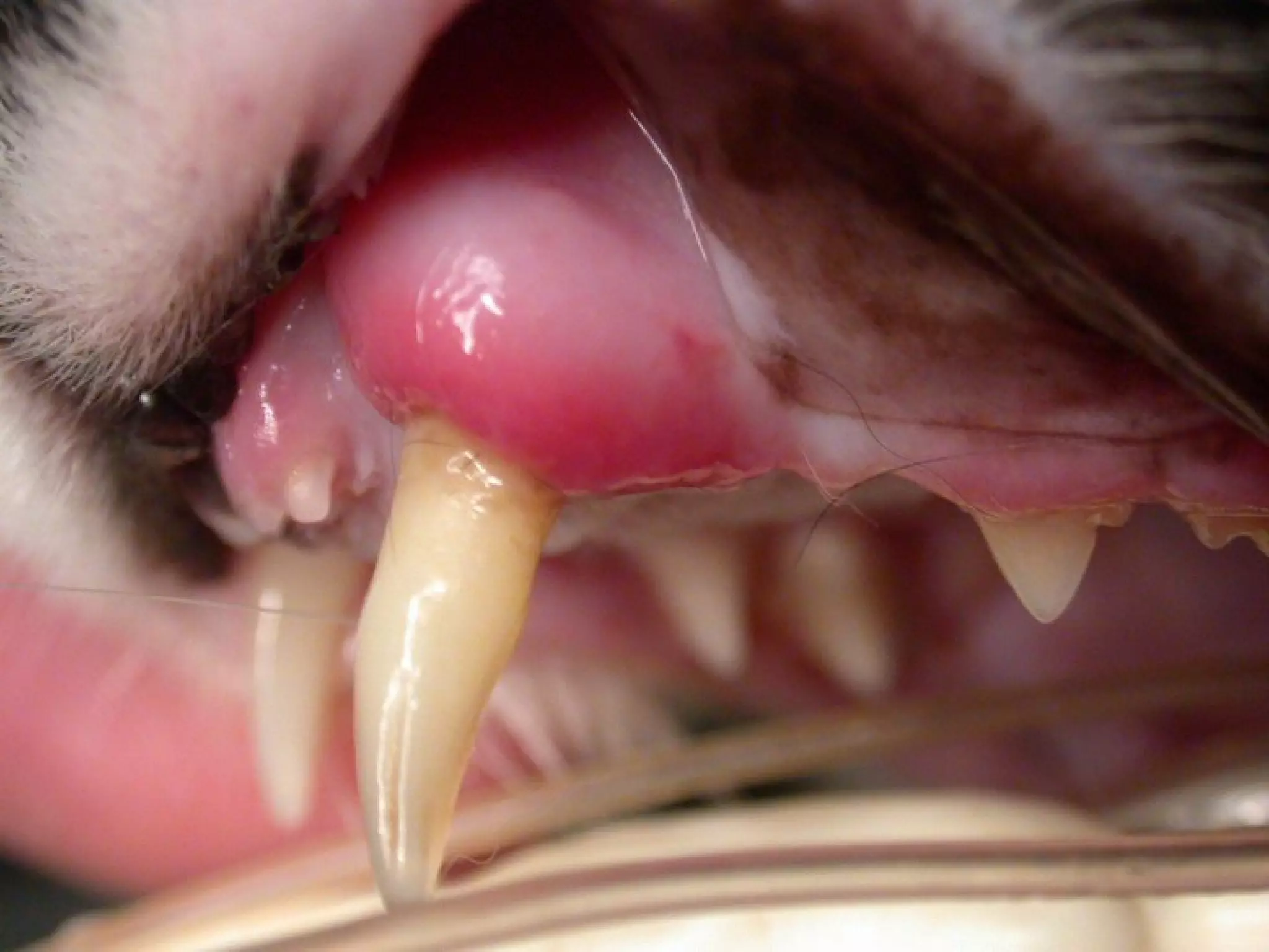
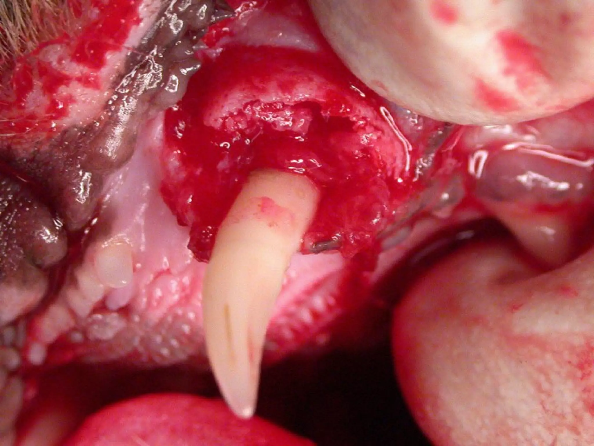
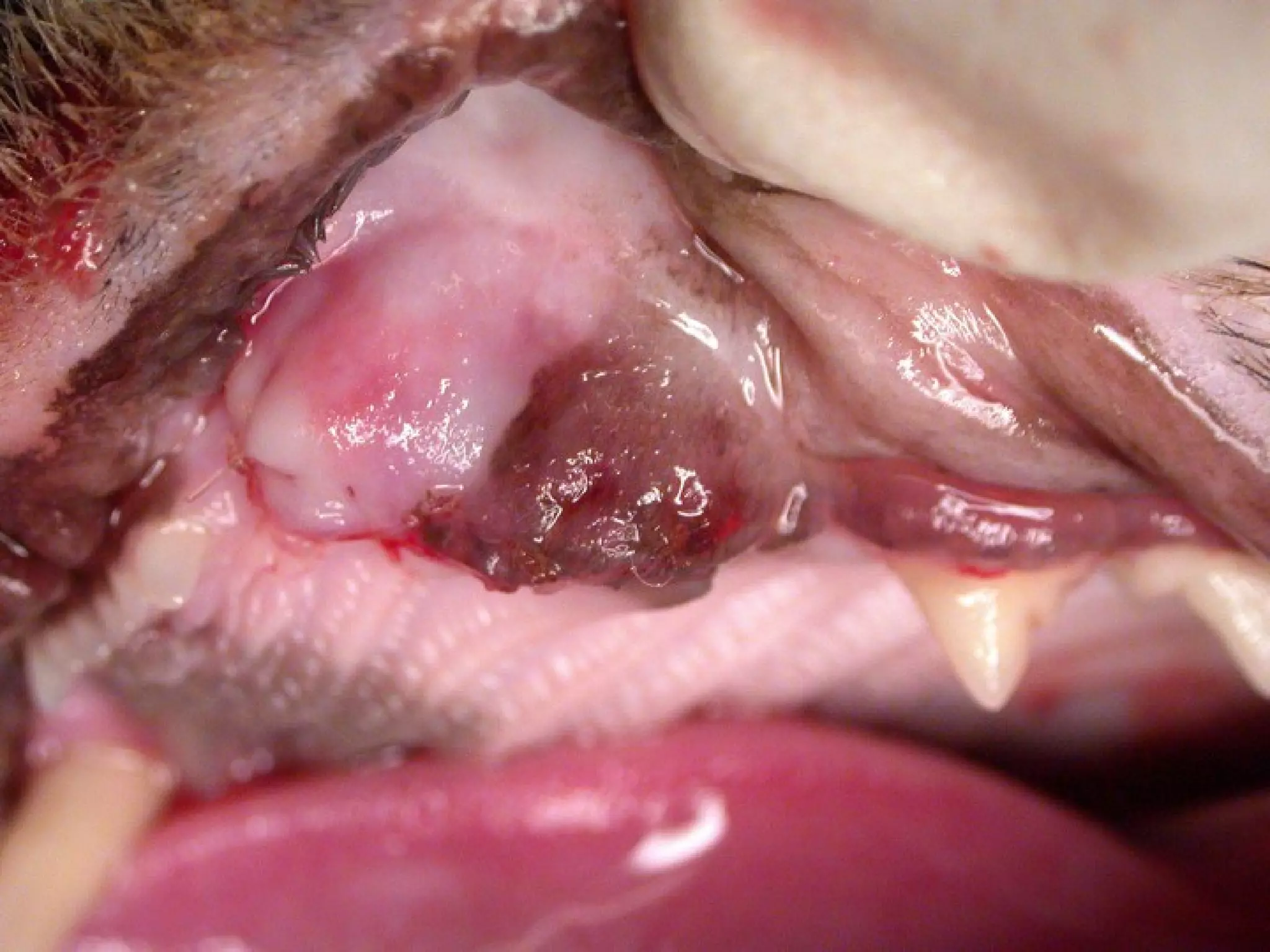
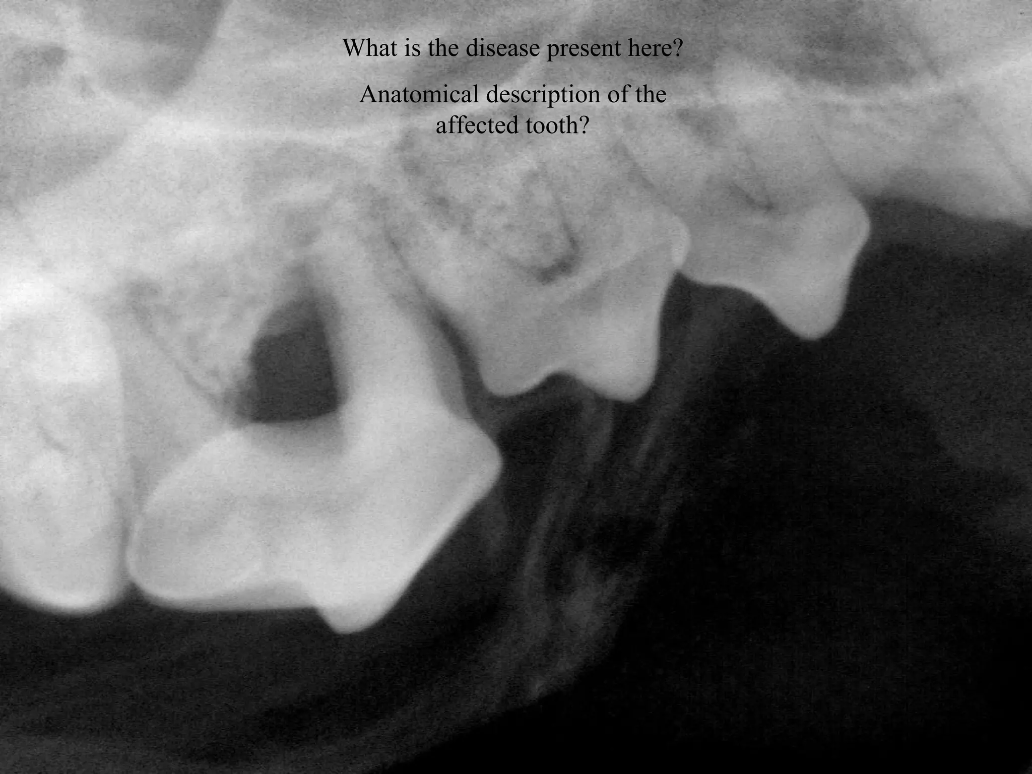
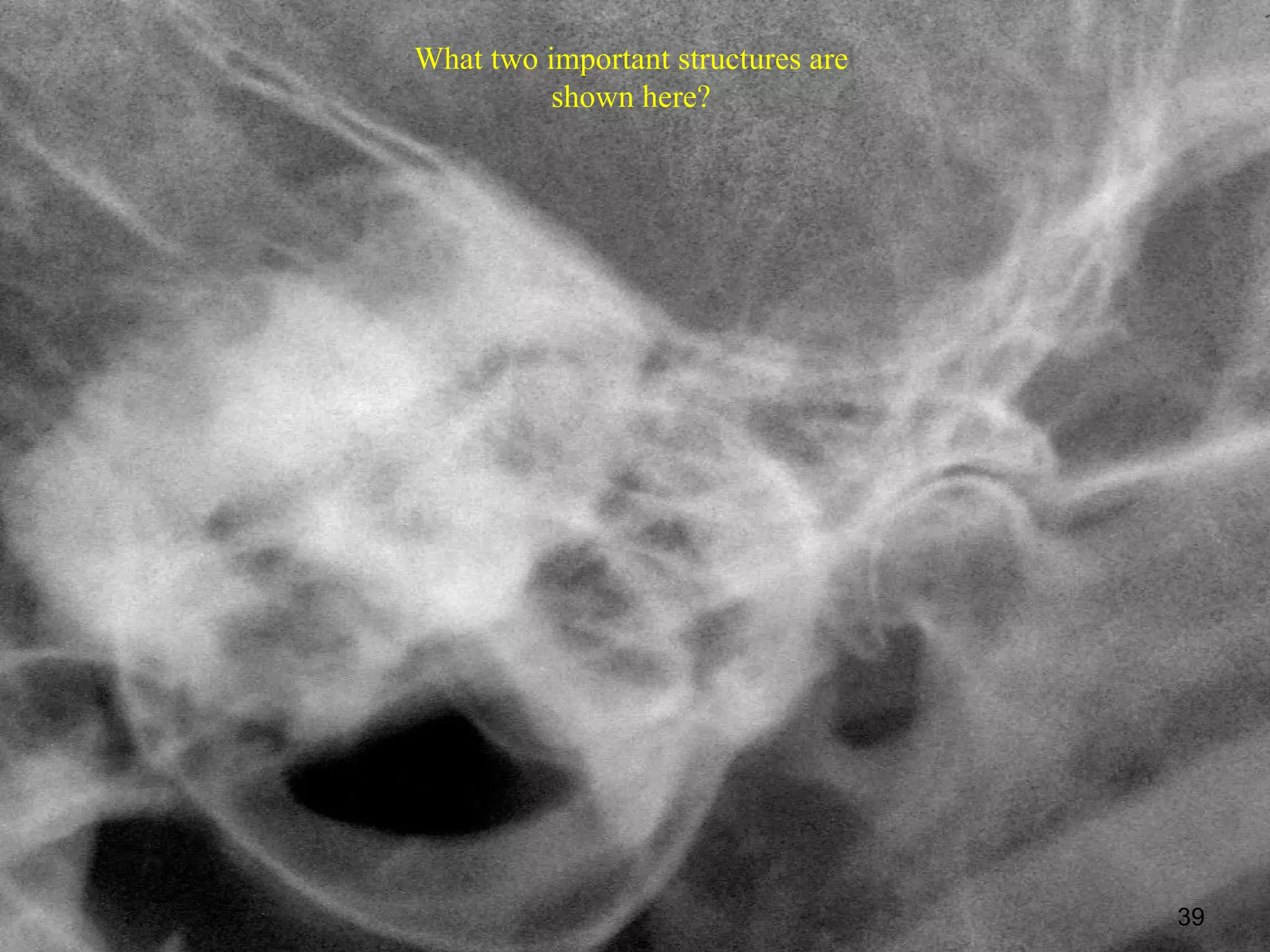
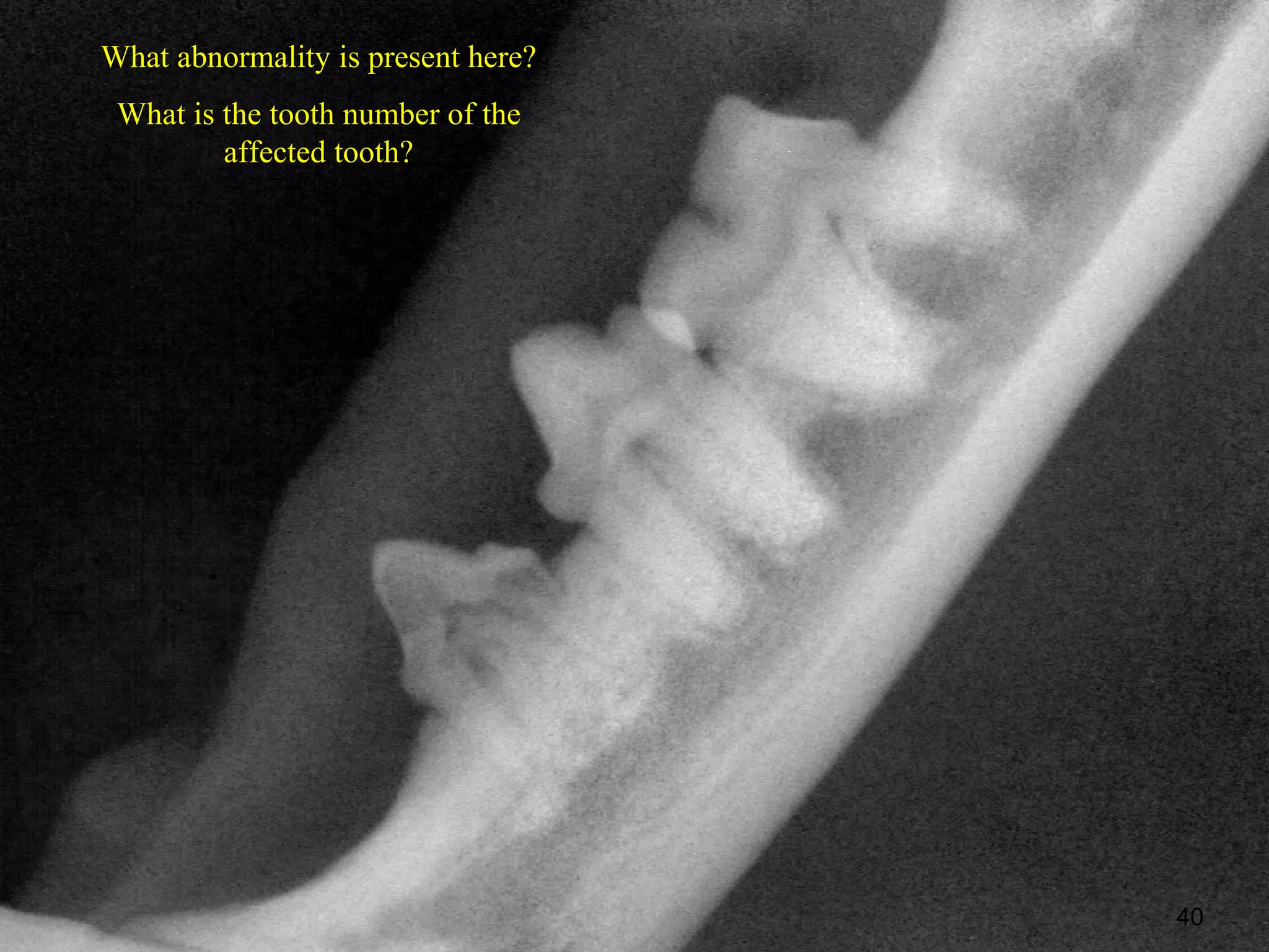
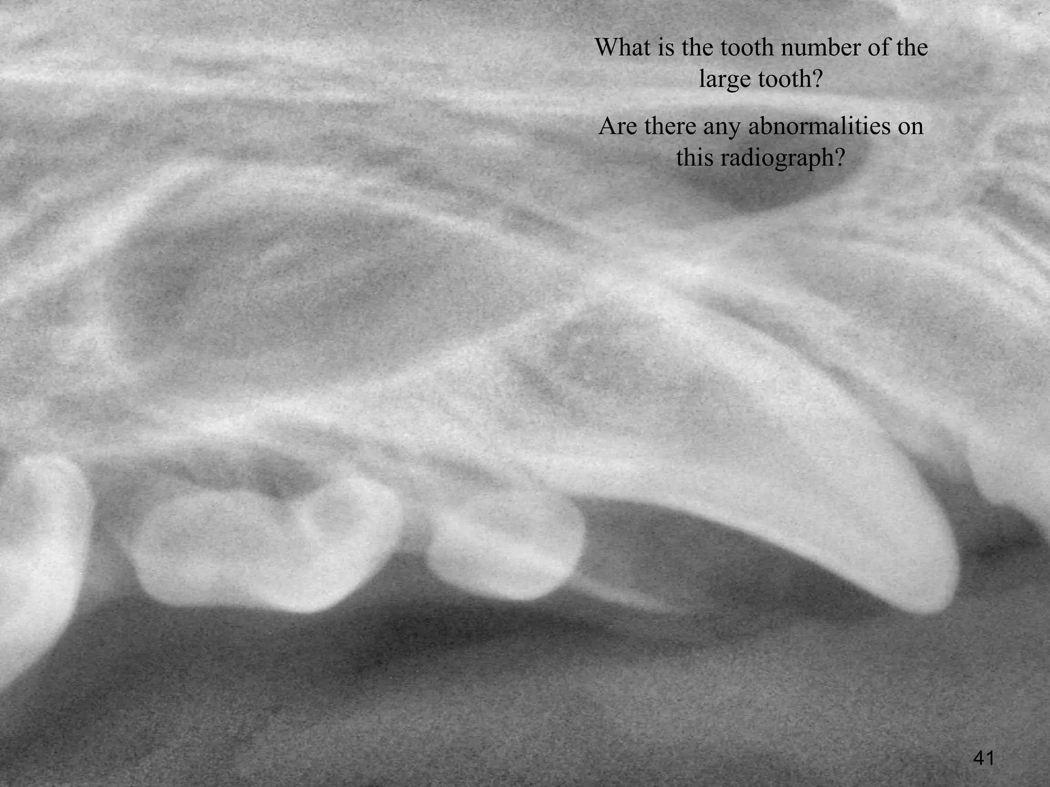
Clinical Radiographic
Anatomy and Interpretation


   Brett Beckman, DVM, FAVD
    Diplomate AVDC, AAPM
What disease is affecting the area
   surrounding these teeth?
 Is this the patient’s right or left
             mandible?
                                       2
What is unusual about
   this radiograph?
Is this the patient’s right
    or left mandible?


                              7
8
Is this the patient’s right or left maxilla?
         What is abnormal here?
What is wrong with the canine tooth on the left of the radiograph?

          Is the affected tooth on the cat’s left or right?


                                                                     18
Take a look at the left mandible on this cat.


                        Text


                                                20
What is the most likely cause of this process
                in this cat?

   What is the only definitive treatment?


                                                21
What is the process affecting
        these teeth?
Tooth number for the tooth to
          your left?


                                27
What is the white density seen
   throughout this tooth?
       Tooth number?


                                 28
What is the tooth number in triadan?
What is the relative age of this patient?


                                            29
What is the process affecting the abnormal tooth?
 What is the name for the radiographic lucency that surrounds the
entire root of a normal tooth and seen here surrounding the tooth on
                               the left?


                                                                  30
What is the anatomic name or description of the
                 large tooth?
What abnormality is present in this radiograph?


                                            31
What are the dark areas or
 lucencies in the bone?


                             32
Rostral Mandibular
    (Mental)


    Beckman BW, Legendre L: Regional Nerve Blocks
    for Oral Surgery in Companion Animals. Comp
    Cont Ed Prac Vet 2002 June; 24 (6):439-44
What is the large tooth number in
            Triadan?
What process is affecting the area
    surrounding this tooth?


                                     34
What is the disease present here?
 Anatomical description of the
       affected tooth?


                                    38
What two important structures are
          shown here?


                                    39
What abnormality is present here?
 What is the tooth number of the
         affected tooth?


                                    40
What is the tooth number of the
          large tooth?
Are there any abnormalities on
       this radiograph?


                                  41
Brett Beckman, DVM, FAVD
   Diplomate AVDC, AAPM

        Orlando Referrals:
Affiliated Veterinary Specialists
           Maitland, FL
                                    42
43
Brett Beckman, DVM, FAVD
   Diplomate AVDC, AAPM

        Orlando Referrals:
Affiliated Veterinary Specialists
45

Veterinary Dental Radiographic Interpretation Quiz

  • 1.
    Clinical Radiographic Anatomy andInterpretation Brett Beckman, DVM, FAVD Diplomate AVDC, AAPM
  • 2.
    What disease isaffecting the area surrounding these teeth? Is this the patient’s right or left mandible? 2
  • 7.
    What is unusualabout this radiograph? Is this the patient’s right or left mandible? 7
  • 8.
  • 15.
    Is this thepatient’s right or left maxilla? What is abnormal here?
  • 18.
    What is wrongwith the canine tooth on the left of the radiograph? Is the affected tooth on the cat’s left or right? 18
  • 20.
    Take a lookat the left mandible on this cat. Text 20
  • 21.
    What is themost likely cause of this process in this cat? What is the only definitive treatment? 21
  • 27.
    What is theprocess affecting these teeth? Tooth number for the tooth to your left? 27
  • 28.
    What is thewhite density seen throughout this tooth? Tooth number? 28
  • 29.
    What is thetooth number in triadan? What is the relative age of this patient? 29
  • 30.
    What is theprocess affecting the abnormal tooth? What is the name for the radiographic lucency that surrounds the entire root of a normal tooth and seen here surrounding the tooth on the left? 30
  • 31.
    What is theanatomic name or description of the large tooth? What abnormality is present in this radiograph? 31
  • 32.
    What are thedark areas or lucencies in the bone? 32
  • 33.
    Rostral Mandibular (Mental) Beckman BW, Legendre L: Regional Nerve Blocks for Oral Surgery in Companion Animals. Comp Cont Ed Prac Vet 2002 June; 24 (6):439-44
  • 34.
    What is thelarge tooth number in Triadan? What process is affecting the area surrounding this tooth? 34
  • 38.
    What is thedisease present here? Anatomical description of the affected tooth? 38
  • 39.
    What two importantstructures are shown here? 39
  • 40.
    What abnormality ispresent here? What is the tooth number of the affected tooth? 40
  • 41.
    What is thetooth number of the large tooth? Are there any abnormalities on this radiograph? 41
  • 42.
    Brett Beckman, DVM,FAVD Diplomate AVDC, AAPM Orlando Referrals: Affiliated Veterinary Specialists Maitland, FL 42
  • 43.
  • 44.
    Brett Beckman, DVM,FAVD Diplomate AVDC, AAPM Orlando Referrals: Affiliated Veterinary Specialists
  • 45.

Editor's Notes

  • #7 August 04
  • #34 ROSTRAL MANDIBULAR (MENTAL) REGIONAL BLOCK The rostral mandibular block infiltrates the rostral extent of the inferior alveolar nerve just before it exits the middle mental foramen. (Figures 6 and 7) The structures anesthetized include the incisors, the canine and the first three premolars. The adjacent bone and soft tissue are also affected. The middle mental foramen is located about 1/3 of the distance from the ventral border to the dorsal border of the mandible at the level of the mesial root of the second premolar. The landmark for infiltration is the mandibular labial frenulum. The frenulum is retracted ventrally. The needle is inserted at the rostral aspect of the frenulum and advanced along the mandibular bone to just enter the canal. If bone is encountered the needle should be backed out and redirected until the needle passes freely into the foramen. Placement can be confirmed by moving the syringe laterally to encounter the lateral aspect of the canal. The patient’s jaw, rather than the alveolar mucosa, will move slightly if the needle is within the canal. The mental nerve block gets the mandibular bone, teeth and soft tissue from the second premolar to the midline. The mandibular labial frenulum is retracted ventrally. The needle is directed into the mesial aspect of the frenulum and advanced against the bone distally to enter the middle mental foramen at the level of the distal frenulum.
  • #36 Whats under whats underneath the gum. Can’t leave that mess