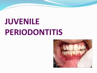
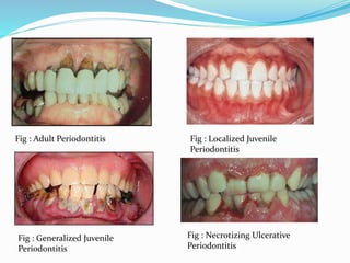
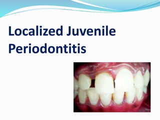
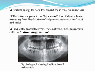
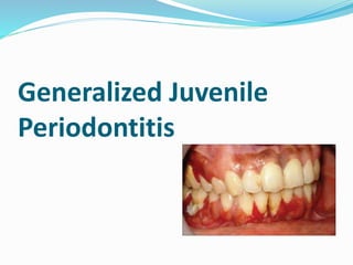
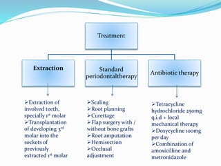
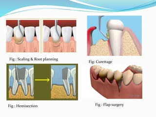
JUVENILE
PERIODONTITIS
What is Periodontitis ?
 Periodontitis is
 Inflammatory disease of the supporting tissue of the teeth
 Caused by specific microorganism
 Resulting in progressive destruction of the periodontal
legament and alveolar bone with pocket formation ,
recession or both
Periodontitis
Adult
periodontitis
Early onset
periodontitis
Prepubertal
. Localized
. Generalized
Juvenile
. Localized
. Generalized
Periodontitis
associated with
systemic disease
. Down’s syndrome
. Diabetes type 1
. AIDS and other
disease
Necrotizing
ulcerative
periodontitis
Refractory
periodontitis
Fig : Adult Periodontitis Fig : Localized Juvenile
Periodontitis
Fig : Generalized Juvenile
Periodontitis
Fig : Necrotizing Ulcerative
Periodontitis
What is Juvenile or Aggressive
Periodontitis?
 Juvenile or Aggressive periodontitis is
 Rapid loss of attachment and bone loss
 Occurring in an otherwise clinicially healthy patients with
the amount of microbial deposits in consistent with disease
severity
 Familial aggregation of diseased individual
Aggressive/
Juvenile
Periodontitis
Localized Generalized
Rapidly
Progressing
Stages in the
development of disease
• Degenaration of principle fibers of PDL
• Cessation of cementum formation
• Resorption of alveolar bone
• Tooth migration without detectable inflammation
Stage I
• Rapid proliferation of the junctional epithelium
along the root
• Earliest sings of inflammation appear
Stage II
• Progressive inflammation and development of
deep, infrabony periodontal pocket
Stage
III
Etiology of juvenile periodontitis
Micro organisms
 Actinobacillus actinomycetemcomitans
 Porphyromonas gingivalis
 Eikenella corrodens
 Caprocytophaga
 Spirocheates
 Bacillus
 Defective neutrophil or monocyte function
 Poor serum antibody response to infecting agent
Localized Juvenile
Periodontitis
What is localized juvenile
periodontitis ?
 Localized juvenile periodontitis is
Occuring in otherwise healthy individual under the
age of 30 years
Destructive periodontitis localized to the 1st
permanent molars and incisors not involving more
than two other teeth
Clinical features
 Age and sex distribution
 Both sexes ( slight female predilection )
 Seen mostly between 20 years
 Distribution of lesion
 1st molar and/or incisors
1st molar and/or incisors + additional teeth ( not more than 2
teeth other than 1st molars and incisors )
 Lack of clinical inflammation despite the presence of deep
periodontal pocket
 Small amount of plaque, forms a thin film rarely
mineralized
 Mobility and migration of 1st molars and incisors
 Classically distolabial migration of maxillary incisors with
diastema
 Root surface sensitivity
 Deep dull radiating pain
 Periodontal abscess
 Enlargement of regional lymphnode
Radiographic findings
 Vertical or angular bone loss around the 1st molars and incisors
 The pattern appears to be “Arc shaped” loss of alveolar bone
extending from distal surface of 2nd premolar to mesial surface of
2nd molar
 Frequently bilaterally symmetrical pattern of bone loss occurs
called as “ mirror image pattern”
Fig : Radiograph showing localized juvenile
periodontitis
Histopathological
features
 Ulcerated pocket epithelium
 Accumulation of various inflammatory cells in the
connective tissue mainly leukocytes, plasma cells and
small number of lymphocytes and macrophages
 Bacterial invasion of connective tissue
 The flora involves A. actinomycetemcomitans ,
Capnocytophaga sputigena and others
Bacteriology
 A. actinomycetemcomitans
 short
 Facultatively anaerobic
 Non motile
 Gram negative rod
 Caprocytophaga
Fig : Showing colony of A.a
Leucotoxin Destroys polymorphonuclear
leukocytes and macrophages
Endotoxin Activates host cells to secret
inflammatory mediators
Bacteriocin Inhibit IgG and IgM production
Collagenase Degradation of collagen
Chemotactic
Inhibition factor
Inhibit neutrophil chemotaxis
Virulence factor associated with
A. actinomycetemcomitans
Immunology
Functionals defects of polymorphoneuclear leukocytes
or monocytes, impairs the chemotactic attraction of
these cells to the site of infection
Generalized Juvenile
Periodontitis
What is Generalized juvenile
periodontitis ?
 Generalized juvenile periodontitis is
 Generalized interproximal attachment loss
 Affecting atleast three permanent teeth other than the
1st molar and incisors
Clinical
features
Age and sex distribution
Affects between puberty and 35 years
No sex discrimination
Distritubiton of lesion
All or most of the teeth are affected, no specific pattern is
observed
 There are two phases of lesion
Destructive phase Non destructive phase
Tissue appears severely
inflammed, ulcerated
and fiery red
Bleeding with or
without stimulation
Suppuration
Attachment and bone
loss
Tissue appears pink with some
stippling
Lack of inflammation
Probling will reveal deep pocket
Bone attachment levels relatively
stable
 Some patients may exhibit
Weight loss
Mental depression
General malaise
Systemic condition may predispose patient to
generalized juvenile periodontitis, these includes
 chornic neutrophil defect
Leukocyte adherence deficiency
Radiographic findings
 No define pattern of distribution
Range from severe bone loss associated with
minimal number of teeth to advanced bone loss
affecting the majority of teeth in the dentition
Fig : showing radiograph of GJP
Treatment
Extraction Standard
periodontaltherapy
Antibiotic therapy
Extraction of
involved teeth,
specially 1st molar
Transplantation
of developing 3rd
molar into the
sockets of
previously
extracted 1st molar
Scaling
Root planning
Curettage
Flap surgery with /
without bone grafts
Root amputation
Hemisection
Occlusal
adjustment
Tetracycline
hydrochloride 250mg
q.i.d + local
mechanical therapy
Doxycycline 100mg
per day
Combination of
amoxicilline and
metronidazole
Fig : Scaling & Root planning
Fig: Curettage
Fig : Hemisection Fig : Flap surgery
Periodontal disease accounts for a majority of
missing teeth in adults and results in tremendous
economic and social burdens both to the individual
and the society
Periodontal disease is so prevalent that only
possible solution to the problem is its prevention by
maintaining the good oral hygiene
Conclusion
Reference
 Essential of Clinical Periodontology And Periodontics
; 4th edition ; Shantipriya Reddy
Carranza’s Clinical Periodontology ; 11th edition ;
Newman ;Takei ; Klokkevold ; Carranza
Cawson’s Essentials of Oral Pathology And Oral
Medicine ; 8th edition ; R. A. Cawson ; E. W. Odell
Juvenile Periodontitis

Juvenile Periodontitis

  • 1.
  • 2.
    What is Periodontitis?  Periodontitis is  Inflammatory disease of the supporting tissue of the teeth  Caused by specific microorganism  Resulting in progressive destruction of the periodontal legament and alveolar bone with pocket formation , recession or both
  • 3.
    Periodontitis Adult periodontitis Early onset periodontitis Prepubertal . Localized .Generalized Juvenile . Localized . Generalized Periodontitis associated with systemic disease . Down’s syndrome . Diabetes type 1 . AIDS and other disease Necrotizing ulcerative periodontitis Refractory periodontitis
  • 4.
    Fig : AdultPeriodontitis Fig : Localized Juvenile Periodontitis Fig : Generalized Juvenile Periodontitis Fig : Necrotizing Ulcerative Periodontitis
  • 5.
    What is Juvenileor Aggressive Periodontitis?  Juvenile or Aggressive periodontitis is  Rapid loss of attachment and bone loss  Occurring in an otherwise clinicially healthy patients with the amount of microbial deposits in consistent with disease severity  Familial aggregation of diseased individual
  • 6.
  • 7.
  • 8.
    • Degenaration ofprinciple fibers of PDL • Cessation of cementum formation • Resorption of alveolar bone • Tooth migration without detectable inflammation Stage I • Rapid proliferation of the junctional epithelium along the root • Earliest sings of inflammation appear Stage II • Progressive inflammation and development of deep, infrabony periodontal pocket Stage III
  • 9.
    Etiology of juvenileperiodontitis Micro organisms  Actinobacillus actinomycetemcomitans  Porphyromonas gingivalis  Eikenella corrodens  Caprocytophaga  Spirocheates  Bacillus  Defective neutrophil or monocyte function  Poor serum antibody response to infecting agent
  • 10.
  • 11.
    What is localizedjuvenile periodontitis ?  Localized juvenile periodontitis is Occuring in otherwise healthy individual under the age of 30 years Destructive periodontitis localized to the 1st permanent molars and incisors not involving more than two other teeth
  • 12.
  • 13.
     Age andsex distribution  Both sexes ( slight female predilection )  Seen mostly between 20 years  Distribution of lesion  1st molar and/or incisors 1st molar and/or incisors + additional teeth ( not more than 2 teeth other than 1st molars and incisors )
  • 14.
     Lack ofclinical inflammation despite the presence of deep periodontal pocket  Small amount of plaque, forms a thin film rarely mineralized  Mobility and migration of 1st molars and incisors  Classically distolabial migration of maxillary incisors with diastema
  • 15.
     Root surfacesensitivity  Deep dull radiating pain  Periodontal abscess  Enlargement of regional lymphnode
  • 16.
  • 17.
     Vertical orangular bone loss around the 1st molars and incisors  The pattern appears to be “Arc shaped” loss of alveolar bone extending from distal surface of 2nd premolar to mesial surface of 2nd molar  Frequently bilaterally symmetrical pattern of bone loss occurs called as “ mirror image pattern” Fig : Radiograph showing localized juvenile periodontitis
  • 18.
  • 19.
     Ulcerated pocketepithelium  Accumulation of various inflammatory cells in the connective tissue mainly leukocytes, plasma cells and small number of lymphocytes and macrophages  Bacterial invasion of connective tissue  The flora involves A. actinomycetemcomitans , Capnocytophaga sputigena and others
  • 20.
    Bacteriology  A. actinomycetemcomitans short  Facultatively anaerobic  Non motile  Gram negative rod  Caprocytophaga Fig : Showing colony of A.a
  • 21.
    Leucotoxin Destroys polymorphonuclear leukocytesand macrophages Endotoxin Activates host cells to secret inflammatory mediators Bacteriocin Inhibit IgG and IgM production Collagenase Degradation of collagen Chemotactic Inhibition factor Inhibit neutrophil chemotaxis Virulence factor associated with A. actinomycetemcomitans
  • 22.
    Immunology Functionals defects ofpolymorphoneuclear leukocytes or monocytes, impairs the chemotactic attraction of these cells to the site of infection
  • 23.
  • 24.
    What is Generalizedjuvenile periodontitis ?  Generalized juvenile periodontitis is  Generalized interproximal attachment loss  Affecting atleast three permanent teeth other than the 1st molar and incisors
  • 25.
  • 26.
    Age and sexdistribution Affects between puberty and 35 years No sex discrimination Distritubiton of lesion All or most of the teeth are affected, no specific pattern is observed  There are two phases of lesion
  • 27.
    Destructive phase Nondestructive phase Tissue appears severely inflammed, ulcerated and fiery red Bleeding with or without stimulation Suppuration Attachment and bone loss Tissue appears pink with some stippling Lack of inflammation Probling will reveal deep pocket Bone attachment levels relatively stable
  • 29.
     Some patientsmay exhibit Weight loss Mental depression General malaise Systemic condition may predispose patient to generalized juvenile periodontitis, these includes  chornic neutrophil defect Leukocyte adherence deficiency
  • 30.
  • 31.
     No definepattern of distribution Range from severe bone loss associated with minimal number of teeth to advanced bone loss affecting the majority of teeth in the dentition Fig : showing radiograph of GJP
  • 32.
    Treatment Extraction Standard periodontaltherapy Antibiotic therapy Extractionof involved teeth, specially 1st molar Transplantation of developing 3rd molar into the sockets of previously extracted 1st molar Scaling Root planning Curettage Flap surgery with / without bone grafts Root amputation Hemisection Occlusal adjustment Tetracycline hydrochloride 250mg q.i.d + local mechanical therapy Doxycycline 100mg per day Combination of amoxicilline and metronidazole
  • 33.
    Fig : Scaling& Root planning Fig: Curettage Fig : Hemisection Fig : Flap surgery
  • 34.
    Periodontal disease accountsfor a majority of missing teeth in adults and results in tremendous economic and social burdens both to the individual and the society Periodontal disease is so prevalent that only possible solution to the problem is its prevention by maintaining the good oral hygiene Conclusion
  • 35.
    Reference  Essential ofClinical Periodontology And Periodontics ; 4th edition ; Shantipriya Reddy Carranza’s Clinical Periodontology ; 11th edition ; Newman ;Takei ; Klokkevold ; Carranza Cawson’s Essentials of Oral Pathology And Oral Medicine ; 8th edition ; R. A. Cawson ; E. W. Odell