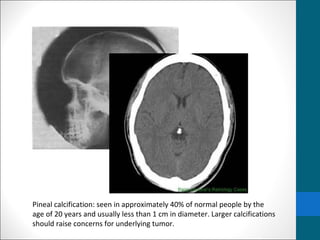
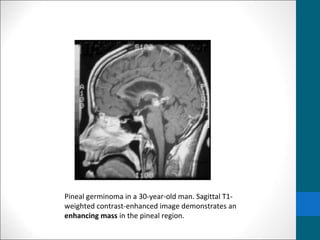
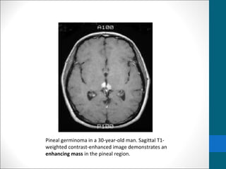
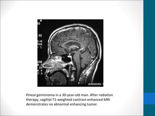
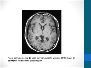
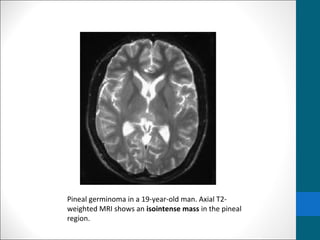
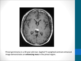
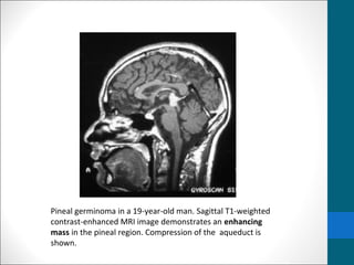
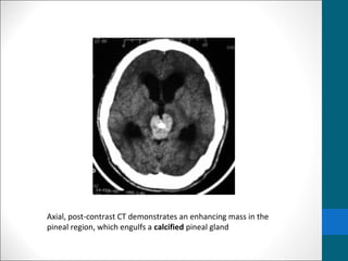
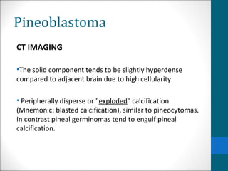
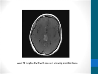
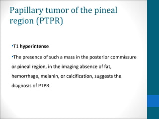
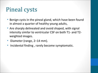
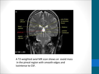
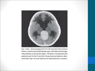
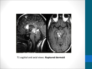
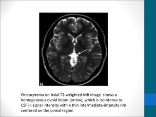
Pineal Gland 
Tumors
Introduction 
 Pineal Gland is midline structure located in the 
epithalamus, near the centre of the brain, between the 
two hemispheres, in a groove where the two halves of 
the thalamus join. 
 Pineal region neoplasm's constitute only 0.3-2.7% of 
intracranial tumors. 
 They are considered an important clinical entity because 
of their strategic location.
Clinical features 
• hydrocephalus and increased intracranial pressure 
• headaches 
• nausea 
• vomiting 
• lethargy 
• altered mental function. 
• Visual Problems 
• Parinaud's syndrome, which includes inability to look 
upward, double vision and nystagmus
Differential diagnosis of pineal 
region mass 
• Pineal parenchymal tumours 
• Pineocytoma 
• Pineal parenchymal tumour with intermediate differentiation 
• Papillary tumour of the pineal region 
• Pineoblastoma 
• Germ cell tumours 
• Pineal germinoma (most common; ~ 50% of all tumours) 
• Pineal embryonal carcinoma 
• Pineal choriocarcinoma 
• Pineal yolk sac carcinoma - endodermal sinus tumour 
• Pineal teratoma
• astrocytoma of pineal gland 
• pineal metastasis 
• pineal cyst 
• meningioma near pineal region 
• cavernoma in pineal region 
• aneurysm in pineal region
Pineal calcification: seen in approximately 40% of normal people by the 
age of 20 years and usually less than 1 cm in diameter. Larger calcifications 
should raise concerns for underlying tumor.
MRI 
•MRI is the preferred imaging modality 
•enables the accurate delineation of pineal masses before 
surgery. 
•allows true pineal masses to be distinguished from 
parapineal masses that impinge on the pineal gland
Limitations 
• Computed tomography (CT) scanning may be needed to 
evaluate a calcified pineal gland that is associated with a 
pineal germinoma or tumor calcification associated with 
other neoplasms in the pineal region.
Radiography 
• Germinomas are associated with a high incidence of 
pineal gland calcification. 
• The tumor does not calcify, but it may engulf a calcified 
pineal gland. 
• A calcified pineal gland on plain radiographs in a boy 
younger than 10 years suggests pineal germinoma.
Computed Tomography 
• Nonenhanced CT scans 
• Typically demonstrate a slightly hyperattenuating 
mass that engulfs a prominent calcified pineal gland. 
• Contrast-enhanced CT scanning 
• demonstrates homogeneous and intense 
enhancement.
• More sensitive than plain radiography in depicting pineal 
calcification. 
• Small speckles of calcification can be seen in children 6 
years and are considered normal 
• Pineal calcification in children younger than 6 years is 
suggestive of pineal neoplasm. 
• Pineal calcifications should be less than 1 cm in diameter 
in size.
Germinoma …..MRI findings 
•T1/T2: 
• Usually isointense relative to cerebral gray matter 
•Occasionally 
• T1:hypointense 
• T2:hyperintense 
• I/V injection of gadolinium-based contrast material, 
• homogeneous and intense enhancement is seen. 
• contrast-enhanced MRI (subarachnoid seeding of 
germinomas.)
Pineal germinoma in a 30-year-old man. Axial T2-weighted 
MRI shows a mildly hyperintense lesion involving the pineal 
region.
Pineal germinoma in a 30-year-old man. Sagittal T1- 
weighted contrast-enhanced image demonstrates an 
enhancing mass in the pineal region.
Pineal germinoma in a 30-year-old man. Sagittal T1- 
weighted contrast-enhanced image demonstrates an 
enhancing mass in the pineal region.
Pineal germinoma in a 30-year-old man. After radiation 
therapy, sagittal T1-weighted contrast-enhanced MRI 
demonstrates no abnormal enhancing tumor.
Pineal germinoma in a 19-year-old man. Axial T1-weighted MRI shows an 
isointense lesion in the pineal region.
Pineal germinoma in a 19-year-old man. Axial T2- 
weighted MRI shows an isointense mass in the pineal 
region.
Pineal germinoma in a 30-year-old man. Sagittal T1-weighted contrast-enhanced 
image demonstrates an enhancing mass in the pineal region.
Pineal germinoma in a 19-year-old man. Sagittal T1-weighted 
contrast-enhanced MRI image demonstrates an enhancing 
mass in the pineal region. Compression of the aqueduct is 
shown.
Axial, post-contrast CT demonstrates an enhancing mass in the 
pineal region, which engulfs a calcified pineal gland
Pineoblastoma 
CT IMAGING 
•The solid component tends to be slightly hyperdense 
compared to adjacent brain due to high cellularity. 
• Peripherally disperse or "exploded" calcification 
(Mnemonic: blasted calcification), similar to pineocytomas. 
In contrast pineal germinomas tend to engulf pineal 
calcification.
MRI IMAGING 
•T1 - isointense to hypointense to adjacent brain 
•T2 isointense to adjacent brain 
•Areas of cyst formation or necrosis my be present 
•T1 C+ (Gd) - vivid heterogeneous enhancement
T1 weighted post contrast axial views showing an enhancing mass in pineal region 
and areas of necrosis within it after treatment
Axial T1 weighted MRI with contrast showing pineoblastoma
Papillary tumor of the pineal 
region (PTPR) 
•T1 hyperintense 
•The presence of such a mass in the posterior commissure 
or pineal region, in the imaging absence of fat, 
hemorrhage, melanin, or calcification, suggests the 
diagnosis of PTPR.
Pineal cysts 
• Benign cysts in the pineal gland, which have been found 
in almost a quarter of healthy young adults, 
• Are sharply delineated and ovoid shaped, with signal 
intensity similar to ventricular CSF on both T1- and T2- 
weighted images. 
• Diameter (range, 2–14 mm). 
• Incidental finding , rarely become symptomatic.
A T2-weighted axial MR scan shows an ovoid mass 
in the pineal region with smooth edges and 
isointense to CSF.
T1 sagittal and axial views :Ruptured dermoid
Pineocytoma on precontrast Sagittal T1-weighted MR shows an 
ovoid lesion that is hypointense to white matter and close to 
CSF in signal intensity
Pineocytoma on Axial T2-weighted MR image shows a 
homogeneous ovoid lesion (arrow), which is isointense to 
CSF in signal intensity with a thin intermediate-intensity rim 
centered on the pineal region.
Pineocytoma on postcontrast Axial postcontrast T1-weighted MR 
image shows a homogeneously enhancing mass (arrow) centered 
on the pineal recess
THANK YOU

Pineal gland tumors

  • 1.
  • 2.
    Introduction  PinealGland is midline structure located in the epithalamus, near the centre of the brain, between the two hemispheres, in a groove where the two halves of the thalamus join.  Pineal region neoplasm's constitute only 0.3-2.7% of intracranial tumors.  They are considered an important clinical entity because of their strategic location.
  • 5.
    Clinical features •hydrocephalus and increased intracranial pressure • headaches • nausea • vomiting • lethargy • altered mental function. • Visual Problems • Parinaud's syndrome, which includes inability to look upward, double vision and nystagmus
  • 6.
    Differential diagnosis ofpineal region mass • Pineal parenchymal tumours • Pineocytoma • Pineal parenchymal tumour with intermediate differentiation • Papillary tumour of the pineal region • Pineoblastoma • Germ cell tumours • Pineal germinoma (most common; ~ 50% of all tumours) • Pineal embryonal carcinoma • Pineal choriocarcinoma • Pineal yolk sac carcinoma - endodermal sinus tumour • Pineal teratoma
  • 7.
    • astrocytoma ofpineal gland • pineal metastasis • pineal cyst • meningioma near pineal region • cavernoma in pineal region • aneurysm in pineal region
  • 8.
    Pineal calcification: seenin approximately 40% of normal people by the age of 20 years and usually less than 1 cm in diameter. Larger calcifications should raise concerns for underlying tumor.
  • 9.
    MRI •MRI isthe preferred imaging modality •enables the accurate delineation of pineal masses before surgery. •allows true pineal masses to be distinguished from parapineal masses that impinge on the pineal gland
  • 10.
    Limitations • Computedtomography (CT) scanning may be needed to evaluate a calcified pineal gland that is associated with a pineal germinoma or tumor calcification associated with other neoplasms in the pineal region.
  • 11.
    Radiography • Germinomasare associated with a high incidence of pineal gland calcification. • The tumor does not calcify, but it may engulf a calcified pineal gland. • A calcified pineal gland on plain radiographs in a boy younger than 10 years suggests pineal germinoma.
  • 12.
    Computed Tomography •Nonenhanced CT scans • Typically demonstrate a slightly hyperattenuating mass that engulfs a prominent calcified pineal gland. • Contrast-enhanced CT scanning • demonstrates homogeneous and intense enhancement.
  • 13.
    • More sensitivethan plain radiography in depicting pineal calcification. • Small speckles of calcification can be seen in children 6 years and are considered normal • Pineal calcification in children younger than 6 years is suggestive of pineal neoplasm. • Pineal calcifications should be less than 1 cm in diameter in size.
  • 14.
    Germinoma …..MRI findings •T1/T2: • Usually isointense relative to cerebral gray matter •Occasionally • T1:hypointense • T2:hyperintense • I/V injection of gadolinium-based contrast material, • homogeneous and intense enhancement is seen. • contrast-enhanced MRI (subarachnoid seeding of germinomas.)
  • 15.
    Pineal germinoma ina 30-year-old man. Axial T2-weighted MRI shows a mildly hyperintense lesion involving the pineal region.
  • 16.
    Pineal germinoma ina 30-year-old man. Sagittal T1- weighted contrast-enhanced image demonstrates an enhancing mass in the pineal region.
  • 17.
    Pineal germinoma ina 30-year-old man. Sagittal T1- weighted contrast-enhanced image demonstrates an enhancing mass in the pineal region.
  • 18.
    Pineal germinoma ina 30-year-old man. After radiation therapy, sagittal T1-weighted contrast-enhanced MRI demonstrates no abnormal enhancing tumor.
  • 19.
    Pineal germinoma ina 19-year-old man. Axial T1-weighted MRI shows an isointense lesion in the pineal region.
  • 20.
    Pineal germinoma ina 19-year-old man. Axial T2- weighted MRI shows an isointense mass in the pineal region.
  • 21.
    Pineal germinoma ina 30-year-old man. Sagittal T1-weighted contrast-enhanced image demonstrates an enhancing mass in the pineal region.
  • 22.
    Pineal germinoma ina 19-year-old man. Sagittal T1-weighted contrast-enhanced MRI image demonstrates an enhancing mass in the pineal region. Compression of the aqueduct is shown.
  • 23.
    Axial, post-contrast CTdemonstrates an enhancing mass in the pineal region, which engulfs a calcified pineal gland
  • 24.
    Pineoblastoma CT IMAGING •The solid component tends to be slightly hyperdense compared to adjacent brain due to high cellularity. • Peripherally disperse or "exploded" calcification (Mnemonic: blasted calcification), similar to pineocytomas. In contrast pineal germinomas tend to engulf pineal calcification.
  • 25.
    MRI IMAGING •T1- isointense to hypointense to adjacent brain •T2 isointense to adjacent brain •Areas of cyst formation or necrosis my be present •T1 C+ (Gd) - vivid heterogeneous enhancement
  • 26.
    T1 weighted postcontrast axial views showing an enhancing mass in pineal region and areas of necrosis within it after treatment
  • 27.
    Axial T1 weightedMRI with contrast showing pineoblastoma
  • 28.
    Papillary tumor ofthe pineal region (PTPR) •T1 hyperintense •The presence of such a mass in the posterior commissure or pineal region, in the imaging absence of fat, hemorrhage, melanin, or calcification, suggests the diagnosis of PTPR.
  • 29.
    Pineal cysts •Benign cysts in the pineal gland, which have been found in almost a quarter of healthy young adults, • Are sharply delineated and ovoid shaped, with signal intensity similar to ventricular CSF on both T1- and T2- weighted images. • Diameter (range, 2–14 mm). • Incidental finding , rarely become symptomatic.
  • 30.
    A T2-weighted axialMR scan shows an ovoid mass in the pineal region with smooth edges and isointense to CSF.
  • 32.
    T1 sagittal andaxial views :Ruptured dermoid
  • 33.
    Pineocytoma on precontrastSagittal T1-weighted MR shows an ovoid lesion that is hypointense to white matter and close to CSF in signal intensity
  • 34.
    Pineocytoma on AxialT2-weighted MR image shows a homogeneous ovoid lesion (arrow), which is isointense to CSF in signal intensity with a thin intermediate-intensity rim centered on the pineal region.
  • 35.
    Pineocytoma on postcontrastAxial postcontrast T1-weighted MR image shows a homogeneously enhancing mass (arrow) centered on the pineal recess
  • 36.

Editor's Notes

  • #5 Sagittal t1 mri
  • #12 A normal pineal gland can become calcified In children younger than 10 years, physiologic calcification of a healthy pineal gland is usually not detectable on plain radiographs. Pineal calcification can be seen in patients with pineal cell tumors.
  • #18 Pineal germinoma in a 30-year-old man. Axial T1-weighted contrast-enhanced image shows an enhancing mass in the pineal region with bilateral extension into the posterior thalami.
  • #20 Pineal germinoma in a 19-year-old man. Axial T1-weighted MRI shows an isointense lesion in the pineal region.
  • #21 Pineal germinoma in a 19-year-old man. Axial T2-weighted MRI shows an isointense mass in the pineal region.
  • #22 Pineal germinoma in a 19-year-old man. Axial T1-weighted contrast-enhanced MRI shows a homogeneously enhancing mass in the pineal region.
  • #23 Pineal germinoma in a 19-year-old man. Sagittal T1-weighted contrast-enhanced MRI image demonstrates an enhancing mass in the pineal region. Compression of the quadrigeminal plate and aqueduct is shown.
  • #28 Axial T1 weighted MRI with contrast showing pineoblastoma
  • #31 A T2-weighted axial MR scan shows an ovoid mass in the pineal region with smooth edges and isointense to CSF.
  • #32 meningioma