SlideShare a Scribd company logo
Classification of brain tumours
Department of Neurosurgery
Dr RE Anto
18/03/2022
Contents
• Introduction
• Epidemiology
• Risk factors
• Etiology
• Hereditary sx
• Clinical presentation
• Classification systems
• Benign vs Malignant
• Tumour locations
• WHO Classification System
• Main changes
• References
Introduction
• Brain tumour is an abnormal mass of tissue
intracranially, in which cells grow and multiply
uncontrollably, seemingly unchecked by the
mechanisms that control normal cells.
• Multiple classification systems
• Primary vs. Secondary
• Type of cell origin
• Location
• Grading (Benign or Malignant)
Epidemiology
• Primary brain tumours are relatively rare: 2% of all cancers in
adults
• NOTE: In paeds: 15-20 % of paediatric cancers
• Great variance in degree of incidence between 1st world and
developing countries
• May be due to differences in resources like imaging modalities and better
registration and data capture systems
• Incidence rises with age
• Males: Female (1.5:1) – except meningiomas (female > male)
Davis FG, McCarthy BJ. Current epidemiological trends and surveillance issues in brain tumours. Expert Rev
Anticancer Ther2001;1:395–401.
Risk factors
• Develop as a consequence of accumulated genetic alterations
that permit cells to evade normal regulatory mechanisms and
destruction by the immune system
• Seizures may herald development of cerebral tumours by years!
• Risk for any cerebral tumour after first admission for epilepsy is
increased 20-fold
• Highlights the need for continued surveillance of patients with new-
onset seizures
Etiology
• Some epidemiological studies suggest increased incidence of astrocytomas
• Petrochemicals eg vinyl chloride
• Electromagnetic radiation
• High dose ionizing radiation
• 10 times risk of meningiomas
• 2.5 times risk for gliomas
• Primary CNS lymphoma
• Viruses eg EBV
• Immunocompromised (transplant patient, AIDS)
Hereditary syndromes
• < 5% of all primary CNS tumours
• Neurofibromatosis I and II (MPNST, Optic n glioma, Meningiomas, Vestibular schwannoma)
• Von-Hippel Lindau sx (Haemangioblastoma)
• Tuberous sclerosis (SEGA, Cortical tubers)
• Li-Fraumeni sx (Malignant gliomas, PNET, Medulloblastoma)
• MEN I (Multiple endocrine neoplasia) ( Pituitary adenomas, Malignant schwannoma)
• Turcot sx (GBM, Medulloblastoma)
• Werner’s sx (Meningioma)
Clinical Presentation
• Generalized or focal signs.
• Epilepsy: focal or generalized tonic clonic seizures
• Frontal lobe:
• Anosmia
• Dementia
• Urinary incontinence
• Conjugate deviation of eyes
• Aphasia
Clinical Presentation
• Temporal lobe:
• Temporal lobe epilepsy
• Auditory hallucinations
• Memory difficulties
• Parietal lobe:
• Hemisensory changes
• Sensory apraxia
• Occipital lobe:
• Visual field changes
• Chiasmatic lesions:
• Visual field changes
Clinical Presentation
• Posterior fossa lesions:
• CN Palsies
• Long tract signs
• Obstructive HCP
• Sellar lesions
• Hormone dysfunction
• Acromegaly
• Hyperprolactinemia
• Hypopituitarism
• Hypocortisolemia
• Visual disturbances
Classification systems
• Primary:
• Arise from the brain tissue or surrounding
meninges itself
• Glial
• Non-glial (in structures in the brain including
nerves, vessels, glands)
• Secondary:
• Arises from other tumours in the body and
migrate to the brain
• 5-10 times more common than primary CNS
tumours (most common tumours in adults)
Benign vs Malignant
• This distinction is less significant than for other systems
• Benign
• Slow-growing tumours
• Low cellularity
• Few mitosis
• No necrosis
• No vascular proliferation
• However, location of these tumours can have lethal consequences
despite its histological classification
• Eg: FM meningioma causing compression of the medulla and causing
cardiorespiratoy arrest
Malignant
• Not malignant “metastasize” out of
CNS (rare)
• Morphological Features:
• 1. Nuclear atypia
• 2. Mitosis
• 3. Endothelial Proliferation
• 4. Necrosis
Daumas-Duport C, Scheithauer B, O'Fallon J, Kelly P (1988).
Grading of astrocytomas. A simple and reproducible method.
Cancer 62: 2152-2165.
WHO Classification
• 1979: 1st edition – Histological typing of CNS tumours
• 1993: 2nd edition – Reflected advances in immunohistochemistry
• 2000: 3rd edition – Started introduction pathology and genetics
• 2007: 4th edition – used up until 2015 (Youmans 6th edition)
• 2016: Revised 4th edition – substantial revision of 4th edition
WHO Classifications (2007/2016)
• Formulating concept of how CNS tumours diagnoses are
structured in the molecular era
• Restructured diffuse gliomas, medulloblastomas and
incorporated genetically identified entities.
• Embryonal tumours (removal of PNET)
• Ependymoma genetic variants introduced
WHO Classifications (2007/2016)
• Addition of newly recognized entities, variants and patterns:
• IDH-wildtype and IDH-mutant glioblastoma (entities)
• Diffuse midline glioma, H3 K27M–mutant (entity)
• Embryonal tumour with multilayered rosettes, C19MC-altered
(entity)
• Ependymoma, RELA fusion–positive (entity)
• Diffuse leptomeningeal glioneuronal tumor (entity)
• Anaplastic PXA (entity)
• Epithelioid glioblastoma (variant)
• Glioblastoma with primitive neuronal component (pattern)
• Multinodular and vacuolated pattern of ganglion cell tumor (pattern)
WHO Classifications (2007/2016)
• Deletion of former entities, variants and terms:
• Gliomatosis cerebri
• Protoplasmic and fibrillary astrocytoma variants
• Cellular ependymoma variant
• “Primitive neuroectodermal tumor”
• Addition of brain invasion as a criteria for atypical meningioma
• Restructuring of solitary fibrous tumour and hemangiopericytoma (SFT/HPC) as one entity and
adapting a grading system to accommodate this change
• Expansion and clarification of entities included in nerve sheath tumors, with addition of hybrid
nerve sheath tumors and separation of melanotic schwannoma from other schwannomas
• Expansion of entities included in hematopoietic/lymphoid tumors of the CNS (lymphomas and
histiocytic tumors
2021 WHO Classification
• 5th edition of WHO Classification of Tumours of the Central
Nervous System (CNS)
• Sixth version of the international standard for the
classification of brain and spinal cord tumours
• Build on the 2016 updated fourth edition and the work of the
Consortium to Inform Molecular and Practical Approaches to
CNS Tumour Taxonomy (cIMPACT-NOW)
cIMPACT-NOW
• Consortium to Inform Molecular and Practical Approaches to
CNS Tumour Taxonomy – Not Officially WHO
• Seven updates from 2016-2020
Gliomas, glioneuronal tumors, and neuronal
tumours
• Adult-type diffuse gliomas
• Astrocytoma, IDH-mutant
• Oligodendroglioma, IDH-mutant, and 1p/19q-codeleted
• Glioblastoma, IDH-wildtype
• Pediatric-type diffuse low-grade gliomas
• Diffuse astrocytoma, MYB- or MYBL1-altered
• Angiocentric glioma
• Polymorphous low-grade neuroepithelial tumor of the young
• Diffuse low-grade glioma, MAPK pathway-altered
• Pediatric-type diffuse high-grade gliomas
• Diffuse midline glioma, H3 K27-altered
• Diffuse hemispheric glioma, H3 G34-mutant
• Diffuse pediatric-type high-grade glioma, H3-wildtype and IDH-wildtype
• Infant-type hemispheric glioma
Gliomas, glioneuronal tumors, and neuronal
tumours
• Circumscribed astrocytic gliomas
• Pilocytic astrocytoma
• High-grade astrocytoma with piloid features
• Pleomorphic xanthoastrocytoma
• Subependymal giant cell astrocytoma
• Chordoid glioma
• Astroblastoma, MN1-altered
Glioneuronal and neuronal tumours
• Ganglioglioma
• Desmoplastic infantile ganglioglioma (DIG) / desmoplastic infantile astrocytoma
• Dysembryoplastic neuroepithelial tumor
• Diffuse glioneuronal tumor with oligodendroglioma-like features and nuclear clusters (provisional
inclusion)
• Papillary glioneuronal tumor
• Rosette-forming glioneuronal tumor
• Myxoid glioneuronal tumor
• Diffuse leptomeningeal glioneuronal tumor
• Gangliocytoma
• Multinodular and vacuolating neuronal tumor
• Dysplastic cerebellar gangliocytoma (lhermitte-duclos disease)
• Central neurocytoma
• Extraventricular neurocytoma
• Cerebellar liponeurocytoma
Ependymal tumours
• Supratentorial ependymoma
• Supratentorial ependymoma, ZFTA fusion-positive
• Supratentorial ependymoma, YAP1 fusion-positive
• Posterior fossa ependymoma
• Posterior fossa ependymoma, Group PFA
• Posterior fossa ependymoma, Group PFB
• Spinal ependymoma
• Spinal ependymoma, MYCN-amplified
• Myxopapillary ependymoma
• Subependymoma
Choroid Plexus tumours
• Choroid plexus papilloma
• Atypical choroid plexus papilloma
• Choroid plexus carcinoma
Embryonal tumours - Medulloblastoma
• Medulloblastomas, molecularly defined
• Medulloblastoma, WNT-activated
• Medulloblastoma, SHH-activated and TP53-wildtype
• Medulloblastoma, SHH-activated and TP53-mutant
• Medulloblastoma, NON-WNT/NON-SHH
• Medulloblastomas, histologically defined
Other CNS Embryonal tumours
• Atypical teratoid/rhabdoid tumor
• Cribriform neuroepithelial tumor (provisional inclusion)
• Embryonal tumor with multilayered rosettes (ETMR)
• CNS neuroblastoma, FOXR2-activated
• CNS tumor with BCOR internal tandem duplication
• CNS embryonal tumor
Pineal tumours
• Pineocytoma
• Pineal parenchymal tumor of intermediate differentiation
• Pineoblastoma
• Papillary tumor of the pineal region
• Desmoplastic myxoid tumor of the pineal region, smarcb1-
mutant
Cranial and Paraspinal nerve tumours
• Schwannoma
• Neurofibroma
• Perineurioma
• Hybrid nerve sheath tumor
• Malignant melanotic nerve sheath tumor
• Malignant peripheral nerve sheath tumor
• Paraganglioma
Meningioma
• Meningothelial
• Fibrous
• Transitional
• Psammomatous
• Angiomatous
• Microcystic
• Secretory
• Lymphoplasmacyte-rich
• Metaplastic
• Chordoid
• Clear Cell
• Atypical
• Papillary
• Rhabdoid
• Anaplastic
Mesenchymal, non-meningothelial tumours –
Soft tissue tumours
• Fibroblastic and myofibroblastic tumors
• Solitary fibrous tumor
• Vascular tumors
• Hemangiomas and vascular malformations
• Hemangioblastoma
• Skeletal muscle tumors
• Rhabdomyosarcoma
• Uncertain differentiation
• Intracranial mesenchymal tumor, FET-CREB fusion-positive (provisional inclusion)
• CIC-rearranged sarcoma
• Primary intracranial sarcoma, dicer1-mutant
• Ewing sarcoma
Chondro-osseous tumours
• Chondrogenic tumors
• Mesenchymal chondrosarcoma
• Chondrosarcoma
• Notochordal tumors
• Chordoma (including poorly differentiated chordoma)
Melanocytic tumours
• Diffuse meningeal melanocytic neoplasms
• Meningeal melanocytosis
• Meningeal melanomatosis
• Circumscribed meningeal melanocytic neoplasms
• Meningeal melanocytoma
• Meningeal melanoma
Hematolymphoid tumours - Lymphomas
• CNS lymphomas
• Primary diffuse large b-cell lymphoma of the CNS
• Immunodeficiency-associated CNS lymphoma
• Lymphomatoid granulomatosis
• Intravascular large b-cell lymphoma
• Miscellaneous rare lymphomas in the CNS
• MALT lymphoma of the dura
• Other low-grade b-cell lymphomas of the CNS
• Anaplastic large cell lymphoma (ALK+/ALK−)
• T-cell and NK/t-cell lymphomas
Germ cell tumors
• Mature teratoma
• Immature teratoma
• Teratoma with somatic-type malignancy
• Germinoma
• Embryonal carcinoma
• Yolk sac tumor
• Choriocarcinoma
• Mixed germ cell tumor
Tumors of the sellar region
• Adamantinomatous craniopharyngioma
• Papillary craniopharyngioma
• Pituicytoma, granular cell tumor of the sellar region, and spindle
cell oncocytoma
• Pituitary adenoma (pitNET)
• Pituitary blastoma
Metastases to the CNS
• Metastases to the brain and spinal cord parenchyma
• Metastases to the meninges
Main changes in 5th edition
• Builds on prior version by placing greater emphasis on molecular
markers in terms of classification and grading
• Heterogenous classification
• Layered report structure
• Integrated diagnosis
• Histopathological classification
• CNS WHO Grade
• Molecular information
Grading
• Within tumour types
• Unlike other WHO classification systems that graded each tumor based on its
own features (i.e. the most low-grade version of a particular tumor was given
grade 1)
• This approach has been abandoned, in favour of grading tumours within “type”
• Example:
• No Grade 1 Diffuse Astrocytoma, IDH Mutant (only 2,3 or 4)
• Glioblastoma, IDH-wildtype – can only be Grade 4
• Arabic numerals
• Previously – I,II,III,IV (Roman numerals)
• Replaced by Arabic Numerals – 1,2,3,4
• Example: Meningioma, CNS WHO Grade 1
Grading
• Anaplastic modifier
• The term anaplastic, used extensively in the prior classifications has been
dropped in favour of grading only.
• Thus what was previously known as an "anaplastic astrocytoma" is now
referred to as an "astrocytoma, IDH-mutant, CNS WHO grade 3
• Molecular grading
• For the first time, molecular features have been explicitly added to the grading
schema
• May supersede histological features.
• Example: an IDH-wildtype astrocytoma with low-grade histologic features can
be considered Grade 4 (Glioblastoma) in the presence of EGFR amplification,
TERT promoter mutation or the combined gain of chromosome 7 and loss of
chromosome 10
Other changes
• NOS (Not Otherwise Specified)
• Diagnostic information (histological or molecular) needed to assign a specific
WHO diagnosis is not available
• Example: not enough tissue available or no resources available
• NEC (Not Elsewhere Classified)
• Results do not readily allow for a WHO diagnosis
• Clinical, histological, or molecular mismatch
References
• Louis D, Perry A, Wesseling P et al. The 2021 WHO Classification of Tumors of the Central
Nervous System: A Summary. Neuro-Oncology. 2021;23(8):1231-51.
doi:10.1093/neuonc/noab106
• Louis D, Perry A, Reifenberger G et al. The 2016 World Health Organization Classification
of Tumors of the Central Nervous System: A Summary. Acta Neuropathol.
2016;131(6):803-20. doi:10.1007/s00401-016-1545-1 – Pubmed
• International Agency for Research on Cancer, Otmar D. Wiestler. WHO Classification of
Tumours of the Central Nervous System. (2016) ISBN: 9789283244929
• GreenBerg 9th edition
• Davis FG, McCarthy BJ. Current epidemiological trends and surveillance issues in brain
tumours. Expert Rev Anticancer Ther2001;1:395–401.
Thank you

More Related Content

What's hot

WHO CNS Tumor Classification 2021.pptx
WHO CNS Tumor Classification 2021.pptxWHO CNS Tumor Classification 2021.pptx
WHO CNS Tumor Classification 2021.pptx
Dr Sachin Biradar MS MCh Neurosurgery
 
Presentation2.pptx. posterior fossa tumour
Presentation2.pptx. posterior fossa tumourPresentation2.pptx. posterior fossa tumour
Presentation2.pptx. posterior fossa tumourAbdellah Nazeer
 
Management of high grade glioma
Management of high grade gliomaManagement of high grade glioma
Management of high grade glioma
Shreya Singh
 
Paediatric Ependymoma (p.o)
Paediatric Ependymoma (p.o)Paediatric Ependymoma (p.o)
Paediatric Ependymoma (p.o)
Abhilash Gavarraju
 
Brain metastasis
Brain metastasis Brain metastasis
Brain metastasis
Kiran Ramakrishna
 
Meningioma of brain
Meningioma of brainMeningioma of brain
Meningioma of brain
suresh Bishokarma
 
Brain Cancer and Metastases
Brain Cancer and MetastasesBrain Cancer and Metastases
Brain Cancer and Metastases
Robert J Miller MD
 
Diagnosis, Treatment & Management of Medulloblastoma
Diagnosis, Treatment & Management of Medulloblastoma Diagnosis, Treatment & Management of Medulloblastoma
Diagnosis, Treatment & Management of Medulloblastoma
Dr Vandana Singh Kushwaha
 
Cancer of unknown primary: Knowing the unknown
Cancer of unknown primary: Knowing the unknownCancer of unknown primary: Knowing the unknown
Cancer of unknown primary: Knowing the unknown
Mary Ondinee Manalo Igot
 
High Grade Glioma
High Grade GliomaHigh Grade Glioma
High Grade Glioma
DrAyush Garg
 
Approach to CNS tumors Dr. Muhammad Bin Zulfiqar
Approach to CNS tumors Dr. Muhammad Bin ZulfiqarApproach to CNS tumors Dr. Muhammad Bin Zulfiqar
Approach to CNS tumors Dr. Muhammad Bin Zulfiqar
Dr. Muhammad Bin Zulfiqar
 
Overview of brain tumors
Overview of brain tumorsOverview of brain tumors
Overview of brain tumors
DrAyush Garg
 
Intra axial posterior fossa tumor
Intra axial posterior fossa tumorIntra axial posterior fossa tumor
Intra axial posterior fossa tumor
Dr Praveen kumar tripathi
 
medulloblastoma
medulloblastomamedulloblastoma
medulloblastoma
Dr Vijay Raturi
 
Brain tumors 2020 by Assist.Prof. Ines Strenja MD, PhD Clinical hospital R...
Brain tumors  2020 by Assist.Prof. Ines Strenja  MD, PhD  Clinical hospital R...Brain tumors  2020 by Assist.Prof. Ines Strenja  MD, PhD  Clinical hospital R...
Brain tumors 2020 by Assist.Prof. Ines Strenja MD, PhD Clinical hospital R...
Neurology Dpt, Clinical Hospital Rijeka
 
Primary cns lymphoma ppt
Primary cns lymphoma pptPrimary cns lymphoma ppt
Primary cns lymphoma ppt
Shashank Bansal
 
Presentation1, radiological imaging of pediatric leukodystrophy.
Presentation1, radiological imaging of pediatric leukodystrophy.Presentation1, radiological imaging of pediatric leukodystrophy.
Presentation1, radiological imaging of pediatric leukodystrophy.
Abdellah Nazeer
 
Primary CNS lymphoma
Primary CNS lymphomaPrimary CNS lymphoma
Primary CNS lymphoma
NeurologyKota
 
Newly Diagnosed Glioblastoma Multiforme: Recent updates; evidence-based medic...
Newly Diagnosed Glioblastoma Multiforme: Recent updates; evidence-based medic...Newly Diagnosed Glioblastoma Multiforme: Recent updates; evidence-based medic...
Newly Diagnosed Glioblastoma Multiforme: Recent updates; evidence-based medic...
Osama Elzaafarany, MD.
 

What's hot (20)

WHO CNS Tumor Classification 2021.pptx
WHO CNS Tumor Classification 2021.pptxWHO CNS Tumor Classification 2021.pptx
WHO CNS Tumor Classification 2021.pptx
 
Presentation2.pptx. posterior fossa tumour
Presentation2.pptx. posterior fossa tumourPresentation2.pptx. posterior fossa tumour
Presentation2.pptx. posterior fossa tumour
 
Management of high grade glioma
Management of high grade gliomaManagement of high grade glioma
Management of high grade glioma
 
Paediatric Ependymoma (p.o)
Paediatric Ependymoma (p.o)Paediatric Ependymoma (p.o)
Paediatric Ependymoma (p.o)
 
Brain metastasis
Brain metastasis Brain metastasis
Brain metastasis
 
Meningioma of brain
Meningioma of brainMeningioma of brain
Meningioma of brain
 
Brain Cancer and Metastases
Brain Cancer and MetastasesBrain Cancer and Metastases
Brain Cancer and Metastases
 
Diagnosis, Treatment & Management of Medulloblastoma
Diagnosis, Treatment & Management of Medulloblastoma Diagnosis, Treatment & Management of Medulloblastoma
Diagnosis, Treatment & Management of Medulloblastoma
 
Cancer of unknown primary: Knowing the unknown
Cancer of unknown primary: Knowing the unknownCancer of unknown primary: Knowing the unknown
Cancer of unknown primary: Knowing the unknown
 
High Grade Glioma
High Grade GliomaHigh Grade Glioma
High Grade Glioma
 
Approach to CNS tumors Dr. Muhammad Bin Zulfiqar
Approach to CNS tumors Dr. Muhammad Bin ZulfiqarApproach to CNS tumors Dr. Muhammad Bin Zulfiqar
Approach to CNS tumors Dr. Muhammad Bin Zulfiqar
 
Overview of brain tumors
Overview of brain tumorsOverview of brain tumors
Overview of brain tumors
 
Intra axial posterior fossa tumor
Intra axial posterior fossa tumorIntra axial posterior fossa tumor
Intra axial posterior fossa tumor
 
medulloblastoma
medulloblastomamedulloblastoma
medulloblastoma
 
Brain tumors 2020 by Assist.Prof. Ines Strenja MD, PhD Clinical hospital R...
Brain tumors  2020 by Assist.Prof. Ines Strenja  MD, PhD  Clinical hospital R...Brain tumors  2020 by Assist.Prof. Ines Strenja  MD, PhD  Clinical hospital R...
Brain tumors 2020 by Assist.Prof. Ines Strenja MD, PhD Clinical hospital R...
 
Primary cns lymphoma ppt
Primary cns lymphoma pptPrimary cns lymphoma ppt
Primary cns lymphoma ppt
 
Glioblastoma
GlioblastomaGlioblastoma
Glioblastoma
 
Presentation1, radiological imaging of pediatric leukodystrophy.
Presentation1, radiological imaging of pediatric leukodystrophy.Presentation1, radiological imaging of pediatric leukodystrophy.
Presentation1, radiological imaging of pediatric leukodystrophy.
 
Primary CNS lymphoma
Primary CNS lymphomaPrimary CNS lymphoma
Primary CNS lymphoma
 
Newly Diagnosed Glioblastoma Multiforme: Recent updates; evidence-based medic...
Newly Diagnosed Glioblastoma Multiforme: Recent updates; evidence-based medic...Newly Diagnosed Glioblastoma Multiforme: Recent updates; evidence-based medic...
Newly Diagnosed Glioblastoma Multiforme: Recent updates; evidence-based medic...
 

Similar to 2021 WHO Classification of brain tumours.pptx

Brain tumor dr. abeer elsayed
Brain tumor dr. abeer elsayedBrain tumor dr. abeer elsayed
Brain tumor dr. abeer elsayed
Abeer Ibrahim
 
Brain tumor in children
Brain tumor in childrenBrain tumor in children
Brain tumor in children
SharminNaharNabila
 
Brain-tumors.pdf
Brain-tumors.pdfBrain-tumors.pdf
Brain-tumors.pdf
KushalathaShetty
 
solid.pptx
solid.pptxsolid.pptx
solid.pptx
AmareDejene
 
PRIMARY TUMOUR OF CNS IN ADLUT.pptx
PRIMARY TUMOUR OF CNS IN ADLUT.pptxPRIMARY TUMOUR OF CNS IN ADLUT.pptx
PRIMARY TUMOUR OF CNS IN ADLUT.pptx
NeurologyKota
 
PRIMARY TUMOUR OF CNS IN ADULT
PRIMARY TUMOUR OF CNS IN ADULTPRIMARY TUMOUR OF CNS IN ADULT
PRIMARY TUMOUR OF CNS IN ADULT
NeurologyKota
 
Classification of brain tumors AND MANAGEMENT OG LOW GRADE GLIOMA
Classification of brain tumors AND MANAGEMENT OG LOW GRADE GLIOMAClassification of brain tumors AND MANAGEMENT OG LOW GRADE GLIOMA
Classification of brain tumors AND MANAGEMENT OG LOW GRADE GLIOMA
radiation oncology
 
CNS malignancies in childhood.pptx
CNS malignancies  in childhood.pptxCNS malignancies  in childhood.pptx
CNS malignancies in childhood.pptx
Ahmed Moharram
 
Meningioma final
Meningioma finalMeningioma final
Meningioma final
DrAyush Garg
 
Recent updates in cns tumors
Recent updates in cns tumorsRecent updates in cns tumors
Recent updates in cns tumors
dhanya89
 
Lecture 4 CNS TUMOR 1 2.ppt
Lecture 4 CNS TUMOR 1 2.pptLecture 4 CNS TUMOR 1 2.ppt
Lecture 4 CNS TUMOR 1 2.ppt
ugonnanwoke
 
Pathology of intracranial tumors lecture
Pathology of intracranial tumors   lecturePathology of intracranial tumors   lecture
Pathology of intracranial tumors lecture
Effiong Akang
 
Neuroblastoma
NeuroblastomaNeuroblastoma
Neuroblastoma
shockarz
 
Brain tumors.ppt
Brain tumors.pptBrain tumors.ppt
Brain tumors.ppt
TWINKLEBANSAL7
 
Neuroblastoma
NeuroblastomaNeuroblastoma
Neuroblastoma
Sreekanth Nallam
 
201 medulloblastoma
201 medulloblastoma201 medulloblastoma
201 medulloblastoma
Neurosurgery Vajira
 
Neuroblastoma
NeuroblastomaNeuroblastoma
Neuroblastoma
Shasidhar Reddy
 
Brain tumors
Brain tumorsBrain tumors
Brain tumors
Ronde Maklago
 
Bethesda updates with Thyroid path 2023.pptx
Bethesda updates with Thyroid path 2023.pptxBethesda updates with Thyroid path 2023.pptx
Bethesda updates with Thyroid path 2023.pptx
DivyaTiwari722899
 
Embryonal tumors in children
Embryonal tumors in childrenEmbryonal tumors in children
Embryonal tumors in children
s_khadija
 

Similar to 2021 WHO Classification of brain tumours.pptx (20)

Brain tumor dr. abeer elsayed
Brain tumor dr. abeer elsayedBrain tumor dr. abeer elsayed
Brain tumor dr. abeer elsayed
 
Brain tumor in children
Brain tumor in childrenBrain tumor in children
Brain tumor in children
 
Brain-tumors.pdf
Brain-tumors.pdfBrain-tumors.pdf
Brain-tumors.pdf
 
solid.pptx
solid.pptxsolid.pptx
solid.pptx
 
PRIMARY TUMOUR OF CNS IN ADLUT.pptx
PRIMARY TUMOUR OF CNS IN ADLUT.pptxPRIMARY TUMOUR OF CNS IN ADLUT.pptx
PRIMARY TUMOUR OF CNS IN ADLUT.pptx
 
PRIMARY TUMOUR OF CNS IN ADULT
PRIMARY TUMOUR OF CNS IN ADULTPRIMARY TUMOUR OF CNS IN ADULT
PRIMARY TUMOUR OF CNS IN ADULT
 
Classification of brain tumors AND MANAGEMENT OG LOW GRADE GLIOMA
Classification of brain tumors AND MANAGEMENT OG LOW GRADE GLIOMAClassification of brain tumors AND MANAGEMENT OG LOW GRADE GLIOMA
Classification of brain tumors AND MANAGEMENT OG LOW GRADE GLIOMA
 
CNS malignancies in childhood.pptx
CNS malignancies  in childhood.pptxCNS malignancies  in childhood.pptx
CNS malignancies in childhood.pptx
 
Meningioma final
Meningioma finalMeningioma final
Meningioma final
 
Recent updates in cns tumors
Recent updates in cns tumorsRecent updates in cns tumors
Recent updates in cns tumors
 
Lecture 4 CNS TUMOR 1 2.ppt
Lecture 4 CNS TUMOR 1 2.pptLecture 4 CNS TUMOR 1 2.ppt
Lecture 4 CNS TUMOR 1 2.ppt
 
Pathology of intracranial tumors lecture
Pathology of intracranial tumors   lecturePathology of intracranial tumors   lecture
Pathology of intracranial tumors lecture
 
Neuroblastoma
NeuroblastomaNeuroblastoma
Neuroblastoma
 
Brain tumors.ppt
Brain tumors.pptBrain tumors.ppt
Brain tumors.ppt
 
Neuroblastoma
NeuroblastomaNeuroblastoma
Neuroblastoma
 
201 medulloblastoma
201 medulloblastoma201 medulloblastoma
201 medulloblastoma
 
Neuroblastoma
NeuroblastomaNeuroblastoma
Neuroblastoma
 
Brain tumors
Brain tumorsBrain tumors
Brain tumors
 
Bethesda updates with Thyroid path 2023.pptx
Bethesda updates with Thyroid path 2023.pptxBethesda updates with Thyroid path 2023.pptx
Bethesda updates with Thyroid path 2023.pptx
 
Embryonal tumors in children
Embryonal tumors in childrenEmbryonal tumors in children
Embryonal tumors in children
 

More from RejoyceAnto

Classification and mechanism of subaxial cervical spine injuries.pptx
Classification and mechanism of subaxial cervical spine injuries.pptxClassification and mechanism of subaxial cervical spine injuries.pptx
Classification and mechanism of subaxial cervical spine injuries.pptx
RejoyceAnto
 
Overview of Cervical Disc Herniation.pptx
Overview of Cervical Disc Herniation.pptxOverview of Cervical Disc Herniation.pptx
Overview of Cervical Disc Herniation.pptx
RejoyceAnto
 
Spina Bifida.pptx
Spina Bifida.pptxSpina Bifida.pptx
Spina Bifida.pptx
RejoyceAnto
 
Trigeminal Neuralgia.pptx
Trigeminal Neuralgia.pptxTrigeminal Neuralgia.pptx
Trigeminal Neuralgia.pptx
RejoyceAnto
 
Surgical approaches to the CCJ.pptx
Surgical approaches to the CCJ.pptxSurgical approaches to the CCJ.pptx
Surgical approaches to the CCJ.pptx
RejoyceAnto
 
Spinal infections
Spinal infectionsSpinal infections
Spinal infections
RejoyceAnto
 
Traumatic Brain Injury (TBI)
Traumatic Brain Injury (TBI)Traumatic Brain Injury (TBI)
Traumatic Brain Injury (TBI)
RejoyceAnto
 
IV Fluids in Neurosurgery
IV Fluids in NeurosurgeryIV Fluids in Neurosurgery
IV Fluids in Neurosurgery
RejoyceAnto
 
External Ventricular Drain (EVD)
External Ventricular Drain (EVD)External Ventricular Drain (EVD)
External Ventricular Drain (EVD)
RejoyceAnto
 
Craniopharyngioma
CraniopharyngiomaCraniopharyngioma
Craniopharyngioma
RejoyceAnto
 
Carotid Cavernous Fistulas
Carotid Cavernous FistulasCarotid Cavernous Fistulas
Carotid Cavernous Fistulas
RejoyceAnto
 

More from RejoyceAnto (11)

Classification and mechanism of subaxial cervical spine injuries.pptx
Classification and mechanism of subaxial cervical spine injuries.pptxClassification and mechanism of subaxial cervical spine injuries.pptx
Classification and mechanism of subaxial cervical spine injuries.pptx
 
Overview of Cervical Disc Herniation.pptx
Overview of Cervical Disc Herniation.pptxOverview of Cervical Disc Herniation.pptx
Overview of Cervical Disc Herniation.pptx
 
Spina Bifida.pptx
Spina Bifida.pptxSpina Bifida.pptx
Spina Bifida.pptx
 
Trigeminal Neuralgia.pptx
Trigeminal Neuralgia.pptxTrigeminal Neuralgia.pptx
Trigeminal Neuralgia.pptx
 
Surgical approaches to the CCJ.pptx
Surgical approaches to the CCJ.pptxSurgical approaches to the CCJ.pptx
Surgical approaches to the CCJ.pptx
 
Spinal infections
Spinal infectionsSpinal infections
Spinal infections
 
Traumatic Brain Injury (TBI)
Traumatic Brain Injury (TBI)Traumatic Brain Injury (TBI)
Traumatic Brain Injury (TBI)
 
IV Fluids in Neurosurgery
IV Fluids in NeurosurgeryIV Fluids in Neurosurgery
IV Fluids in Neurosurgery
 
External Ventricular Drain (EVD)
External Ventricular Drain (EVD)External Ventricular Drain (EVD)
External Ventricular Drain (EVD)
 
Craniopharyngioma
CraniopharyngiomaCraniopharyngioma
Craniopharyngioma
 
Carotid Cavernous Fistulas
Carotid Cavernous FistulasCarotid Cavernous Fistulas
Carotid Cavernous Fistulas
 

Recently uploaded

Alcohol_Dr. Jeenal Mistry MD Pharmacology.pdf
Alcohol_Dr. Jeenal Mistry MD Pharmacology.pdfAlcohol_Dr. Jeenal Mistry MD Pharmacology.pdf
Alcohol_Dr. Jeenal Mistry MD Pharmacology.pdf
Dr Jeenal Mistry
 
Evaluation of antidepressant activity of clitoris ternatea in animals
Evaluation of antidepressant activity of clitoris ternatea in animalsEvaluation of antidepressant activity of clitoris ternatea in animals
Evaluation of antidepressant activity of clitoris ternatea in animals
Shweta
 
NVBDCP.pptx Nation vector borne disease control program
NVBDCP.pptx Nation vector borne disease control programNVBDCP.pptx Nation vector borne disease control program
NVBDCP.pptx Nation vector borne disease control program
Sapna Thakur
 
ARTIFICIAL INTELLIGENCE IN HEALTHCARE.pdf
ARTIFICIAL INTELLIGENCE IN  HEALTHCARE.pdfARTIFICIAL INTELLIGENCE IN  HEALTHCARE.pdf
ARTIFICIAL INTELLIGENCE IN HEALTHCARE.pdf
Anujkumaranit
 
New Drug Discovery and Development .....
New Drug Discovery and Development .....New Drug Discovery and Development .....
New Drug Discovery and Development .....
NEHA GUPTA
 
Superficial & Deep Fascia of the NECK.pptx
Superficial & Deep Fascia of the NECK.pptxSuperficial & Deep Fascia of the NECK.pptx
Superficial & Deep Fascia of the NECK.pptx
Dr. Rabia Inam Gandapore
 
How STIs Influence the Development of Pelvic Inflammatory Disease.pptx
How STIs Influence the Development of Pelvic Inflammatory Disease.pptxHow STIs Influence the Development of Pelvic Inflammatory Disease.pptx
How STIs Influence the Development of Pelvic Inflammatory Disease.pptx
FFragrant
 
Lung Cancer: Artificial Intelligence, Synergetics, Complex System Analysis, S...
Lung Cancer: Artificial Intelligence, Synergetics, Complex System Analysis, S...Lung Cancer: Artificial Intelligence, Synergetics, Complex System Analysis, S...
Lung Cancer: Artificial Intelligence, Synergetics, Complex System Analysis, S...
Oleg Kshivets
 
New Directions in Targeted Therapeutic Approaches for Older Adults With Mantl...
New Directions in Targeted Therapeutic Approaches for Older Adults With Mantl...New Directions in Targeted Therapeutic Approaches for Older Adults With Mantl...
New Directions in Targeted Therapeutic Approaches for Older Adults With Mantl...
i3 Health
 
Flu Vaccine Alert in Bangalore Karnataka
Flu Vaccine Alert in Bangalore KarnatakaFlu Vaccine Alert in Bangalore Karnataka
Flu Vaccine Alert in Bangalore Karnataka
addon Scans
 
How to Give Better Lectures: Some Tips for Doctors
How to Give Better Lectures: Some Tips for DoctorsHow to Give Better Lectures: Some Tips for Doctors
How to Give Better Lectures: Some Tips for Doctors
LanceCatedral
 
KDIGO 2024 guidelines for diabetologists
KDIGO 2024 guidelines for diabetologistsKDIGO 2024 guidelines for diabetologists
KDIGO 2024 guidelines for diabetologists
د.محمود نجيب
 
Triangles of Neck and Clinical Correlation by Dr. RIG.pptx
Triangles of Neck and Clinical Correlation by Dr. RIG.pptxTriangles of Neck and Clinical Correlation by Dr. RIG.pptx
Triangles of Neck and Clinical Correlation by Dr. RIG.pptx
Dr. Rabia Inam Gandapore
 
Non-respiratory Functions of the Lungs.pdf
Non-respiratory Functions of the Lungs.pdfNon-respiratory Functions of the Lungs.pdf
Non-respiratory Functions of the Lungs.pdf
MedicoseAcademics
 
heat stroke and heat exhaustion in children
heat stroke and heat exhaustion in childrenheat stroke and heat exhaustion in children
heat stroke and heat exhaustion in children
SumeraAhmad5
 
Thyroid Gland- Gross Anatomy by Dr. Rabia Inam Gandapore.pptx
Thyroid Gland- Gross Anatomy by Dr. Rabia Inam Gandapore.pptxThyroid Gland- Gross Anatomy by Dr. Rabia Inam Gandapore.pptx
Thyroid Gland- Gross Anatomy by Dr. Rabia Inam Gandapore.pptx
Dr. Rabia Inam Gandapore
 
Ocular injury ppt Upendra pal optometrist upums saifai etawah
Ocular injury  ppt  Upendra pal  optometrist upums saifai etawahOcular injury  ppt  Upendra pal  optometrist upums saifai etawah
Ocular injury ppt Upendra pal optometrist upums saifai etawah
pal078100
 
For Better Surat #ℂall #Girl Service ❤85270-49040❤ Surat #ℂall #Girls
For Better Surat #ℂall #Girl Service ❤85270-49040❤ Surat #ℂall #GirlsFor Better Surat #ℂall #Girl Service ❤85270-49040❤ Surat #ℂall #Girls
For Better Surat #ℂall #Girl Service ❤85270-49040❤ Surat #ℂall #Girls
Savita Shen $i11
 
Ozempic: Preoperative Management of Patients on GLP-1 Receptor Agonists
Ozempic: Preoperative Management of Patients on GLP-1 Receptor Agonists  Ozempic: Preoperative Management of Patients on GLP-1 Receptor Agonists
Ozempic: Preoperative Management of Patients on GLP-1 Receptor Agonists
Saeid Safari
 
Basavarajeeyam - Ayurvedic heritage book of Andhra pradesh
Basavarajeeyam - Ayurvedic heritage book of Andhra pradeshBasavarajeeyam - Ayurvedic heritage book of Andhra pradesh
Basavarajeeyam - Ayurvedic heritage book of Andhra pradesh
Dr. Madduru Muni Haritha
 

Recently uploaded (20)

Alcohol_Dr. Jeenal Mistry MD Pharmacology.pdf
Alcohol_Dr. Jeenal Mistry MD Pharmacology.pdfAlcohol_Dr. Jeenal Mistry MD Pharmacology.pdf
Alcohol_Dr. Jeenal Mistry MD Pharmacology.pdf
 
Evaluation of antidepressant activity of clitoris ternatea in animals
Evaluation of antidepressant activity of clitoris ternatea in animalsEvaluation of antidepressant activity of clitoris ternatea in animals
Evaluation of antidepressant activity of clitoris ternatea in animals
 
NVBDCP.pptx Nation vector borne disease control program
NVBDCP.pptx Nation vector borne disease control programNVBDCP.pptx Nation vector borne disease control program
NVBDCP.pptx Nation vector borne disease control program
 
ARTIFICIAL INTELLIGENCE IN HEALTHCARE.pdf
ARTIFICIAL INTELLIGENCE IN  HEALTHCARE.pdfARTIFICIAL INTELLIGENCE IN  HEALTHCARE.pdf
ARTIFICIAL INTELLIGENCE IN HEALTHCARE.pdf
 
New Drug Discovery and Development .....
New Drug Discovery and Development .....New Drug Discovery and Development .....
New Drug Discovery and Development .....
 
Superficial & Deep Fascia of the NECK.pptx
Superficial & Deep Fascia of the NECK.pptxSuperficial & Deep Fascia of the NECK.pptx
Superficial & Deep Fascia of the NECK.pptx
 
How STIs Influence the Development of Pelvic Inflammatory Disease.pptx
How STIs Influence the Development of Pelvic Inflammatory Disease.pptxHow STIs Influence the Development of Pelvic Inflammatory Disease.pptx
How STIs Influence the Development of Pelvic Inflammatory Disease.pptx
 
Lung Cancer: Artificial Intelligence, Synergetics, Complex System Analysis, S...
Lung Cancer: Artificial Intelligence, Synergetics, Complex System Analysis, S...Lung Cancer: Artificial Intelligence, Synergetics, Complex System Analysis, S...
Lung Cancer: Artificial Intelligence, Synergetics, Complex System Analysis, S...
 
New Directions in Targeted Therapeutic Approaches for Older Adults With Mantl...
New Directions in Targeted Therapeutic Approaches for Older Adults With Mantl...New Directions in Targeted Therapeutic Approaches for Older Adults With Mantl...
New Directions in Targeted Therapeutic Approaches for Older Adults With Mantl...
 
Flu Vaccine Alert in Bangalore Karnataka
Flu Vaccine Alert in Bangalore KarnatakaFlu Vaccine Alert in Bangalore Karnataka
Flu Vaccine Alert in Bangalore Karnataka
 
How to Give Better Lectures: Some Tips for Doctors
How to Give Better Lectures: Some Tips for DoctorsHow to Give Better Lectures: Some Tips for Doctors
How to Give Better Lectures: Some Tips for Doctors
 
KDIGO 2024 guidelines for diabetologists
KDIGO 2024 guidelines for diabetologistsKDIGO 2024 guidelines for diabetologists
KDIGO 2024 guidelines for diabetologists
 
Triangles of Neck and Clinical Correlation by Dr. RIG.pptx
Triangles of Neck and Clinical Correlation by Dr. RIG.pptxTriangles of Neck and Clinical Correlation by Dr. RIG.pptx
Triangles of Neck and Clinical Correlation by Dr. RIG.pptx
 
Non-respiratory Functions of the Lungs.pdf
Non-respiratory Functions of the Lungs.pdfNon-respiratory Functions of the Lungs.pdf
Non-respiratory Functions of the Lungs.pdf
 
heat stroke and heat exhaustion in children
heat stroke and heat exhaustion in childrenheat stroke and heat exhaustion in children
heat stroke and heat exhaustion in children
 
Thyroid Gland- Gross Anatomy by Dr. Rabia Inam Gandapore.pptx
Thyroid Gland- Gross Anatomy by Dr. Rabia Inam Gandapore.pptxThyroid Gland- Gross Anatomy by Dr. Rabia Inam Gandapore.pptx
Thyroid Gland- Gross Anatomy by Dr. Rabia Inam Gandapore.pptx
 
Ocular injury ppt Upendra pal optometrist upums saifai etawah
Ocular injury  ppt  Upendra pal  optometrist upums saifai etawahOcular injury  ppt  Upendra pal  optometrist upums saifai etawah
Ocular injury ppt Upendra pal optometrist upums saifai etawah
 
For Better Surat #ℂall #Girl Service ❤85270-49040❤ Surat #ℂall #Girls
For Better Surat #ℂall #Girl Service ❤85270-49040❤ Surat #ℂall #GirlsFor Better Surat #ℂall #Girl Service ❤85270-49040❤ Surat #ℂall #Girls
For Better Surat #ℂall #Girl Service ❤85270-49040❤ Surat #ℂall #Girls
 
Ozempic: Preoperative Management of Patients on GLP-1 Receptor Agonists
Ozempic: Preoperative Management of Patients on GLP-1 Receptor Agonists  Ozempic: Preoperative Management of Patients on GLP-1 Receptor Agonists
Ozempic: Preoperative Management of Patients on GLP-1 Receptor Agonists
 
Basavarajeeyam - Ayurvedic heritage book of Andhra pradesh
Basavarajeeyam - Ayurvedic heritage book of Andhra pradeshBasavarajeeyam - Ayurvedic heritage book of Andhra pradesh
Basavarajeeyam - Ayurvedic heritage book of Andhra pradesh
 

2021 WHO Classification of brain tumours.pptx

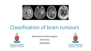
  • 1. Classification of brain tumours Department of Neurosurgery Dr RE Anto 18/03/2022
  • 2. Contents • Introduction • Epidemiology • Risk factors • Etiology • Hereditary sx • Clinical presentation • Classification systems • Benign vs Malignant • Tumour locations • WHO Classification System • Main changes • References
  • 3. Introduction • Brain tumour is an abnormal mass of tissue intracranially, in which cells grow and multiply uncontrollably, seemingly unchecked by the mechanisms that control normal cells. • Multiple classification systems • Primary vs. Secondary • Type of cell origin • Location • Grading (Benign or Malignant)
  • 4. Epidemiology • Primary brain tumours are relatively rare: 2% of all cancers in adults • NOTE: In paeds: 15-20 % of paediatric cancers • Great variance in degree of incidence between 1st world and developing countries • May be due to differences in resources like imaging modalities and better registration and data capture systems • Incidence rises with age • Males: Female (1.5:1) – except meningiomas (female > male)
  • 5. Davis FG, McCarthy BJ. Current epidemiological trends and surveillance issues in brain tumours. Expert Rev Anticancer Ther2001;1:395–401.
  • 6. Risk factors • Develop as a consequence of accumulated genetic alterations that permit cells to evade normal regulatory mechanisms and destruction by the immune system • Seizures may herald development of cerebral tumours by years! • Risk for any cerebral tumour after first admission for epilepsy is increased 20-fold • Highlights the need for continued surveillance of patients with new- onset seizures
  • 7. Etiology • Some epidemiological studies suggest increased incidence of astrocytomas • Petrochemicals eg vinyl chloride • Electromagnetic radiation • High dose ionizing radiation • 10 times risk of meningiomas • 2.5 times risk for gliomas • Primary CNS lymphoma • Viruses eg EBV • Immunocompromised (transplant patient, AIDS)
  • 8. Hereditary syndromes • < 5% of all primary CNS tumours • Neurofibromatosis I and II (MPNST, Optic n glioma, Meningiomas, Vestibular schwannoma) • Von-Hippel Lindau sx (Haemangioblastoma) • Tuberous sclerosis (SEGA, Cortical tubers) • Li-Fraumeni sx (Malignant gliomas, PNET, Medulloblastoma) • MEN I (Multiple endocrine neoplasia) ( Pituitary adenomas, Malignant schwannoma) • Turcot sx (GBM, Medulloblastoma) • Werner’s sx (Meningioma)
  • 9. Clinical Presentation • Generalized or focal signs. • Epilepsy: focal or generalized tonic clonic seizures • Frontal lobe: • Anosmia • Dementia • Urinary incontinence • Conjugate deviation of eyes • Aphasia
  • 10. Clinical Presentation • Temporal lobe: • Temporal lobe epilepsy • Auditory hallucinations • Memory difficulties • Parietal lobe: • Hemisensory changes • Sensory apraxia • Occipital lobe: • Visual field changes • Chiasmatic lesions: • Visual field changes
  • 11. Clinical Presentation • Posterior fossa lesions: • CN Palsies • Long tract signs • Obstructive HCP • Sellar lesions • Hormone dysfunction • Acromegaly • Hyperprolactinemia • Hypopituitarism • Hypocortisolemia • Visual disturbances
  • 12. Classification systems • Primary: • Arise from the brain tissue or surrounding meninges itself • Glial • Non-glial (in structures in the brain including nerves, vessels, glands) • Secondary: • Arises from other tumours in the body and migrate to the brain • 5-10 times more common than primary CNS tumours (most common tumours in adults)
  • 13.
  • 14.
  • 15. Benign vs Malignant • This distinction is less significant than for other systems • Benign • Slow-growing tumours • Low cellularity • Few mitosis • No necrosis • No vascular proliferation • However, location of these tumours can have lethal consequences despite its histological classification • Eg: FM meningioma causing compression of the medulla and causing cardiorespiratoy arrest
  • 16. Malignant • Not malignant “metastasize” out of CNS (rare) • Morphological Features: • 1. Nuclear atypia • 2. Mitosis • 3. Endothelial Proliferation • 4. Necrosis Daumas-Duport C, Scheithauer B, O'Fallon J, Kelly P (1988). Grading of astrocytomas. A simple and reproducible method. Cancer 62: 2152-2165.
  • 17.
  • 18. WHO Classification • 1979: 1st edition – Histological typing of CNS tumours • 1993: 2nd edition – Reflected advances in immunohistochemistry • 2000: 3rd edition – Started introduction pathology and genetics • 2007: 4th edition – used up until 2015 (Youmans 6th edition) • 2016: Revised 4th edition – substantial revision of 4th edition
  • 20. WHO Classifications (2007/2016) • Formulating concept of how CNS tumours diagnoses are structured in the molecular era • Restructured diffuse gliomas, medulloblastomas and incorporated genetically identified entities. • Embryonal tumours (removal of PNET) • Ependymoma genetic variants introduced
  • 21. WHO Classifications (2007/2016) • Addition of newly recognized entities, variants and patterns: • IDH-wildtype and IDH-mutant glioblastoma (entities) • Diffuse midline glioma, H3 K27M–mutant (entity) • Embryonal tumour with multilayered rosettes, C19MC-altered (entity) • Ependymoma, RELA fusion–positive (entity) • Diffuse leptomeningeal glioneuronal tumor (entity) • Anaplastic PXA (entity) • Epithelioid glioblastoma (variant) • Glioblastoma with primitive neuronal component (pattern) • Multinodular and vacuolated pattern of ganglion cell tumor (pattern)
  • 22. WHO Classifications (2007/2016) • Deletion of former entities, variants and terms: • Gliomatosis cerebri • Protoplasmic and fibrillary astrocytoma variants • Cellular ependymoma variant • “Primitive neuroectodermal tumor” • Addition of brain invasion as a criteria for atypical meningioma • Restructuring of solitary fibrous tumour and hemangiopericytoma (SFT/HPC) as one entity and adapting a grading system to accommodate this change • Expansion and clarification of entities included in nerve sheath tumors, with addition of hybrid nerve sheath tumors and separation of melanotic schwannoma from other schwannomas • Expansion of entities included in hematopoietic/lymphoid tumors of the CNS (lymphomas and histiocytic tumors
  • 23.
  • 24. 2021 WHO Classification • 5th edition of WHO Classification of Tumours of the Central Nervous System (CNS) • Sixth version of the international standard for the classification of brain and spinal cord tumours • Build on the 2016 updated fourth edition and the work of the Consortium to Inform Molecular and Practical Approaches to CNS Tumour Taxonomy (cIMPACT-NOW)
  • 25. cIMPACT-NOW • Consortium to Inform Molecular and Practical Approaches to CNS Tumour Taxonomy – Not Officially WHO • Seven updates from 2016-2020
  • 26. Gliomas, glioneuronal tumors, and neuronal tumours • Adult-type diffuse gliomas • Astrocytoma, IDH-mutant • Oligodendroglioma, IDH-mutant, and 1p/19q-codeleted • Glioblastoma, IDH-wildtype • Pediatric-type diffuse low-grade gliomas • Diffuse astrocytoma, MYB- or MYBL1-altered • Angiocentric glioma • Polymorphous low-grade neuroepithelial tumor of the young • Diffuse low-grade glioma, MAPK pathway-altered • Pediatric-type diffuse high-grade gliomas • Diffuse midline glioma, H3 K27-altered • Diffuse hemispheric glioma, H3 G34-mutant • Diffuse pediatric-type high-grade glioma, H3-wildtype and IDH-wildtype • Infant-type hemispheric glioma
  • 27. Gliomas, glioneuronal tumors, and neuronal tumours • Circumscribed astrocytic gliomas • Pilocytic astrocytoma • High-grade astrocytoma with piloid features • Pleomorphic xanthoastrocytoma • Subependymal giant cell astrocytoma • Chordoid glioma • Astroblastoma, MN1-altered
  • 28. Glioneuronal and neuronal tumours • Ganglioglioma • Desmoplastic infantile ganglioglioma (DIG) / desmoplastic infantile astrocytoma • Dysembryoplastic neuroepithelial tumor • Diffuse glioneuronal tumor with oligodendroglioma-like features and nuclear clusters (provisional inclusion) • Papillary glioneuronal tumor • Rosette-forming glioneuronal tumor • Myxoid glioneuronal tumor • Diffuse leptomeningeal glioneuronal tumor • Gangliocytoma • Multinodular and vacuolating neuronal tumor • Dysplastic cerebellar gangliocytoma (lhermitte-duclos disease) • Central neurocytoma • Extraventricular neurocytoma • Cerebellar liponeurocytoma
  • 29. Ependymal tumours • Supratentorial ependymoma • Supratentorial ependymoma, ZFTA fusion-positive • Supratentorial ependymoma, YAP1 fusion-positive • Posterior fossa ependymoma • Posterior fossa ependymoma, Group PFA • Posterior fossa ependymoma, Group PFB • Spinal ependymoma • Spinal ependymoma, MYCN-amplified • Myxopapillary ependymoma • Subependymoma
  • 30. Choroid Plexus tumours • Choroid plexus papilloma • Atypical choroid plexus papilloma • Choroid plexus carcinoma
  • 31. Embryonal tumours - Medulloblastoma • Medulloblastomas, molecularly defined • Medulloblastoma, WNT-activated • Medulloblastoma, SHH-activated and TP53-wildtype • Medulloblastoma, SHH-activated and TP53-mutant • Medulloblastoma, NON-WNT/NON-SHH • Medulloblastomas, histologically defined
  • 32. Other CNS Embryonal tumours • Atypical teratoid/rhabdoid tumor • Cribriform neuroepithelial tumor (provisional inclusion) • Embryonal tumor with multilayered rosettes (ETMR) • CNS neuroblastoma, FOXR2-activated • CNS tumor with BCOR internal tandem duplication • CNS embryonal tumor
  • 33. Pineal tumours • Pineocytoma • Pineal parenchymal tumor of intermediate differentiation • Pineoblastoma • Papillary tumor of the pineal region • Desmoplastic myxoid tumor of the pineal region, smarcb1- mutant
  • 34. Cranial and Paraspinal nerve tumours • Schwannoma • Neurofibroma • Perineurioma • Hybrid nerve sheath tumor • Malignant melanotic nerve sheath tumor • Malignant peripheral nerve sheath tumor • Paraganglioma
  • 35. Meningioma • Meningothelial • Fibrous • Transitional • Psammomatous • Angiomatous • Microcystic • Secretory • Lymphoplasmacyte-rich • Metaplastic • Chordoid • Clear Cell • Atypical • Papillary • Rhabdoid • Anaplastic
  • 36. Mesenchymal, non-meningothelial tumours – Soft tissue tumours • Fibroblastic and myofibroblastic tumors • Solitary fibrous tumor • Vascular tumors • Hemangiomas and vascular malformations • Hemangioblastoma • Skeletal muscle tumors • Rhabdomyosarcoma • Uncertain differentiation • Intracranial mesenchymal tumor, FET-CREB fusion-positive (provisional inclusion) • CIC-rearranged sarcoma • Primary intracranial sarcoma, dicer1-mutant • Ewing sarcoma
  • 37. Chondro-osseous tumours • Chondrogenic tumors • Mesenchymal chondrosarcoma • Chondrosarcoma • Notochordal tumors • Chordoma (including poorly differentiated chordoma)
  • 38. Melanocytic tumours • Diffuse meningeal melanocytic neoplasms • Meningeal melanocytosis • Meningeal melanomatosis • Circumscribed meningeal melanocytic neoplasms • Meningeal melanocytoma • Meningeal melanoma
  • 39. Hematolymphoid tumours - Lymphomas • CNS lymphomas • Primary diffuse large b-cell lymphoma of the CNS • Immunodeficiency-associated CNS lymphoma • Lymphomatoid granulomatosis • Intravascular large b-cell lymphoma • Miscellaneous rare lymphomas in the CNS • MALT lymphoma of the dura • Other low-grade b-cell lymphomas of the CNS • Anaplastic large cell lymphoma (ALK+/ALK−) • T-cell and NK/t-cell lymphomas
  • 40. Germ cell tumors • Mature teratoma • Immature teratoma • Teratoma with somatic-type malignancy • Germinoma • Embryonal carcinoma • Yolk sac tumor • Choriocarcinoma • Mixed germ cell tumor
  • 41. Tumors of the sellar region • Adamantinomatous craniopharyngioma • Papillary craniopharyngioma • Pituicytoma, granular cell tumor of the sellar region, and spindle cell oncocytoma • Pituitary adenoma (pitNET) • Pituitary blastoma
  • 42. Metastases to the CNS • Metastases to the brain and spinal cord parenchyma • Metastases to the meninges
  • 43. Main changes in 5th edition • Builds on prior version by placing greater emphasis on molecular markers in terms of classification and grading • Heterogenous classification • Layered report structure • Integrated diagnosis • Histopathological classification • CNS WHO Grade • Molecular information
  • 44. Grading • Within tumour types • Unlike other WHO classification systems that graded each tumor based on its own features (i.e. the most low-grade version of a particular tumor was given grade 1) • This approach has been abandoned, in favour of grading tumours within “type” • Example: • No Grade 1 Diffuse Astrocytoma, IDH Mutant (only 2,3 or 4) • Glioblastoma, IDH-wildtype – can only be Grade 4 • Arabic numerals • Previously – I,II,III,IV (Roman numerals) • Replaced by Arabic Numerals – 1,2,3,4 • Example: Meningioma, CNS WHO Grade 1
  • 45. Grading • Anaplastic modifier • The term anaplastic, used extensively in the prior classifications has been dropped in favour of grading only. • Thus what was previously known as an "anaplastic astrocytoma" is now referred to as an "astrocytoma, IDH-mutant, CNS WHO grade 3 • Molecular grading • For the first time, molecular features have been explicitly added to the grading schema • May supersede histological features. • Example: an IDH-wildtype astrocytoma with low-grade histologic features can be considered Grade 4 (Glioblastoma) in the presence of EGFR amplification, TERT promoter mutation or the combined gain of chromosome 7 and loss of chromosome 10
  • 46. Other changes • NOS (Not Otherwise Specified) • Diagnostic information (histological or molecular) needed to assign a specific WHO diagnosis is not available • Example: not enough tissue available or no resources available • NEC (Not Elsewhere Classified) • Results do not readily allow for a WHO diagnosis • Clinical, histological, or molecular mismatch
  • 47. References • Louis D, Perry A, Wesseling P et al. The 2021 WHO Classification of Tumors of the Central Nervous System: A Summary. Neuro-Oncology. 2021;23(8):1231-51. doi:10.1093/neuonc/noab106 • Louis D, Perry A, Reifenberger G et al. The 2016 World Health Organization Classification of Tumors of the Central Nervous System: A Summary. Acta Neuropathol. 2016;131(6):803-20. doi:10.1007/s00401-016-1545-1 – Pubmed • International Agency for Research on Cancer, Otmar D. Wiestler. WHO Classification of Tumours of the Central Nervous System. (2016) ISBN: 9789283244929 • GreenBerg 9th edition • Davis FG, McCarthy BJ. Current epidemiological trends and surveillance issues in brain tumours. Expert Rev Anticancer Ther2001;1:395–401.